

Loading, please wait...
Loading, please wait...

Urologists often encounter giant vesical calculi in patients presenting with long-standing lower urinary tract symptoms, macroscopic hematuria, and chronic pelvic discomfort. However, clinicians rarely anticipate an underlying sarcomatoid urothelial carcinoma masked by extensive calculous disease. Massive calculi cause severe mechanical irritation that induces diffuse urothelial inflammation, reactive mucosal edema, and substantial detrusor hypertrophy. Consequently, clinicians frequently attribute bilateral hydronephrosis and profound bladder wall thickening solely to chronic outflow obstruction or longstanding lithiasis. This cognitive diagnostic trap can cause substantial surgical delay. Patients commonly present in late stages because mechanical stone symptoms overshadow ominous neoplastic warning signs. Furthermore, chronic stone formation often distracts the initial clinical evaluation away from malignant transformation. When surgeons perform open cystolithotomy, they must maintain immense suspicion. In particular, they should inspect any abnormal mucosal friability, irregular mural induration, or atypical exophytic tissue. Timely tissue retrieval prevents missed diagnoses in patients presenting with seemingly benign calculus complications. Therefore, every clinician treating complex lithiasis must thoroughly evaluate persistent unexplained hematuria.
Pathologists recognize sarcomatoid urothelial carcinoma as a scarce, biologically aggressive malignancy. It features a biphasic morphology combining malignant epithelial and spindle-cell elements. Molecular evidence demonstrates that these divergent phenotypic components arise from a monoclonal stem cell population through complete epithelial-mesenchymal transition. During this dedifferentiation cascade, neoplastic epithelial cells downregulate essential junctional proteins like E-cadherin. Concurrently, these cells vigorously upregulate mesenchymal drivers, particularly vimentin, SNAIL, and ZEB1. Consequently, tumor cells acquire an elongated spindle morphology, extraordinary invasive velocity, and high resistance to conventional chemotherapeutic regimens. Chronic physical trauma from a giant vesical stone promotes sustained microenvironmental cytokine release. In addition, chronic local inflammation creates an ideal stromal niche that accelerates genomic instability and malignant dedifferentiation. The resulting neoplasm exhibits profound cytological pleomorphism, abundant atypical mitotic figures, and destructive transmural invasion. Furthermore, sarcomatoid variants possess an inherent biological propensity for rapid lymphatic dissemination and vascular invasion compared to conventional urothelial neoplasms. Because this histological transformation generates an aggressive clinical course, understanding epithelial-mesenchymal transition helps clinicians grasp the rapid progression seen in affected individuals.
Preoperative diagnostic imaging poses formidable challenges when giant calculi coexist with bladder wall neoplasms. Computed tomography routinely demonstrates marked, irregular mural thickening surrounding large intravesical stones. However, radiologists frequently interpret these structural abnormalities as benign reactive hyperplasia or chronic cystitis secondary to intense decubitus irritation. Acoustic shadowing and beam-hardening artifacts from dense calcified masses further obscure direct visualization of mucosal lesions. Additionally, bilateral hydroureteronephrosis often results from mechanical obstruction at the ureterovesical junction rather than direct tumor infiltration. Therefore, cross-sectional imaging cannot reliably differentiate simple chronic inflammation from invasive carcinoma in the vicinity of a giant stone. Clinicians must actively consider advanced multiparametric magnetic resonance imaging using dynamic contrast enhancement and diffusion-weighted sequences. These specialized sequences help identify focal thickening that appears disproportionate to stone contact. Moreover, asymmetric extravesical soft tissue extension should immediately raise clinical suspicion for malignant infiltration. Relying exclusively on routine computed tomography descriptions can delay life-saving oncological intervention. Consequently, surgical teams should establish standardized protocols ensuring thorough cystoscopic inspection whenever large stones obscure underlying detrusor architecture.
Histopathological evaluation forms the cornerstone of accurate diagnosis. However, identifying sarcomatoid urothelial carcinoma presents immense diagnostic dilemmas. The predominant mesenchymal spindle-cell pattern frequently mimics true mesenchymal sarcomas, inflammatory myofibroblastic tumors, or benign pseudosarcomatous stromal proliferations. Therefore, pathologists must utilize an extensive immunohistochemical panel to establish correct lineage attribution. Pathologists primarily confirm epithelial differentiation within the atypical spindle cells by demonstrating immunoreactivity for cytokeratin cocktails, high-molecular-weight cytokeratin, and epithelial membrane antigen. Furthermore, modern diagnostic algorithms employ GATA3 and p63 to confirm urothelial lineage. Nevertheless, dedifferentiated spindle cells can lose these urothelial markers during epithelial-mesenchymal transition. In contrast, pure sarcomas lack consistent epithelial marker expression and display specific lineage signatures like desmin or myogenin. Pathologists must also examine deep detrusor muscle to assess invasive depth accurately, because superficial biopsies frequently miss high-grade sarcomatoid components. Inadequate muscularis propria sampling severely hampers clinical staging and therapeutic decision-making. Thus, diligent immunohistochemical validation protects oncologists against misdiagnosing primary vesical sarcomas, which require completely different systemic protocols.
Managing high-grade sarcomatoid carcinoma demands an aggressive, coordinated multidisciplinary strategy combining radical surgery and systemic therapy. Because these tumors carry a dismal prognosis, clinical guidelines advocate prompt radical cystectomy with bilateral pelvic lymphadenectomy. Early surgical extirpation remains crucial for resectable muscle-invasive disease. Transurethral resection alone serves purely as a diagnostic tool and fails to provide durable oncological control. In addition, medical oncologists frequently recommend neoadjuvant cisplatin-based chemotherapy to eradicate micrometastatic disease prior to definitive surgery. However, sarcomatoid variants often exhibit significant chemoresistance, necessitating close radiographic surveillance during neoadjuvant cycles. When patients present with severe medical comorbidities or obstructive renal failure, salvage systemic options become crucial. Emerging genomic data reveal that sarcomatoid tumors frequently harbor basal molecular subtypes with heightened PD-L1 expression. Consequently, immune checkpoint inhibitors provide valuable therapeutic alternatives for refractory or unresectable cases. Nevertheless, median overall survival remains poor, rarely exceeding one year in advanced presentations. Multidisciplinary tumor boards must therefore balance aggressive intervention against patient performance status. Teams must prioritize timely surgical intervention before systemic deterioration precludes definitive treatment.
Pathologists distinguish sarcomatoid urothelial carcinoma from primary sarcomas by demonstrating epithelial lineage markers within the atypical spindle-cell population. While true sarcomas remain consistently negative for epithelial antigens, sarcomatoid carcinomas express pancytokeratin, cytokeratin cocktails, and epithelial membrane antigen. Furthermore, diagnostic panels evaluate p63 and GATA3 to corroborate urothelial histogenesis. Positivity for these markers confirms an epithelial origin undergoing mesenchymal transition, thereby excluding primary mesenchymal sarcomas and guiding appropriate oncological staging and definitive therapy.
Preoperative cross-sectional imaging frequently misinterprets malignant neoplasms because massive calculi generate pronounced secondary inflammatory changes within the bladder wall. Computed tomography scans often demonstrate symmetric or irregular detrusor thickening, which radiologists readily attribute to mechanical decubitus trauma or severe chronic cystitis. In addition, intense acoustic shadowing and dense calcification artifacts severely obscure underlying soft-tissue masses. Consequently, clinicians fail to detect invasive malignancies until direct intraoperative visualization reveals suspicious tissue beneath the stone bed.
The standard therapeutic paradigm for resectable muscle-invasive disease centers on prompt radical cystectomy combined with bilateral pelvic lymphadenectomy. Because sarcomatoid tumors demonstrate aggressive local infiltration and early metastasis, multidisciplinary guidelines recommend neoadjuvant cisplatin-based chemotherapy whenever renal function permits. However, if disease progression occurs or chemoresistance develops, clinicians consider immune checkpoint inhibitors targeting programmed death-ligand 1 pathways. Overall, timely surgical extirpation remains the only curative modality, making early histopathological confirmation and rapid intervention crucial for survival.
Disclaimer: This content is for informational and educational purposes only and should not be construed as medical advice, diagnosis, or treatment. It is intended to support, not replace, the relationship between patients and their healthcare providers. Healthcare professionals must exercise their independent clinical judgment when evaluating and treating patients. While we strive to provide accurate and up-to-date medical information, medical knowledge is constantly evolving, and individual patient circumstances may vary. Refer to the latest local and national guidelines for clinical practice.
References

Read summarized clinical updates, watch expert medical content, and earn CME certifications right from your smartphone.
Open your smartphone camera to scan and install.





A case report highlights a giant vesical calculus concealing high-grade sarcomatoid urothelial carcinoma, emphasizing diagnostic pitfalls on imaging, the critical role of immunohistochemistry, and aggressive multidisciplinary management.
Today
Nonadherence to immunosuppressive therapy after lung transplantation threatens graft and patient survival. This review highlights evidence-based digital, behavioral, and therapy-related interventions to enhance adherence and emphasizes the need for tailored, multifactorial management strategies.
Today

A landmark study reveals that physiologic subtypes of gestational glucose intolerance, including insulin resistance and insulin deficiency, independently raise long-term risks of maternal prediabetes and diabetes, underscoring the urgent need for structured postpartum surveillance.
Today

A systematic review and component network meta-analysis evaluated the incremental benefits of drug classes in HFmrEF and HFpEF. SGLT-2 inhibitors and MRAs provided the most robust reductions in heart failure hospitalizations, while mortality benefits remained elusive across all investigated drug classes.
Today
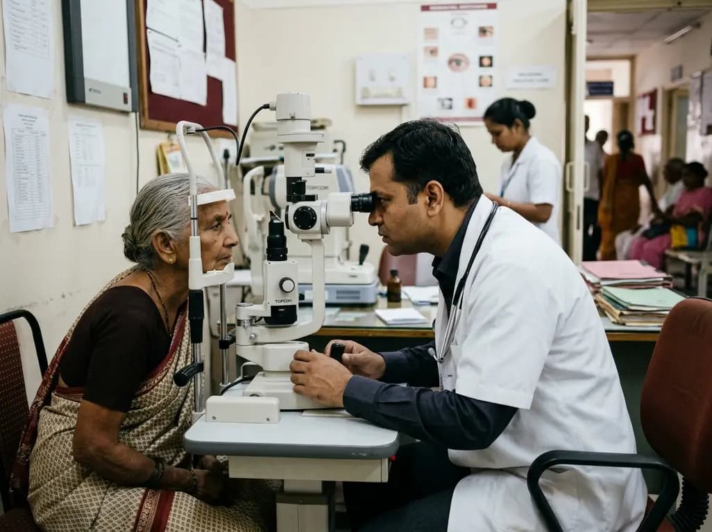
A meta-analysis of 19 studies reveals that 69% of ophthalmologists suffer from work-related musculoskeletal disorders. Slit lamps, microscopes, and high surgical loads cause severe cervical and lumbar strain, underscoring the urgent need for ergonomic workstations, micro-breaks, and institutional reforms.
Today

Mayo Clinic trials and real-world datasets show GLP-1 receptor agonists improve left ventricular remodeling, reduce intracardiac filling pressures, decrease hospitalizations by 34%, and lower geriatric fall risks in patients with obesity and heart failure, delivering notable clinical and economic benefits.
Today
