

Loading, please wait...
Loading, please wait...

Ophthalmology offers remarkable clinical rewards, yet the physical toll of daily practice poses serious occupational challenges. Recent global evidence reveals an alarming prevalence of musculoskeletal disorders among ophthalmologists, solidifying this problem as an urgent specialty-wide concern. A comprehensive systematic review and meta-analysis examining nineteen international studies with 6,770 participants established a pooled prevalence of 69%. Consequently, nearly seven out of ten eye care specialists suffer from recurring physical pain throughout their careers. Furthermore, this epidemiological burden equals or surpasses rates documented in orthopedic surgery and interventional cardiology. However, formal training in ergonomics remains remarkably absent from medical school and residency curricula worldwide. In high-volume clinical settings, such as tertiary outpatient departments across India, doctors examine dozens of patients daily without scheduled physical breaks. In fact, repetitive slit lamp screenings and indirect ophthalmoscopy force practitioners into static postures for prolonged durations. As a result, continuous mechanical load triggers early tissue fatigue and chronic inflammation. Therefore, recognizing these epidemiological facts is crucial for promoting systemic workplace reforms and protecting clinicians.
Ophthalmic practice presents unique biomechanical stressors that stem directly from specialized examination and operative equipment. For instance, routine slit lamp biomicroscopy obliges the doctor to maintain an unnatural forward tilt. Specifically, clinicians sustain cervical spine flexion exceeding twenty degrees while hunching awkwardly toward the oculars during consultations. Additionally, indirect ophthalmoscopy requires bearing heavy head-mounted light units while contorting the cervical and thoracic spine into extreme angles to examine the peripheral retina. During microscopic intraocular surgery, practitioners remain seated in fixed positions for extensive intervals. Furthermore, foot-pedal activation for phacoemulsification or vitrectomy induces asymmetric pelvic tilt, transferring harmful rotational torque to the lumbar spine. Unfortunately, standard examination rooms rarely possess motorized height-adjustable desks or contoured seating with proper lumbar support. When clinicians continually conform their bodies to static instruments rather than configuring tools to suit individual biomechanics, tissue breakdown accelerates. Consequently, repetitive strain injury develops into an unavoidable occupational hazard without dedicated workstation adjustments.
Surveys assessing eye specialists reveal distinctive anatomical patterns of structural injury. In particular, the cervical spine serves as the primary focal point of pain, with more than sixty percent of ophthalmologists reporting persistent, chronic neck soreness. Additionally, low back pain affects nearly half of surveyed practitioners, representing another dominant clinical manifestation. Shoulder discomfort and scapular pain also occur with alarming frequency among surgeons who operate extensively. Because intraocular surgery requires extreme hand stability, practitioners frequently suspend their forearms without adequate armrests. As a result, static contraction overworks the trapezius, levator scapulae, and rotator cuff tendon insertions. Similarly, repetitive finger pinch grips while manipulating micro-forceps or handheld diagnostic lenses promote forearm tendinitis and carpal tunnel syndrome. Over years of uncorrected biomechanical stress, these initial muscular strains evolve into irreversible structural issues, such as cervical spondylosis and degenerative disc herniation. Therefore, clinicians must recognize these localized somatic warning signs before minor aches progress toward severe neurological deficits.
The ramifications of persistent musculoskeletal dysfunction reach far beyond temporary daily soreness. Specifically, severe pain directly threatens surgical precision, clinical productivity, and overall career longevity. Published studies demonstrate that up to forty-four percent of symptomatic eye surgeons actively modify their clinical volume or curtail surgical sessions due to chronic discomfort. Moreover, more than ten percent of practitioners contemplate early retirement or experience permanent work disability attributable to unaddressed spine disease. In addition, chronic somatic pain significantly fuels physician burnout, anxiety, and diminished job satisfaction. When surgeons contend with unceasing neck stiffness or sciatica, their cognitive bandwidth and procedural stamina decline. In fast-paced healthcare environments with towering patient volumes, losing an experienced ophthalmic surgeon compromises patient access to essential sight-saving surgeries. Unfortunately, many practitioners downplay their physical distress, resorting to self-medication with anti-inflammatory analgesics instead of instituting root-cause ergonomic corrections. Consequently, this persistent neglect exacerbates tissue injury and compromises long-term clinical excellence.
Reversing this occupational epidemic requires comprehensive workstation redesign and active physical conditioning. First, healthcare facilities must install motorized examination tables and fully adjustable operator stools that provide dynamic lumbar support. Clinicians must adjust their seating so that feet rest flat on the floor, hips bend at ninety degrees, and oculars meet the eyes naturally. Furthermore, surgical suites should adopt tiltable binocular microscope heads, which allow operators to maintain a neutral vertical cervical posture. Recently, innovative three-dimensional digital visualization systems have gained widespread traction in retinal and anterior segment procedures. By projecting high-definition stereoscopic imagery onto large displays, heads-up visualization permits surgeons to operate while sitting straight without bending their necks. In addition, surgeons must introduce structured micro-breaks into daily practice routines. Pausing for thirty seconds between heavy surgical cases to perform scapular retractions and gentle spinal extensions effectively resets muscle spindle tension. Therefore, pairing technological innovation with diligent postural hygiene safeguards the surgeon's physical health throughout decades of surgical practice.
Ophthalmologists face extreme physical vulnerability because clinical examinations and delicate microsurgeries demand static, awkward postures for hours every day. Equipment such as slit lamps and surgical microscopes forces prolonged forward neck flexion, asymmetric shoulder elevation, and restricted pelvic alignment. Furthermore, surgeons perform repetitive, high-precision manual tasks under substantial cognitive stress. Without structured micro-breaks and formal ergonomic training, this chronic mechanical strain accumulates rapidly, causing progressive soft tissue degeneration and debilitating axial pain.
Eye clinics can significantly minimize spinal stress by investing in independently adjustable, motorized tables and clinician chairs. Clinicians must adjust the slit lamp oculars directly to their eye level while sitting back with firm lumbar support, rather than leaning forward across the table. In addition, practitioners should position the patient's forehead firmly against the rest by adjusting patient chair height, which eliminates unnecessary doctor bending and prevents chronic cervical ligament fatigue.
Eye surgeons benefit most from targeted mobility exercises that counteract forward head carriage and protracted shoulders. Key exercises include chin tucks for cervical retraction, scapular pinches to strengthen the middle trapezius, and pectoral doorway stretches to open tight anterior chest muscles. Additionally, performing thoracic extensions, gentle hamstring stretches, and standing lumbar extensions between operating cases relieves intradiscal pressure. Engaging regularly in core stabilization exercises like planks further bolsters spinal resilience against long surgical strain.
Disclaimer: This content is for informational and educational purposes only... Refer to the latest local and national guidelines for clinical practice.
References

Read summarized clinical updates, watch expert medical content, and earn CME certifications right from your smartphone.
Open your smartphone camera to scan and install.





A meta-analysis of 19 studies reveals that 69% of ophthalmologists suffer from work-related musculoskeletal disorders. Slit lamps, microscopes, and high surgical loads cause severe cervical and lumbar strain, underscoring the urgent need for ergonomic workstations, micro-breaks, and institutional reforms.
Today
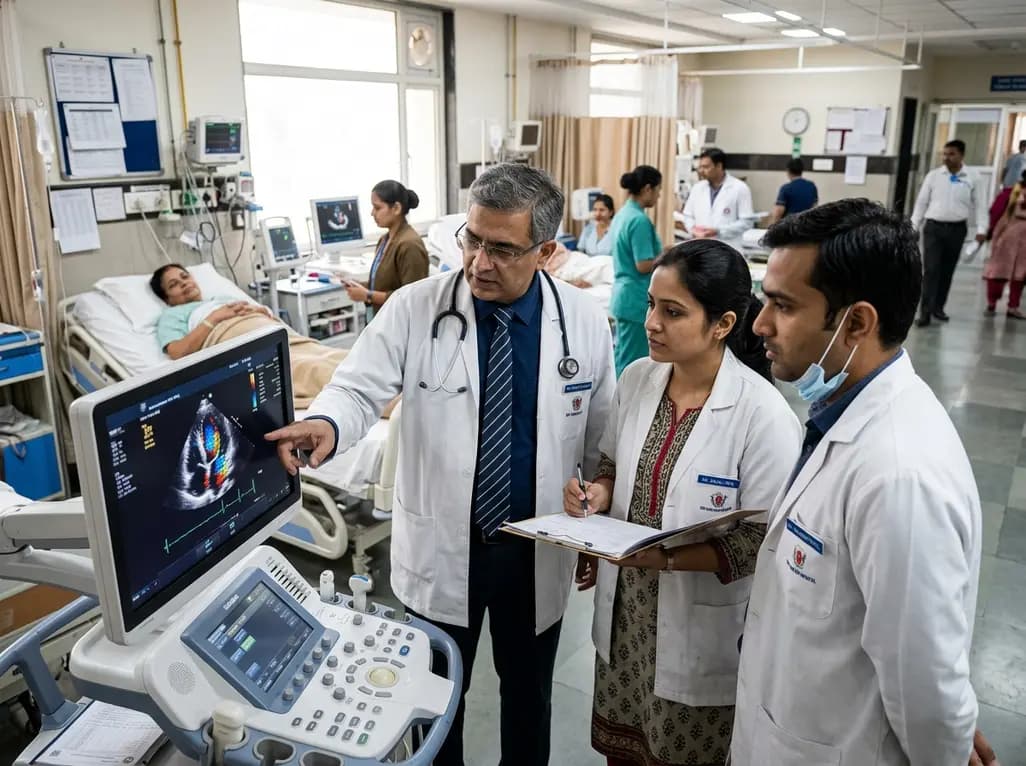
Mayo Clinic trials and real-world datasets show GLP-1 receptor agonists improve left ventricular remodeling, reduce intracardiac filling pressures, decrease hospitalizations by 34%, and lower geriatric fall risks in patients with obesity and heart failure, delivering notable clinical and economic benefits.
Today
Groundbreaking experimental research demonstrates that CX3CR1 deficiency significantly attenuates endometriotic lesion growth by suppressing pro-repair M2 macrophage polarization. This discovery identifies the CX3CL1-CX3CR1 chemokine axis as a promising non-hormonal target for managing chronic endometriosis.
Today
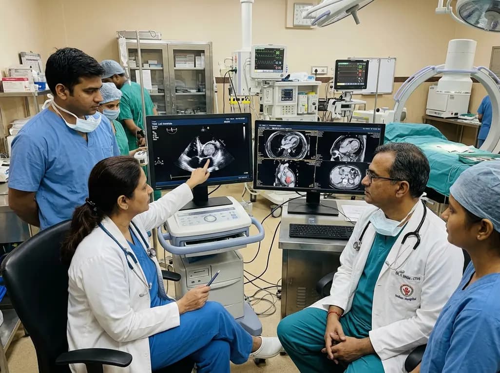
A rare case of an asymptomatic left ventricular hemangioma highlights the vital role of multimodal imaging in differentiating vascular tumors from thrombus. Complete surgical resection under transesophageal echocardiography guidance prevents thromboembolism and delivers optimal outcomes.
Today

An analysis of 1,000 posts by macro- and mega-influencers on Instagram and TikTok revealed that 78.5% contained nutrition misinformation, while only 24% of creators held valid credentials. This article examines the clinical risks of viral dietary fads and provides actionable guidance for healthcare professionals.
Today
