

Loading, please wait...
Loading, please wait...
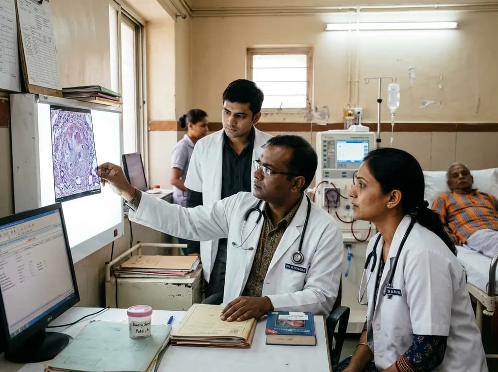
Granulomatous interstitial nephritis represents an uncommon yet clinically critical histological entity in nephrology practice. Clinicians often encounter severe diagnostic dilemmas when systemic disorders masquerade as chronic endemic infections. Sarcoidosis characteristically manifests with bilateral hilar lymphadenopathy and pulmonary parenchymal infiltrates. However, renal involvement remains relatively infrequent, affecting fewer than five percent of all documented cases. When the disease infiltrates renal tissue, granulomatous interstitial nephritis frequently develops and can precipitate dialysis-dependent acute kidney injury. In countries with high tuberculosis burdens, physicians maintain a naturally high index of suspicion for mycobacteria. Consequently, significant radiological and clinical overlaps between sarcoidosis and tuberculosis create frequent diagnostic traps. Patients presenting with constitutional symptoms and thoracic lymph node enlargement often receive empirical anti-tuberculosis treatment. Nevertheless, this empirical approach carries major clinical risks when microbiological tests return negative. Misattributing sterile granulomas to culture-negative tuberculosis delays effective immunosuppressive management. As a result, persistent parenchymal inflammation induces irreversible tubular atrophy and chronic nephron loss. Physicians must therefore pursue comprehensive multiorgan investigations to establish an accurate diagnosis early.
Renal sarcoidosis exhibits diverse clinical presentations ranging from asymptomatic sterile pyuria to life-threatening renal failure. Most frequently, abnormal calcium metabolism causes nephrocalcinosis, nephrolithiasis, and prerenal azotemia. Activated macrophages within sarcoid granulomas independently synthesize 1-alpha-hydroxylase without normal negative feedback controls. Consequently, elevated calcitriol levels accelerate intestinal calcium absorption and induce marked hypercalcemia. This hypercalcemic state causes intrarenal vasoconstriction, reduces glomerular filtration, and impairs tubular concentrating ability. In contrast, direct parenchymal infiltration causing granulomatous interstitial nephritis occurs less commonly. When severe interstitial inflammation develops, patients experience rapidly worsening acute kidney injury that may necessitate emergent hemodialysis. Patients also frequently report systemic symptoms, including non-specific fatigue, unexplained weight loss, and anterior uveitis. Laboratory workups characteristically reveal hypercalcemia alongside suppressed parathyroid hormone and elevated serum angiotensin-converting enzyme. Interestingly, some elderly patients present with isolated renal failure without prominent respiratory complaints. Because these multisystem findings closely resemble disseminated mycobacterial disease, clinicians must evaluate organ involvement systematically. Thus, identifying this metabolic and clinical pattern prevents dangerous diagnostic delays.
Differentiating sarcoidosis from tuberculosis presents formidable challenges in clinical practice. Both entities produce non-caseating or caseating granulomas, mediastinal adenopathy, and constitutional symptoms. When pulmonary tuberculosis is paucibacillary, sputum smears, mycobacterial cultures, and molecular assays often remain entirely negative. Under these circumstances, physicians frequently diagnose culture-negative tuberculosis based on clinical suspicion alone. However, empirical anti-tuberculosis therapy can create misleading impressions of therapeutic efficacy. For example, sarcoidosis lesions can fluctuate spontaneously or improve temporarily during incidental corticosteroid exposure. Clinicians may mistakenly attribute this transient improvement to anti-mycobacterial drugs rather than natural disease fluctuation. Furthermore, continuing anti-tuberculosis therapy fails to halt ongoing autoimmune renal inflammation. In contrast, administering systemic corticosteroids during active, untreated tuberculosis risks catastrophic mycobacterial dissemination. Clinicians must therefore interpret negative sputum tests and negative GeneXpert results with caution. When a patient demonstrates poor microbiological proof and atypical disease trajectories, doctors should actively question the presumptive tuberculosis diagnosis. Re-evaluating the working hypothesis protects patients from prolonged ineffective regimens.
Percutaneous renal biopsy provides the definitive diagnostic method when evaluating unexplained granulomatous kidney injury. Histopathological examination characteristically demonstrates well-demarcated, non-caseating epithelioid cell granulomas within the renal interstitium. These granulomas contain multinucleated Langerhans-type giant cells bordered by lymphocytes and mature plasma cells. In addition, pathologists systematically employ special histochemical stains to exclude infectious microorganisms. Ziehl-Neelsen and auramine-rhodamine staining assess for acid-fast mycobacteria, while silver staining excludes fungal pathogens. Although caseating necrosis strongly implies tuberculosis, mycobacterial interstitial nephritis can occasionally lack overt necrosis. Therefore, performing tissue polymerase chain reaction for mycobacterial DNA provides vital diagnostic reassurance. Pathologists also examine the tissue under polarized light to exclude foreign body reactions. Importantly, a renal biopsy accurately determines the chronicity index by quantifying interstitial fibrosis and tubular atrophy. This histological grading helps clinicians predict the likelihood of functional renal recovery following therapy. Ultimately, early biopsy confirmation empowers nephrologists to initiate targeted immunosuppression safely and confidently.
Systemic glucocorticoids represent the undisputed cornerstone of treatment for renal sarcoidosis and granulomatous interstitial nephritis. For dialysis-dependent acute kidney injury, clinicians usually prescribe intravenous methylprednisolone pulse therapy for three consecutive days. Subsequently, patients transition to oral prednisolone at a dose of one milligram per kilogram daily. Furthermore, glucocorticoids rapidly suppress macrophage 1-alpha-hydroxylase activity, which normalizes serum calcium and reduces angiotensin-converting enzyme. Concurrently, steroid therapy clears dense interstitial inflammatory infiltrates and halts progressive tubulitis. Clinical evidence shows that timely steroid administration facilitates substantial renal recovery, permitting dialysis discontinuation in most patients. Therefore, physicians typically maintain high-dose oral corticosteroids for one month before initiating a slow, structured taper. This gradual reduction should extend over six to twelve months to prevent disease relapse. In patients with steroid resistance or severe toxicity, clinicians introduce steroid-sparing immunosuppressants such as azathioprine or mycophenolate mofetil. Additionally, biological therapies like infliximab offer valuable rescue options in refractory disease. Routine monitoring of renal parameters ensures durable clinical stability and protects baseline kidney health.
Granulomatous interstitial nephritis develops when activated CD4-positive T-helper cells and macrophages infiltrate the renal parenchyma. These immune cells release pro-inflammatory cytokines, including interferon-gamma and tumor necrosis factor-alpha, triggering organized non-caseating granuloma formation within the tubulointerstitium. Concurrently, dysregulated calcium metabolism from extra-renal calcitriol production leads to hypercalcemia and tubular injury. Left untreated, severe cellular infiltration and ongoing tubulitis cause rapid nephron loss, culminating in acute kidney injury that may necessitate acute dialysis support.
Sarcoidosis and tuberculosis share remarkable clinical, biochemical, and radiological similarities, particularly in regions where mycobacterial infections remain endemic. Both conditions frequently produce bilateral hilar lymphadenopathy, pulmonary infiltrates, fatigue, and systemic granulomatous inflammation. When pulmonary tuberculosis is paucibacillary, microbiological tests frequently yield negative results, leading clinicians to diagnose culture-negative tuberculosis empirically. Consequently, patients receive prolonged anti-tuberculosis therapy while the true underlying autoimmune disorder progresses unrecognized, ultimately causing severe organ complications like granulomatous nephritis.
Therapy centers on prompt, high-dose systemic corticosteroids to arrest parenchymal inflammation and reverse tubular dysfunction. Physicians frequently initiate intravenous pulse methylprednisolone for three consecutive days, followed by oral prednisolone at one milligram per kilogram daily. This regimen rapidly controls 1-alpha-hydroxylase activity, corrects hypercalcemia, and restores glomerular filtration. Following clinical stabilization, clinicians taper the steroid dose gradually over six to twelve months, occasionally adding steroid-sparing immunosuppressants like azathioprine to maintain remission and prevent recurrent nephrotoxicity.
Disclaimer: This content is for informational and educational purposes only... Refer to the latest local and national guidelines for clinical practice.
References
Ding N et al. Sarcoidosis Initially Misdiagnosed as Culture-Negative Pulmonary Tuberculosis Presenting as Dialysis-Requiring Acute Kidney Injury: A Case Report. J Nephrol. 2026 Nov 21. doi: undefined. PMID: 43065300.
Ramachandran R, Kumar V, Kumar A, et al. Etiological diagnosis of granulomatous tubulointerstitial nephritis in the tropics. Clin Kidney J. 2015;8(6):710-715.
Mahévas M, Lescure FX, Boffa JJ, et al. Renal sarcoidosis: clinical, laboratory, and histologic presentation and outcome in 47 patients. Medicine (Baltimore). 2004;83(5):277-283.

Read summarized clinical updates, watch expert medical content, and earn CME certifications right from your smartphone.
Open your smartphone camera to scan and install.





Sarcoidosis presenting as dialysis-requiring acute kidney injury from granulomatous interstitial nephritis is often misdiagnosed as culture-negative tuberculosis. Accurate renal biopsy, calcium assessment, and corticosteroid therapy are essential to ensure renal recovery and prevent permanent parenchymal damage.
Today

Nonadherence to immunosuppressive therapy after lung transplantation threatens graft and patient survival. This review highlights evidence-based digital, behavioral, and therapy-related interventions to enhance adherence and emphasizes the need for tailored, multifactorial management strategies.
Today

A landmark study reveals that physiologic subtypes of gestational glucose intolerance, including insulin resistance and insulin deficiency, independently raise long-term risks of maternal prediabetes and diabetes, underscoring the urgent need for structured postpartum surveillance.
Today

A systematic review and component network meta-analysis evaluated the incremental benefits of drug classes in HFmrEF and HFpEF. SGLT-2 inhibitors and MRAs provided the most robust reductions in heart failure hospitalizations, while mortality benefits remained elusive across all investigated drug classes.
Today
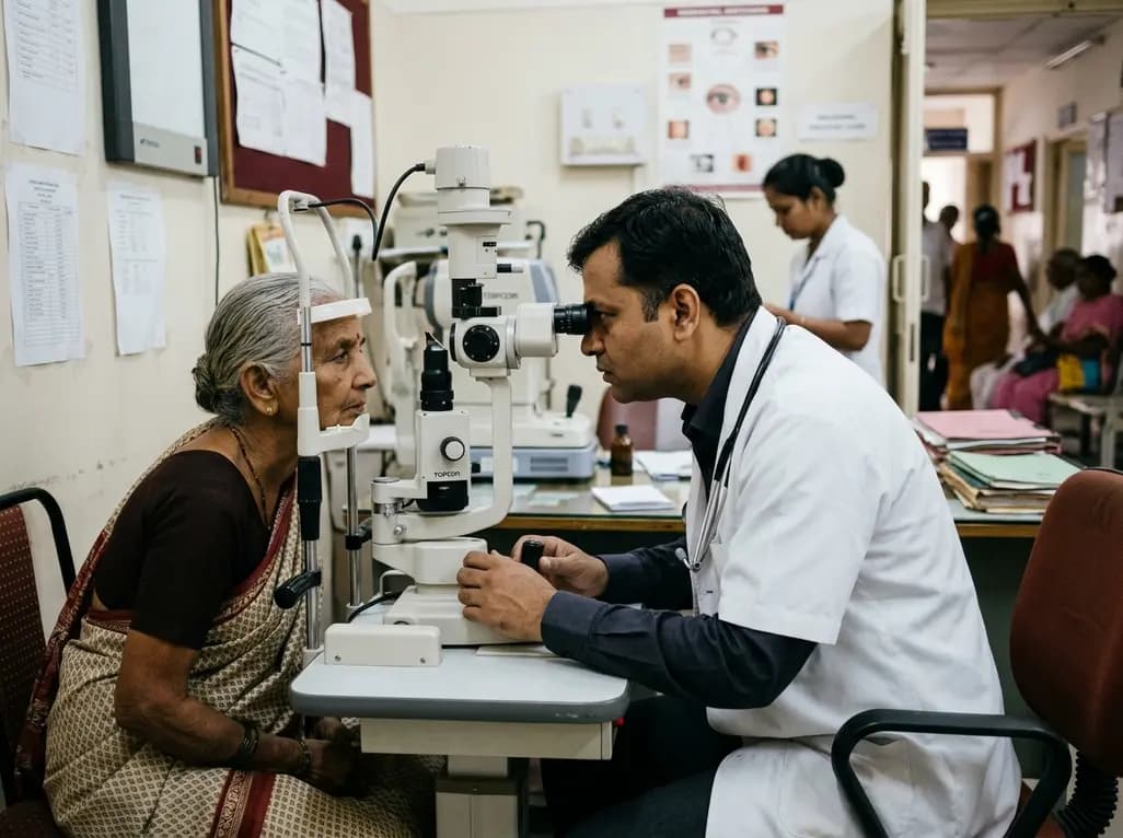
A meta-analysis of 19 studies reveals that 69% of ophthalmologists suffer from work-related musculoskeletal disorders. Slit lamps, microscopes, and high surgical loads cause severe cervical and lumbar strain, underscoring the urgent need for ergonomic workstations, micro-breaks, and institutional reforms.
Today
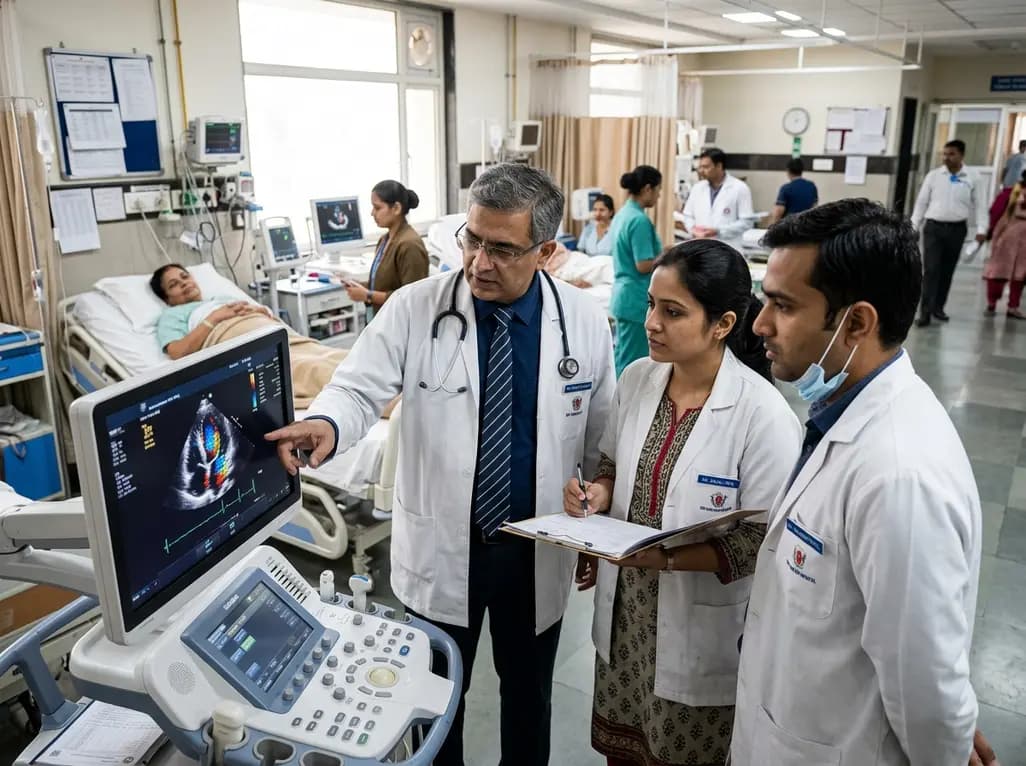
Mayo Clinic trials and real-world datasets show GLP-1 receptor agonists improve left ventricular remodeling, reduce intracardiac filling pressures, decrease hospitalizations by 34%, and lower geriatric fall risks in patients with obesity and heart failure, delivering notable clinical and economic benefits.
Today
