.webp&w=384&q=75)

Loading, please wait...
Loading, please wait...
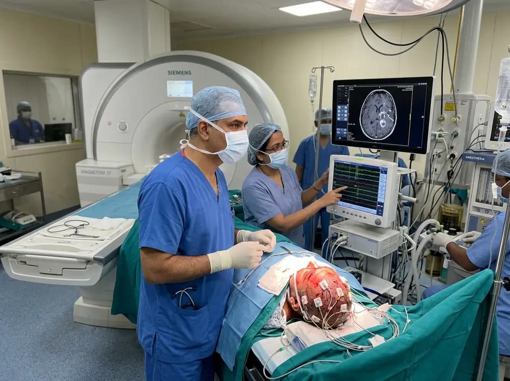
Maximal safe resection forms the cornerstone of modern neurosurgical oncology, particularly for infiltrative high-grade gliomas and glioblastomas. Achieving complete tumor removal while simultaneously preserving critical neurological functions requires advanced real-time tools. Surgeons frequently rely on high-field imaging to evaluate residual pathology before concluding a procedure. However, combining real-time scanning with continuous electrophysiological tracking has historically presented major thermal and electromagnetic risks. A pioneering clinical protocol now demonstrates how neurosurgical teams can seamlessly integrate intraoperative MRI neuromonitoring at 3-Tesla field strength without removing subdermal scalp electrodes. Consequently, this innovation safeguards functional pathways while optimizing cytoreductive goals.
Extending surgical margins significantly improves overall survival for patients with diffuse intracranial tumors. Nevertheless, aggressive resection near eloquent cortical areas and subcortical tracts carries substantial danger of permanent neurological deficits. Intraoperative magnetic resonance imaging effectively identifies residual disease and corrects for intraoperative brain shift caused by cerebrospinal fluid drainage and tissue displacement. In parallel, intraoperative neurophysiological monitoring provides real-time functional surveillance of motor, sensory, visual, and auditory pathways.
Unfortunately, combining these two essential modalities creates an operational dilemma. Traditionally, surgical teams had to remove all neuromonitoring scalp needles before moving the patient into high-field magnetic resonance scanners. This removal prevented radiofrequency heating and severe image artifacts. However, reinserting electrodes after intraoperative scanning consumed substantial operative time and compromised sterile field integrity. Furthermore, accurate electrode replacement was technically challenging under ongoing surgical conditions. As a result, neurosurgeons often hesitated to continue functional resection after scanning. The clinical imperative therefore demanded a reliable protocol that maintains subdermal needle electrodes safely in situ throughout high-field imaging sessions.
Introducing metallic implants into a 3-Tesla magnetic environment introduces distinct biophysical hazards. High-frequency radiofrequency pulses can induce electrical currents along conductive leads, generating localized antenna effects. Consequently, significant resistive heating may occur at electrode tips, creating risks of subdermal thermal injuries. In addition, electromagnetic coupling can distort local magnetic fields, creating susceptibility artifacts that obscure crucial surgical margins.
To resolve these hazards, researchers conducted rigorous preclinical tests evaluating electromagnetic coupling across five distinct electrode lengths. They precisely quantified local changes in the radiofrequency B1 field and tracked temperature elevations around subdermal needle tips. The physical analysis revealed that electrode length profoundly dictates resonant antenna coupling during radiofrequency excitation. Specifically, investigators identified an optimal electrode length of 1360 millimeters. This designated length limited mean temperature elevation to a negligible 0.49 degrees Celsius with an electromagnetic coupling index of 2.25 degrees squared per square centimeter. Moreover, this exact lead length provided sufficient slack to reach the neuromonitoring headbox without creating dangerous cable loops. These preclinical calculations established the biophysical foundation necessary for safe in vivo patient application.
Translating these electromagnetic principles into clinical reality requires strict operational discipline inside the operating suite. The investigators implemented this structured approach across a prospective cohort of twelve consecutive neurosurgical patients. During these complex craniotomies, teams maintained five to nine scalp subdermal needle electrodes in place during 3-Tesla imaging. These electrodes supported continuous monitoring of motor evoked potentials, somatosensory evoked potentials, and brainstem auditory or visual pathways.
Crucially, the surgical team followed precise geometrical positioning rules to prevent radiofrequency resonance. Clinicians carefully oriented all subdermal scalp needles perpendicular to the primary magnetic field axis. Additionally, technicians aligned the electrode leads axially along the central longitudinal axis of the scanner bore. They avoided any wire loops, sharp bends, or lead crossings, because intersecting conductive pathways dramatically amplify radiofrequency induction. Furthermore, the team instituted customized specific absorption rate thresholds. While six patients underwent imaging under standard regulatory thresholds of 2 Watts per kilogram, conservative limits of 1.0 Watt per kilogram were maintained whenever necessary. Consequently, these systematic parameters eliminated radiofrequency spikes and preserved crystal-clear anatomical resolution across all scanning sequences.
The clinical implementation yielded exceptional safety and procedural efficiency across all twelve treated cases. Post-scan cutaneous inspections revealed zero thermal erythema, burns, or blister formation around any electrode insertion site. Furthermore, patients reported no discomfort or pain related to needle placement following emergence from anesthesia. Radiologically, the images showed negligible susceptibility artifacts and complete absence of radiofrequency interference spikes. Therefore, the diagnostic value of the intraoperative scans remained entirely intact.
Most importantly, leaving neuromonitoring leads in place directly influenced surgical decision-making and resection extent. In three patients, the intraoperative scans revealed unanticipated residual tumor volume, prompting the neurosurgical team to resume resection immediately. Because the electrodes remained sterile and properly positioned, surgeons reinstated electrophysiological stimulation without delays. For instance, in a patient undergoing resection of a frontal glioblastoma, the team rapidly reactivated motor pathway monitoring. They successfully extended tumor clearance while continuously confirming functional tract preservation. This streamlined workflow eliminated thirty to forty-five minutes of re-instrumentation time. Consequently, the surgical team maximized cytoreduction without compromising operative momentum or patient safety.
The successful validation of this intraoperative technique marks a transformative shift for image-guided neurosurgery. Historically, high-field intraoperative imaging and continuous neurophysiology functioned as disconnected checkpoints rather than an integrated continuum. By proving that subdermal needle electrodes can safely remain in place inside a 3-Tesla magnet, this protocol bridges a longstanding technical divide. Neurosurgical centers worldwide can adopt these clear geometric and lead-length guidelines to refine their own intraoperative workflows.
Moreover, this unified approach directly benefits patients facing formidable diagnoses such as diffuse astrocytomas and glioblastomas. Maximizing cytoreduction directly correlates with prolonged progression-free survival, yet preserving motor and language networks defines long-term quality of life. As operating theaters increasingly acquire advanced intraoperative imaging suites, integrating standardized electromagnetic safety protocols will become essential practice. In addition, multidisciplinary collaboration among neurosurgeons, electrophysiologists, medical physicists, and neuroradiologists will ensure seamless execution. Ultimately, combining real-time anatomic confirmation with uninterrupted neurophysiological monitoring empowers surgical teams to pursue aggressive, curative-intent resections with unprecedented precision and clinical confidence.
Radiofrequency pulses emitted during magnetic resonance imaging can induce electrical currents along metallic electrode cables through antenna resonance. When these currents concentrate at the sharp tip of a subdermal needle, localized resistive energy dissipates into surrounding tissue. Consequently, this electrical coupling can produce rapid temperature elevations and severe dermal burns unless properly managed.
Positioning scalp needle electrodes perpendicular to the static magnetic field while aligning lead wires strictly parallel along the scanner tunnel center prevents electromagnetic loops. Avoiding crossings, coils, and redundant loops minimizes radiofrequency energy coupling. Consequently, this standardized spatial arrangement prevents current induction and eliminates localized heating at subdermal needle interfaces.
Leaving subdermal scalp electrodes in place during intraoperative scanning eliminates the need to remove and reinsert sterile needles mid-procedure. This preserves the sterile field and saves up to forty-five minutes of operating time. Consequently, neurosurgeons can instantly resume electrophysiological monitoring if post-imaging resection of residual tumor tissue becomes necessary.
Disclaimer: This content is for informational and educational purposes only and does not constitute medical advice, diagnosis, or treatment. Always consult a qualified healthcare professional regarding any medical condition or before making decisions based on this information. The findings and recommendations presented here reflect current evidence and clinical research, but individual clinical circumstances vary. Neither the authors nor the publishers assume any liability for decisions made or actions taken based on this publication. Refer to the latest local and national guidelines for clinical practice.
References

Read summarized clinical updates, watch expert medical content, and earn CME certifications right from your smartphone.
Open your smartphone camera to scan and install.





A new clinical protocol confirms that scalp needle electrodes can safely remain in place during intraoperative 3-T MRI. By eliminating thermal risks and radiofrequency artifacts, this technique allows neurosurgeons to continue neuromonitored glioma resection seamlessly after mid-procedure scanning.
last month

Discover the crucial role of glymphatic dysfunction in stroke, where perivascular clearance failure and neuroinflammation exacerbate cerebral edema and post-stroke brain injury.
Today

A prenatal case highlights severe glutaric acidemia type II presenting as bilateral hyperechoic cystic kidneys at 18 weeks. Whole-exome sequencing revealed a novel homozygous ETFDH frameshift variant, demonstrating the power of rapid genetic testing in fetal renal anomalies.
Today

Review how tailored exercise programs benefit frail older patients with heart failure. Explore pathophysiological mechanisms, individualized training prescriptions, and strategies to overcome adherence barriers in clinical practice.
Yesterday

This comparative study evaluates posterior labral tears with and without glenohumeral instability, highlighting distinct demographic patterns, MRI underestimation in stable shoulders, and successful arthroscopic repair outcomes.
Yesterday

A clinical study demonstrates that early intervention with tocilizumab, either as monotherapy or bridged with corticosteroids, achieves significant proptosis reduction and resolves active inflammation in treatment-naive thyroid eye disease.
Yesterday
