

Loading, please wait...
Loading, please wait...

Managing giant vertebrobasilar aneurysms represents a major clinical challenge in modern cerebrovascular care. These rare vascular lesions measure at least twenty-five millimeters across their widest diameter. Furthermore, posterior circulation aneurysms inherently carry a significantly higher rupture risk than anterior lesions of comparable size. Because the posterior fossa provides minimal anatomical clearance, enlarging aneurysms rapidly compress adjacent neurovascular structures. Unfortunately, patients frequently present with subtle, nonspecific symptoms that mimic peripheral neuropathies or cervical spondylosis. Consequently, clinicians often encounter diagnostic delays that place patients at immense risk. Recent illustrative reports emphasize how rapidly unruptured lesions can progress to fatal subarachnoid hemorrhage. Both documented patients sought medical attention due to acute brainstem compression. However, despite planned surgical intervention, catastrophic rupture occurred within days of presentation, causing rapid neurological death. Therefore, neurosurgeons must recognize that new brainstem deficits reflect severe mechanical instability. In addition, persistent hemodynamic shear stresses weaken the diseased aneurysmal wall continuously. As a result, immediate multidisciplinary assessment is essential whenever teams identify these lesions. Medical teams must prioritize urgent neuroimaging, hemodynamic stabilization, and timely intervention to prevent devastating patient outcomes.
The unique vascular geometry of the vertebrobasilar junction contributes directly to aneurysmal expansion and structural failure. High-velocity blood streams merge from bilateral vertebral arteries into a single basilar trunk. Consequently, intense turbulent flow exerts relentless shear stress against the arterial endothelium. Over time, internal elastic lamina degradation permits progressive ectasia and aneurysmal enlargement. Moreover, giant vertebrobasilar aneurysms frequently develop extensive intramural thrombosis. This intraluminal thrombus is biologically active rather than quiescent. Specifically, inflammatory cascades within the thrombus release matrix metalloproteinases that degrade the adventitial collagen framework. As a result, the arterial wall paradoxically weakens even as external dimensions expand. Because the brainstem sits firmly against the rigid clivus, the expanding sac compresses vital motor tracts and cranial nerve nuclei. Thus, progressive neurological deficits do not merely indicate chronic mass effect. Instead, they signal acute biomechanical remodeling and impending structural rupture. Clinicians must interpret any new focal deficit as an alarming warning sign that demands immediate therapeutic action.
Recognizing early clinical manifestations can prevent fatal diagnostic delays. Patients rarely present with acute thunderclap headache at the outset. Instead, they typically describe progressive complaints that evolve over several weeks. For example, compression of the lower cranial nerves generates hoarseness, dysphagia, or blunted gag reflexes. Similarly, involvement of the sixth cranial nerve produces horizontal diplopia, while trigeminal nerve irritation causes facial numbness or pain. In addition, patients frequently develop progressive gait ataxia, vertigo, and unilateral facial droop. Because these signs resemble common ischemic strokes or vestibulopathies, misdiagnosis occurs frequently. However, progressive long-tract compression causes hyperreflexia, spastic hemiparesis, or sudden gait instability. Furthermore, large posterior fossa lesions can obstruct normal cerebrospinal fluid pathways through the fourth ventricle, causing obstructive hydrocephalus. Consequently, patients may experience dull morning headaches and cognitive slowing. When these compressive features accelerate suddenly, they reflect rapid intra-aneurysmal expansion. Therefore, any patient exhibiting unexplained brainstem signs requires immediate advanced neuroimaging. Clinicians must maintain high clinical suspicion whenever deficits progress rapidly.
Rapid, accurate diagnostic imaging is paramount when physicians suspect complex posterior circulation pathology. Non-contrast computed tomography quickly rules out acute subarachnoid hemorrhage or hydrocephalus. However, magnetic resonance imaging provides superior anatomical characterization of giant vertebrobasilar aneurysms. Specifically, high-resolution sequences delineate the spatial relationship between the aneurysmal wall, distorted brainstem, and critical perforating vessels. T2-weighted and susceptibility-weighted sequences display characteristic laminated thrombus alongside patent arterial flow voids. Furthermore, digital subtraction angiography remains the indispensable gold standard for definitive hemodynamic assessment. Angiographic series map the inflow jet, identify parent vessel fenestrations, and confirm collateral flow through the posterior communicating arteries. In addition, modern vessel wall magnetic resonance imaging allows clinicians to identify localized wall enhancement. Wall enhancement correlates strongly with acute mural inflammation and imminent aneurysmal rupture. Because diagnostic delays directly increase mortality, emergency protocols must expedite these evaluations immediately. Multidisciplinary teams must review vascular anatomy rapidly to formulate an effective treatment strategy before irreversible neurological collapse occurs.
Managing these intricate lesions requires sophisticated neurovascular techniques tailored to patient anatomy. While open microsurgical clipping carries substantial morbidity in the posterior fossa, endovascular reconstruction has emerged as the primary treatment strategy. Specifically, flow diversion represents a major advance for giant posterior circulation lesions. Flow-diverting stents redirect hemodynamics away from the aneurysmal sac while preserving vital brainstem perforators. Consequently, thrombosis develops progressively within the aneurysm, promoting vessel remodeling. However, early post-treatment risks remain substantial, as acute intra-aneurysmal thrombosis can temporarily exacerbate brainstem compression. Alongside definitive intervention, strict neurocritical care is vital. Because elevated transmural pressure precipitates wall rupture, clinicians must continuously regulate blood pressure using intravenous infusions. Furthermore, if obstructive hydrocephalus requires cerebrospinal fluid diversion, neurointensivists must avoid rapid ventricular drainage. Abrupt cerebrospinal fluid depletion lowers surrounding intracranial pressure, creating a dangerous pressure gradient that can trigger fatal aneurysmal rupture. Therefore, surgeons utilize gradual, highly monitored ventricular drainage to maintain intracranial stability until definitive repair concludes.
Giant vertebrobasilar aneurysms occupy critical space within the rigid posterior fossa, where natural expansion room is remarkably minimal. As these aneurysms enlarge beyond twenty-five millimeters, they physically compress delicate brainstem parenchyma, corticospinal tracts, and cranial nerve roots against the clivus. Furthermore, intramural thrombosis triggers local inflammatory cascades and edema, worsening mechanical mass effect. Consequently, patients experience cranial neuropathies, limb weakness, and ataxia, which herald severe structural instability and high rupture risk.
Clinicians require multi-modal neuroimaging to characterize giant posterior circulation aneurysms accurately. Magnetic resonance imaging with magnetic resonance angiography provides outstanding visualization of brainstem compression, intramural thrombosis, and vessel wall inflammation. In addition, digital subtraction angiography serves as the diagnostic gold standard, offering precise spatial resolution of arterial branches, collateral flow pathways, and hemodynamic inflow jets. Together, these complementary modalities enable neurointerventional teams to design targeted flow-diversion or microsurgical strategies safely.
Intervention should occur urgently once clinicians detect symptomatic brainstem compression from a giant aneurysm. Progressive neurological deficits indicate active mechanical expansion, remodeling, and localized tissue distress. Delayed intervention exposes the patient to extreme risk, because catastrophic subarachnoid hemorrhage can strike unpredictably within days of presentation. Therefore, neurosurgical teams must stabilize blood pressure, complete high-resolution diagnostic imaging, and perform definitive flow diversion or parent vessel occlusion without unnecessary administrative or clinical delays.
Disclaimer: This content is for informational and educational purposes only. It should not be used as a substitute for professional medical advice, diagnosis, or treatment. Always seek the advice of your physician or other qualified health provider with any questions you may have regarding a medical condition. Refer to the latest local and national guidelines for clinical practice.
References

Read summarized clinical updates, watch expert medical content, and earn CME certifications right from your smartphone.
Open your smartphone camera to scan and install.





Giant vertebrobasilar aneurysms presenting with brainstem compression carry high mortality and imminent rupture risk. Rapid clinical recognition, blood pressure stabilization, and timely endovascular or surgical interventions are essential to prevent catastrophic outcomes in these vulnerable neurovascular patients.
Today

Despite proven efficacy in preventing severe morbidity and mortality, adult immunization coverage among Indian seniors remains below two percent. Pulmonologists and geriatricians urge widespread adoption of seasonal influenza, pneumococcal conjugate, and recombinant zoster vaccines to curb preventable hospitalizations.
Today

An intracranial EWSR1::CREM fusion sarcoma in a 31-year-old female manifested as a paraneoplastic inflammatory syndrome with anemia and secondary infertility. The dural mass mimicked meningioma, but gross-total resection resolved her systemic inflammation and restored natural fertility within one month.
Today

The DOMINO study protocol presents a multidisciplinary implementation mapping approach to overcome provider, patient, and payer barriers to antiobesity medications, offering vital insights for Indian clinicians managing the growing diabesity burden.
Today

A comprehensive analysis of MAP therapy resistance in osteosarcoma, examining canonical and non-canonical mechanisms across damage avoidance, enhanced DNA repair, and replication stress tolerance to identify novel vulnerabilities.
Today
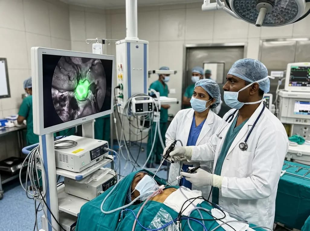
Near-infrared autofluorescence provides real-time label-free optical guidance during endoscopic transsphenoidal pituitary surgery. By highlighting stark signal contrasts between normal pituitary gland and adenomas, this technique aids gross-total tumor resection while safeguarding critical endocrine function.
Today
