

Loading, please wait...
Loading, please wait...

Primary central nervous system mesenchymal neoplasms represent an exceptionally rare group of tumors that present substantial diagnostic dilemmas. Clinicians often confuse these aggressive extra-axial masses with common benign lesions during routine evaluations. Furthermore, dysregulated neoplastic signaling can stimulate profound systemic inflammation through aberrant cytokine release. Notably, an intracranial EWSR1::CREM fusion sarcoma can trigger a systemic paraneoplastic syndrome that causes severe microcytic anemia, systemic malaise, and unexplained secondary infertility in reproductive-aged women.
When clinicians evaluate extra-axial dural-based lesions, they usually place meningiomas and solitary fibrous tumors at the top of their differential diagnosis. However, rare mesenchymal neoplasms harboring FET-CREB gene rearrangements challenge these standard clinical assumptions. Chromosomal translocations involving EWSR1 and CREM generate oncogenic chimeric transcription factors that stimulate abnormal cell growth. Consequently, these malignant tumors frequently arise along the cerebral convexities, skull base, or falx cerebri, closely mimicking benign meningothelial lesions.
In addition, primary central nervous system sarcomas account for less than one percent of all intracranial soft-tissue neoplasms. Because these entities occur so rarely, medical teams seldom consider them during initial patient consultations. Instead, clinicians often investigate non-specific constitutional manifestations before discovering the primary intracranial lesion. These manifestations frequently include persistent fatigue, microcytic anemia, and unexplained fever. Therefore, extensive rheumatologic and hematologic workups often precede definitive neuroimaging. Recognizing this atypical clinical presentation allows physicians to avoid diagnostic delay and expedite definitive intracranial imaging.
Paraneoplastic syndromes driven by intracranial mesenchymal tumors represent a fascinating yet deceptive clinical phenomenon. Neoplastic cells harboring EWSR1 rearrangements produce substantial concentrations of pro-inflammatory cytokines, especially interleukin-6. High circulating interleukin-6 levels induce hepatic overproduction of hepcidin, which directly blocks intestinal iron absorption and sequesters macrophage iron stores. As a direct result, patients develop severe anemia of chronic disease that resists standard oral iron therapy.
Furthermore, persistent systemic inflammation significantly disrupts the delicate hypothalamic-pituitary-ovarian axis. Elevated inflammatory cytokines impair the pulsatile secretion of gonadotropin-releasing hormone from the hypothalamus. Consequently, ovarian follicles fail to mature properly, resulting in anovulation and secondary infertility. Because these systemic complaints overshadow localized intracranial symptoms, physicians may mistakenly diagnose an autoimmune or hematologic disease. Fortunately, successful tumor extirpation halts cytokine production instantly. Thus, both hematologic parameters and ovarian functions normalize rapidly after surgery, permitting natural conception within weeks.
Neuroimaging plays an essential role in initial tumor assessment, yet conventional magnetic resonance imaging presents serious diagnostic traps. On standard scans, these mesenchymal neoplasms appear as well-circumscribed, extra-axial masses attached to the dura mater. Moreover, they exhibit robust, homogeneous contrast enhancement and commonly display a prominent dural tail sign. Because these radiographic features duplicate the classical appearance of benign meningiomas, neurosurgeons frequently anticipate a routine meningioma during surgical planning.
Nevertheless, advanced neuroimaging modalities can provide critical clues suggesting a malignant mesenchymal etiology. On diffusion-weighted imaging, these tumors often demonstrate pronounced diffusion restriction, which indicates high cellularity and elevated nuclear-to-cytoplasmic ratios. Similarly, magnetic resonance spectroscopy reveals elevated choline peaks alongside depressed N-acetylaspartate and creatine levels. Perfusion-weighted sequences also demonstrate significant hyperperfusion driven by brisk neoplastic neoangiogenesis. Despite these modern diagnostic tools, preoperative imaging cannot reliably separate these sarcomas from aggressive meningiomas. Therefore, histopathological and molecular analysis remains necessary for an accurate diagnosis.
Accurate pathological identification of intracranial mesenchymal neoplasms requires comprehensive tissue evaluation and advanced molecular diagnostics. Histologically, these neoplasms demonstrate varied architectural patterns, ranging from hypocellular myxoid matrices to dense sheets of round, spindle, or epithelioid cells. Furthermore, high-grade tumors exhibit frequent atypical mitoses, brisk proliferative activity, and focal areas of tumor necrosis. Routine immunohistochemistry often yields equivocal findings because tumor cells express non-specific markers like CD99, EMA, and vimentin.
Consequently, molecular diagnostic testing provides the indispensable gold standard for establishing the definitive diagnosis. Although break-apart fluorescence in situ hybridization can identify EWSR1 gene rearrangement, it cannot reveal the specific fusion partner. Therefore, next-generation RNA sequencing remains critical to identify the exact in-frame transcript between EWSR1 and CREM. Additionally, genome-wide DNA methylation profiling reliably distinguishes these neoplasms from true meningiomas, clear cell sarcomas, and solitary fibrous tumors. Identifying this specific translocation clarifies the tumor biology and guides tailored oncologic treatment.
Gross-total surgical resection serves as the primary therapeutic strategy for patients with primary intracranial sarcomas. Neurosurgeons must prioritize complete removal of the tumor mass alongside an aggressive excision of involved dural margins. During craniotomy, these lesions frequently display intense vascularity and tough dural adherence, necessitating careful microsurgical dissection and continuous electrophysiological monitoring. Achieving complete gross-total excision substantially reduces the risk of early local recurrence.
Remarkably, complete surgical extirpation provides an immediate cure for the associated paraneoplastic inflammatory syndrome. Once the surgical team removes the cytokine-secreting tumor tissue, circulating levels of interleukin-6 drop abruptly toward normal physiological ranges. As a result, acute-phase reactants decline rapidly, bone marrow erythropoiesis resumes, and endocrine balance returns. Patients achieve resolution of chronic fatigue and recover normal reproductive potential, occasionally conceiving within a single menstrual cycle postoperatively. Nevertheless, because these high-grade tumors retain malignant potential, multidisciplinary neuro-oncology teams must arrange long-term magnetic resonance surveillance.
The tumor produces excessive pro-inflammatory cytokines, specifically interleukin-6, through aberrant chimeric transcription factor signaling. Circulating interleukin-6 triggers hepatic hepcidin production, which locks iron inside cellular storage sites and induces refractory microcytic anemia. Furthermore, this intense inflammatory cascade disrupts hypothalamic gonadotropin-releasing hormone secretion, ultimately arresting normal ovarian follicular development and impairing reproductive capacity. Complete tumor resection eradicates the primary cytokine source, leading to rapid normalization of hematological and endocrine systems.
This mesenchymal neoplasm typically arises in dural-based extra-axial locations along cerebral convexities, mirroring the anatomical distribution of common meningiomas. On contrast-enhanced magnetic resonance imaging, the tumor exhibits avid, uniform contrast enhancement and frequently features a classic dural tail sign. Because these radiological characteristics match standard meningioma findings, radiologists routinely predict a benign diagnosis. Advanced techniques like diffusion-weighted imaging and magnetic resonance spectroscopy suggest malignant behavior, but definitive differentiation requires histopathological and molecular examination.
Following gross-total surgical resection, a multidisciplinary neuro-oncology board evaluates pathological margins, cellular atypia, and mitotic rates to determine whether adjuvant radiation therapy is necessary. Because standardized chemotherapy protocols do not exist for this rare neoplasm, close surveillance with high-resolution brain magnetic resonance imaging every three to six months remains paramount. Furthermore, clinicians must coordinate long-term reproductive and systemic monitoring, offering targeted molecular therapies if surveillance scans detect local or distant disease recurrence.
Disclaimer: This content is for informational and educational purposes only. It is not intended to be a substitute for professional medical advice, diagnosis, or treatment. Always seek the advice of your physician or other qualified health provider with any questions you may have regarding a medical condition. Refer to the latest local and national guidelines for clinical practice.
References

Read summarized clinical updates, watch expert medical content, and earn CME certifications right from your smartphone.
Open your smartphone camera to scan and install.





An intracranial EWSR1::CREM fusion sarcoma in a 31-year-old female manifested as a paraneoplastic inflammatory syndrome with anemia and secondary infertility. The dural mass mimicked meningioma, but gross-total resection resolved her systemic inflammation and restored natural fertility within one month.
Today
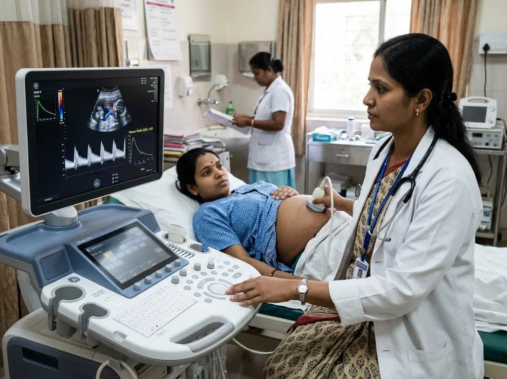
Adaptive spectral analysis of umbilical venous Doppler recordings reveals that an elevated second-trimester maternal-fetal power ratio can distinguish pregnancies at risk of preeclampsia, offering a novel biophysical approach to enhance early vascular risk stratification.
Today

A real-world cohort study of 6,537 patients from the FOUNTAIN platform demonstrates that initiating finerenone significantly reduces albuminuria by 40.7% with minimal eGFR decline and manageable hyperkalemia risk, confirming its practical effectiveness and safety in diabetic kidney disease.
Yesterday

A comprehensive analysis of MAP therapy resistance in osteosarcoma, examining canonical and non-canonical mechanisms across damage avoidance, enhanced DNA repair, and replication stress tolerance to identify novel vulnerabilities.
Today
Near-infrared autofluorescence provides real-time label-free optical guidance during endoscopic transsphenoidal pituitary surgery. By highlighting stark signal contrasts between normal pituitary gland and adenomas, this technique aids gross-total tumor resection while safeguarding critical endocrine function.
Today
