.webp&w=384&q=75)

Loading, please wait...
Loading, please wait...

Pediatric and adult spine surgery often relies on precise spinal instrumentation to stabilize deformities and restore spinal alignment. Consequently, accurate thoracic pedicle screw placement remains essential to prevent devastating neurological and vascular complications. Historically, preoperative computed tomography delivered the high-resolution bony detail required for robotic navigation platforms. However, ionizing radiation exposure poses substantial oncological risks, particularly for adolescent idiopathic scoliosis patients who undergo extensive multilevel spinal fusion. Emerging developments now introduce CT-like three-dimensional magnetic resonance imaging as a viable, radiation-free alternative. A novel cadaveric investigation evaluated whether this MRI sequence could guide robotic navigation platforms effectively without compromising accuracy. The promising results demonstrate high fidelity, suggesting a major paradigm shift toward safer pediatric and adult spinal interventions.
Modern corrective spinal procedures demand exceptional spatial precision because pedicle corridors in the thoracic spine remain notoriously narrow. Furthermore, misplaced thoracic screws can easily injure the adjacent spinal cord, aorta, or parietal pleura. Therefore, surgeons increasingly rely on robotic-assisted navigation systems to achieve flawless implant trajectories and minimize intraoperative complications. Conventional robotic workflows require thin-slice preoperative computed tomography scans to construct accurate anatomical models. Unfortunately, these CT protocols deliver considerable doses of ionizing radiation to vulnerable anatomical areas. Pediatric patients face disproportionate risks from this radiation exposure because their rapidly dividing cells exhibit greater radiosensitivity. Moreover, young individuals with progressive deformities often require multiple scans throughout surveillance and surgical correction. Cumulative exposure significantly elevates lifetime malignancy risks, prompting spine surgeons worldwide to advocate for the ALARA principle. Consequently, establishing a reliable bone-imaging protocol that avoids ionizing radiation represents a crucial clinical priority. By replacing standard preoperative CT scans with specialized MRI protocols, surgical teams can protect pediatric patients while maintaining optimal navigational accuracy.
Magnetic resonance imaging traditionally offers superior soft-tissue resolution but struggles to depict cortical bone margins with sufficient geometric clarity. Recently developed CT-like 3D-MRI sequences overcome this limitation through specialized gradient-echo acquisitions that capture bone-specific contrast. Specifically, these sequences invert voxel signals or employ deep-learning reconstruction algorithms to generate synthetic bone datasets resembling conventional CT scans. As a result, the robotic workstation interprets the processed MRI scan just like standard CT data, enabling seamless trajectory planning. During the preclinical investigation, researchers obtained high-resolution CT-like 3D-MRI thoracic spine datasets from two human cadaveric specimens. Subsequently, the surgical team imported these reconstructed volume renderings directly into a state-of-the-art robotic-assisted navigation workstation. The platform allowed investigators to map 48 individual screw paths across all thoracic vertebrae from T1 through T12 bilaterally. Because the software identified bony margins accurately, planners could select optimal screw dimensions and angulations prior to making surgical incisions. Thus, the advanced MRI acquisition successfully bridged the gap between safe radiation-free imaging and high-definition robotic planning.
Navigating the mid-thoracic vertebrae presents substantial technical challenges because pedicle diameters shrink significantly between T4 and T8. In addition, spinal ribs and adjacent chest wall dynamics complicate manual trajectory verification. However, robotic navigation systems eliminate physiological tremors and maintain rigid alignment along virtually planned pathways. In this study, the robotic arm moved autonomously into position according to the MRI-derived coordinates, allowing the surgeon to drill and tap each thoracic pedicle accurately. In total, the surgical team inserted 48 robotic pedicle screws across bilateral thoracic levels from T1 to T12. Following screw insertion, the researchers performed high-resolution postoperative CT scans to evaluate technical fidelity against the original 3D-MRI trajectory plans. Notably, the median deviation between the planned path and the actual screw position measured merely 0.4 mm in the axial plane. Furthermore, sagittal plane assessments demonstrated a minuscule median deviation of 0.05 mm. These tight interquartile deviations confirm that CT-like 3D-MRI provides exceptional spatial fidelity for robotic guidance, rivaling standard CT-based navigation metrics.
To validate clinical safety, researchers evaluate spinal instrumentation using the standardized Gertzbein-Robbins scale. Under this system, Grade A denotes an ideal screw contained entirely within the pedicular cortex, whereas Grade B indicates a cortical breach under two millimetres. Clinicians universally consider both Grade A and Grade B placements acceptable because minor cortical breaches rarely produce neurovascular injury. In the evaluated cadaveric series, postoperative imaging revealed that 100% of the placed screws achieved clinically acceptable positions. Specifically, 87.5% of the inserted screws met the stringent criteria for Grade A accuracy, exhibiting complete cortical containment. Meanwhile, the remaining 12.5% showed minor breaches under two millimeters, qualifying as Grade B placement. Importantly, no screw breached beyond two millimeters, and zero hardware impinged upon the central spinal canal or major vascular structures. Therefore, the combination of CT-like 3D-MRI and robotic guidance prevented catastrophic cortical breaches across challenging upper and middle thoracic vertebrae. These findings demonstrate that MRI-guided robotics can maintain exemplary safety standards without requiring preoperative radiation.
The ability to achieve flawless pedicle cannulation using radiation-free MRI holds profound clinical significance for pediatric deformity specialists. Adolescent idiopathic scoliosis surgery frequently requires extensive posterior fusion extending across ten or more vertebral segments. Consequently, preoperative CT scans of such long constructs impart substantial radiation doses to young breast and thyroid tissue. In contrast, integrating CT-like 3D-MRI sequences eliminates this diagnostic radiation burden entirely. Furthermore, modern MRI protocols simultaneously display neural elements, allowing surgeons to screen for hidden spinal cord abnormalities like syringomyelia or Chiari malformations during the same scan session. In addition, robotic execution reduces intraoperative fluoroscopy requirements, thereby decreasing radiation exposure for operating room staff and spine surgeons. In developing healthcare environments, including major tertiary centers across India, radiation reduction initiatives have gained tremendous momentum. Although MRI scanner availability and software accessibility present initial implementation hurdles, adopting MRI-based robotic workflows represents a transformative advancement. Ultimately, this approach enhances patient safety while upholding the highest surgical standards in complex spinal deformity reconstruction.
While these preclinical findings are encouraging, translating MRI-navigated robotics into routine operating room practice requires addressing practical clinical variables. For example, physiological motion artifacts from patient respiration and cardiac contractions can alter MRI image quality in live surgical candidates. Therefore, manufacturers must develop accelerated imaging sequences and motion-correction algorithms to ensure uniform image quality in vivo. Moreover, surgeons must validate registration techniques to confirm that intraoperative patient positioning matches preoperative MRI geometry precisely. Future clinical trials should evaluate operative duration, workflow efficiency, and cost-effectiveness across diverse patient populations. In addition, artificial intelligence algorithms may further streamline synthetic CT generation, producing reliable bone reconstructions in minutes. As robotic spine platforms evolve, radiation-free image guidance will likely become the preferred modality for pediatric and young adult spinal surgery. Continued clinical validation will ensure that safe, radiation-free instrumentation transitions seamlessly from experimental cadaveric laboratories to worldwide surgical suites.
In preclinical testing, MRI-guided robotic navigation achieved 100% clinically acceptable placement on the Gertzbein-Robbins scale. Exactly 87.5% of screws achieved perfect cortical containment (Grade A), while 12.5% had minor breaches under two millimeters (Grade B). Median deviations measured 0.4 mm axially and 0.05 mm sagittally.
Pediatric patients possess highly radiosensitive developing tissues, making them susceptible to radiation-induced malignancies later in life. Multilevel spinal deformity surgery historically required high-dose preoperative computed tomography scans. Eliminating preoperative CT scans through specialized CT-like 3D-MRI substantially decreases cumulative lifetime radiation exposure without sacrificing surgical navigation accuracy.
Yes. Specialized CT-like 3D-MRI sequences reconstruct high-contrast cortical bone contours necessary for navigation planning while retaining MRI's intrinsic ability to visualize soft tissue. Consequently, clinicians can evaluate the spinal cord, nerve roots, and disc morphology alongside bony pedicle anatomy within a single comprehensive, radiation-free imaging session.
Disclaimer: This content is for informational and educational purposes only and does not substitute professional medical advice, diagnosis, or treatment. Always seek the advice of your physician or other qualified health provider with any questions you may have regarding a medical condition. Refer to the latest local and national guidelines for clinical practice.
References

Read summarized clinical updates, watch expert medical content, and earn CME certifications right from your smartphone.
Open your smartphone camera to scan and install.





A cadaveric study demonstrates that robotic navigation using CT-like 3D-MRI achieves high accuracy for thoracic pedicle screw placement. This novel radiation-free technique offers a viable alternative to conventional CT scans, significantly reducing radiation exposure in pediatric spinal deformity surgery.
Today
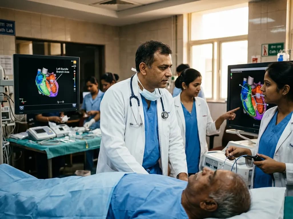
Left bundle branch pacing is an emerging physiological resynchronization strategy for heart failure. While observational data and some trials show benefits over biventricular pacing, divergent trial outcomes highlight the importance of confirmed conduction capture and specialized operator technique.
Today

A retrospective study of 86,012 pregnancies demonstrates that size-selective cfDNA enrichment boosts mean fetal fraction to 18.88% and achieves a 59.20% PPV for fetal copy number variants. However, 85.83% of CNVs showed size discrepancies, underscoring the mandatory role of confirmatory microarray testing.
Today

Radiofrequency catheter ablation of the cavotricuspid isthmus successfully terminates typical atrial flutter. This article reviews the mechanisms driving coronary sinus electrogram changes, wavefront activation pathways, and clinical criteria to verify bidirectional block during electrophysiology studies.
Today

A recent investigation examined load-dependent kinetic and kinematic profiles during two-way ballistic exercise across 30% to 80% 1-RM. Findings highlight load effects on velocity, impulse, and power, offering sports physicians objective guidance for neuromuscular conditioning and rehabilitation.
Today
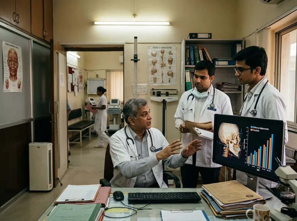
A cross-sectional study demonstrates that increased BMI in adolescents significantly enhances mandibular length and alters salivary adipokines like adiponectin and resistin, providing novel insights into craniofacial growth and orthodontic planning.
Today
