.webp&w=384&q=75)

Loading, please wait...
Loading, please wait...
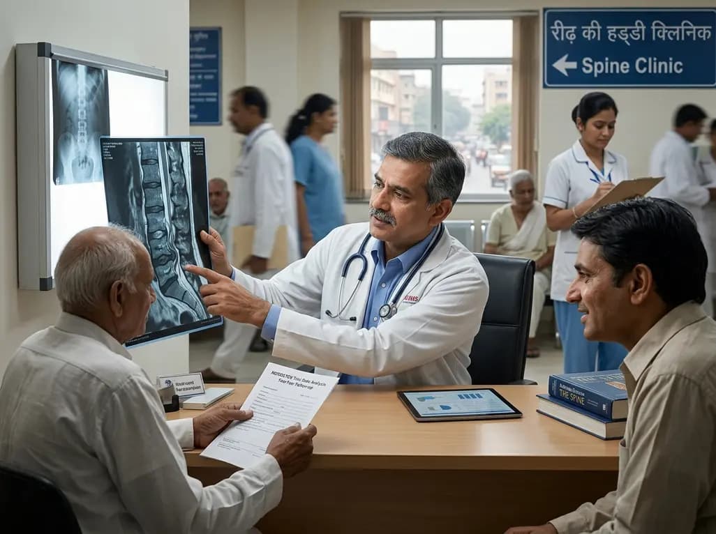
Lumbar spinal stenosis remains a predominant cause of neurogenic claudication and functional disability among older adults worldwide. Magnetic resonance imaging frequently reveals tortuous, elongated cauda equina fibers known as redundant nerve roots within the subarachnoid space above severe spinal constrictions. Historically, spine specialists considered this specific radiological phenomenon as a clear predictor of poor post-surgical recovery. However, recent robust prospective scientific evidence from high-quality clinical investigations is challenging these long-held assumptions regarding surgical prognosis.
The phenomenon of redundant nerve roots represents a distinct morphological change observed on sagittal T2-weighted magnetic resonance imaging. Characteristically, the nerve roots appear serpiginous, coiled, or elongated in the subarachnoid space proximal to a severe lumbar constriction. Pathophysiologically, chronic mechanical compression combined with continuous squeeze forces disrupts the longitudinal sliding movement of cauda equina nerve fibers. Consequently, persistent mechanical restriction, retrograde venous congestion, and localized nerve swelling cause the nerve fibers to lengthen and buckle.
In clinical practice, spine specialists frequently debate whether these altered neurovascular structures retain functional recovery potential after surgical decompression. Early descriptive studies suggested that chronic friction and ischemic stretching lead to irreversible intraneural fibrosis. Consequently, clinicians widely regarded the presence of coiled nerves as an indicator of advanced neurological damage. However, many historical studies lacked standardized prospective cohorts and relied primarily on retrospective data. Therefore, establishing whether this radiological marker truly portends an inferior clinical recovery required rigorous secondary evaluations of large prospective randomized clinical trials. Understanding these mechanical changes helps spine surgeons better interpret preoperative diagnostic imaging and explain disease progression to symptomatic patients.
To determine the true clinical significance of this radiological sign, researchers performed a secondary analysis using prospective data from the NORDSTEN spinal stenosis trial. The study evaluated 416 symptomatic patients undergoing decompressive spine surgery, dividing them into cohorts with and without redundant nerve roots. Surprisingly, baseline comparison revealed no significant clinical differences in self-reported pain levels or physical disability scores between the two cohorts.
Specifically, both groups reported comparable baseline Oswestry Disability Index scores, alongside similar pain intensity ratings on Numerical Rating Scales for both leg and back pain. In addition, baseline patient age, body mass index, smoking habits, and total symptom duration showed remarkable similarity between groups. This observation directly contradicts older retrospective series, which frequently described patients with coiled nerves as older and far more severely disabled at baseline. Nevertheless, objective radiological evaluations demonstrated important anatomical differences. Patients displaying redundant nerve roots exhibited a significantly higher prevalence of severe lumbar stenosis and multi-level spinal canal narrowing. Furthermore, male patients demonstrated a higher frequency of this neurovascular morphological change. These baseline findings highlight that severe anatomical compression on imaging does not automatically translate to worse preoperative clinical symptoms or functional performance.
Surgical decompression aims to eliminate mechanical constriction, restore cerebrospinal fluid flow, and relieve ischemic stress on compromised nerve structures. At the two-year postoperative follow-up, both patient cohorts experienced substantial clinical improvements across all measured outcomes. However, patients presenting preoperatively with redundant nerve roots achieved significantly greater overall functional recovery compared to those without this morphological feature.
Quantitatively, the mean improvement in the Oswestry Disability Index was -22.1 points for the group with redundant nerve roots, compared to -17.4 points in the group without them. This statistically significant difference of 4.7 points in disability reduction highlights a clinically meaningful benefit for patients with preoperative nerve redundancy. Similarly, secondary outcome measures, including the Zurich Claudication Questionnaire and Numerical Rating Scales for back and leg pain, showed significant additional benefits favoring the affected group. Consequently, decompression effectively releases severe mechanical entrapment, allowing coiled nerve fibers to regain function and relieve clinical symptoms. These findings provide reassuring objective evidence for spine surgeons considering decompression for severe lumbar canal narrowing. Consequently, surgical intervention should not be withheld or considered high-risk purely based on the presence of tortuous nerve roots on magnetic resonance imaging.
For decades, medical literature frequently cited redundant nerve roots as an unfavorable prognostic factor following decompressive spine surgery. A comprehensive meta-analysis conducted in 2018 reinforced this perspective, suggesting that patients with coiled nerve roots achieved inferior post-surgical outcomes. However, many early investigations suffered from small sample sizes, heterogeneous surgical techniques, and inconsistent baseline risk adjustments.
The high-quality prospective data from the NORDSTEN trial fundamentally challenges this historical paradigm. Because the NORDSTEN trial utilized standardized surgical protocols, uniform prospective follow-up, and validated outcome measures, its findings carry substantial clinical weight. The robust trial design minimizes common selection biases that plagued earlier retrospective research. Pathophysiologically, severe focal compression creates significant mechanical tension, which resolves once surgical laminectomy or laminotomy removes the bone and ligamentous constriction. Releasing this acute mechanical entrapment appears to allow substantial neural recovery and symptomatic relief, even when nerve roots appear tortuous or elongated on preoperative magnetic resonance scans. Therefore, clinicians must re-evaluate historical assumptions and avoid labeling nerve root redundancy as a marker of irreversible nerve damage or surgical failure.
Integrating these prospective trial findings into daily clinical practice provides clear guidance for orthopaedic surgeons, neurosurgeons, and pain management specialists. When reviewing preoperative lumbar magnetic resonance imaging, clinicians should recognize that tortuous or serpiginous nerve roots indicate severe focal spinal canal narrowing rather than permanent neural dysfunction. Consequently, identifying this radiological feature should encourage appropriate surgical decompression rather than create hesitation.
Furthermore, these findings significantly improve preoperative patient counseling and decision-making. Spine specialists can confidently reassure patients that displaying redundant nerve roots does not limit their potential for clinical improvement following decompression surgery. In fact, patients with this feature often experience greater relative pain relief and functional recovery than those without it, provided effective mechanical decompression is achieved. In addition, surgical planning should focus on achieving thorough decompression across all severe stenotic levels identified on preoperative imaging. Broadening clinical understanding regarding nerve root redundancy prevents unnecessary denial of effective surgical care. As spine care continues to emphasize evidence-based outcomes, these high-quality prospective findings offer actionable clinical insights that optimize patient care and refine surgical prognostic discussions.
Redundant nerve roots represent coiled, elongated, or serpiginous cauda equina nerve fibers seen above severe spinal canal constrictions on sagittal T2-weighted MRI. Chronic mechanical compression and restricted nerve sliding cause these structural changes. Although once considered a sign of irreversible nerve damage, recent studies confirm they represent severe compression that responds well to surgical decompression.
Prospective evidence from the NORDSTEN trial shows that patients with redundant nerve roots have baseline pain and disability scores similar to those without them. Although patients with redundant nerve roots have more severe multi-level canal narrowing on MRI, their subjective pain, age, and duration of symptoms do not differ significantly from other stenosis patients.
No, presence of redundant nerve roots does not lower surgical success rates. Two-year outcomes from high-quality clinical trial data show that patients with redundant nerve roots experience significantly greater improvements in functional disability and pain scores after surgical decompression compared to patients without this radiological feature.
Disclaimer: This content is for informational and educational purposes only and does not constitute medical advice, diagnosis, or treatment. Always seek the advice of a qualified healthcare professional with any questions regarding a medical condition. Refer to the latest local and national guidelines for clinical practice.
References

Read summarized clinical updates, watch expert medical content, and earn CME certifications right from your smartphone.
Open your smartphone camera to scan and install.





A secondary analysis of the NORDSTEN trial shows that patients with redundant nerve roots (RNRs) experiencing symptomatic lumbar spinal stenosis achieve significantly greater functional improvement two years after decompression surgery, challenging historical assumptions of poor prognosis.
2 months ago

Review how tailored exercise programs benefit frail older patients with heart failure. Explore pathophysiological mechanisms, individualized training prescriptions, and strategies to overcome adherence barriers in clinical practice.
Today

Leucine Rich Bio has introduced South Asia's first comprehensive vaginal microbiome test, BugSpeaks. By profiling microbial dysbiosis, pathogens, and antimicrobial resistance, this next-generation diagnostic platform provides clinicians with deeper insights into recurrent infections, fertility, and reproductive health.
Today

In high-risk pulmonary embolism complicated by severe anemia and active uterine bleeding, systemic thrombolysis is contraindicated. Large-bore aspiration thrombectomy provides rapid hemodynamic rescue without fibrinolysis, supported by inferior vena cava filter placement and multidisciplinary critical care.
Today

This comparative study evaluates posterior labral tears with and without glenohumeral instability, highlighting distinct demographic patterns, MRI underestimation in stable shoulders, and successful arthroscopic repair outcomes.
Today

A clinical study demonstrates that early intervention with tocilizumab, either as monotherapy or bridged with corticosteroids, achieves significant proptosis reduction and resolves active inflammation in treatment-naive thyroid eye disease.
Today
