.webp&w=384&q=75)

Loading, please wait...
Loading, please wait...
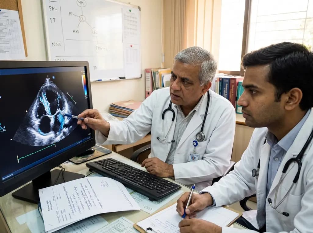
Restrictive cardiomyopathy encompasses a diverse spectrum of myocardial disorders characterized by impaired ventricular relaxation and markedly elevated filling pressures. Among its rarest and most challenging etiologies is Löffler endocarditis, a severe cardiovascular manifestation of hypereosinophilic syndrome. In this condition, sustained eosinophil activation triggers acute tissue necrosis, progressive intracardiac thrombosis, and irreversible endomyocardial fibrosis. Recognizing this rare pathology early remains vital because timely immunosuppressive and medical interventions can prevent permanent cardiac damage and catastrophic thromboembolic complications.
The pathogenesis of Löffler endocarditis evolves through three distinct chronological phases. Initially, the acute necrotic stage develops as eosinophils infiltrate the myocardium and release toxic granular proteins, including eosinophil cationic protein and major basic protein. Consequently, these cytotoxic mediators cause direct myocardial necrosis, microvascular injury, and marked endocardial inflammation. Patients in this early phase frequently present with subtle constitutional symptoms or acute myocarditis.
Subsequently, the disease enters the thrombotic stage over several weeks to months. Endothelial disruption exposes the underlying thrombogenic subendothelial matrix. As a result, layered mural thrombi form along the ventricular apices and subvalvular structures. These thrombi frequently entrap chordae tendineae, which leads to severe atrioventricular valve regurgitation. In addition, apical mural thrombi pose an ongoing risk of systemic and cerebral thromboembolism.
Finally, the chronic fibrotic stage emerges after prolonged inflammation. During this phase, dense fibrous tissue gradually replaces organized thrombi and damaged myocardium. This widespread endomyocardial fibrosis stiffens ventricular chambers and restricts diastolic relaxation. Therefore, patients develop classic restrictive physiology with preserved systolic function but significantly elevated filling pressures.
Clinicians often struggle to identify hypereosinophilic cardiac disease because its initial features mimic more common cardiac conditions. Patients typically present with progressive exertional dyspnea, extreme fatigue, orthopnea, and peripheral edema. Furthermore, chest discomfort, palpitations, and non-specific systemic symptoms such as low-grade fever, weight loss, or myalgias may precede overt heart failure.
Laboratory evaluation provides crucial diagnostic clues. An elevated absolute eosinophil count exceeding 1500 cells per microliter strongly suggests hypereosinophilic syndrome. However, clinicians must recognize that peripheral eosinophil counts do not always correlate linearly with myocardial damage. In fact, severe cardiac infiltration can occasionally occur with only moderate peripheral eosinophilia. Serum cardiac biomarkers, including troponin and B-type natriuretic peptide, frequently rise during active inflammation and chamber stretch.
Moreover, clinicians must thoroughly exclude secondary causes of hypereosinophilia. These potential triggers include parasitic infections, drug-induced hypersensitivity reactions, autoimmune vasculitides like eosinophilic granulomatosis with polyangiitis, and hematologic neoplasms. Thrombocytopenia or anemia may also appear, reflecting bone marrow involvement or microvascular consumption. Consequently, comprehensive hematologic testing, including bone marrow biopsy and cytogenetic analysis, remains indispensable.
Diagnostic imaging plays a pivotal role in confirming myocardial involvement and tracking disease progression. Transthoracic echocardiography usually serves as the first-line modality. It frequently demonstrates apical obliteration of the ventricles, subvalvular thickening, and restrictive transmitral filling patterns. Additionally, biatrial enlargement with normal or mildly increased ventricular wall thickness commonly indicates chronic diastolic restriction.
Nevertheless, cardiac magnetic resonance imaging has emerged as the gold standard for non-invasive evaluation of Löffler endocarditis. Cardiac MRI offers superior tissue characterization, clearly delineating endomyocardial inflammation, necrosis, fibrosis, and intracardiac thrombi. Specifically, T2-weighted imaging detects active myocardial edema during the acute necrotic phase. Early gadolinium enhancement accurately identifies vascularity and distinguishes avascular thrombi from hypervascular myocardium.
Furthermore, late gadolinium enhancement displays characteristic subendocardial enhancement patterns that do not follow coronary vascular distributions. This subendocardial rim of enhancement reflects extensive fibrosis or active endocardial inflammation. Cardiac MRI also evaluates ventricular volumes, diastolic filling dynamics, and ejection fraction with high reproducibility. Therefore, serial imaging provides an objective method to assess treatment response and detect recurrent subclinical inflammation.
Medical management of Löffler endocarditis aims to rapidly reduce circulating eosinophils, suppress cardiac inflammation, and prevent thromboembolic complications. High-dose systemic corticosteroids, such as oral prednisone or intravenous methylprednisolone, represent the standard first-line therapy. Glucocorticoids rapidly induce eosinophil apoptosis and suppress cytokine production. As a result, clinical symptoms and peripheral eosinophil counts often improve promptly after treatment initiation.
However, some patients exhibit steroid-refractory disease or experience symptom relapse during steroid tapering. In such instances, clinicians must introduce second-line immunosuppressive or cytotoxic agents. Hydroxyurea, azathioprine, and cyclophosphamide provide effective disease control in refractory idiopathic cases. In addition, targeted biologic therapies have transformed contemporary treatment. For example, mepolizumab, an anti-interleukin-5 monoclonal antibody, selectively inhibits eosinophil proliferation and offers a valuable steroid-sparing alternative.
Similarly, identifying specific genetic mutations directs precision therapy. Patients harboring the FIP1L1-PDGFRA fusion gene respond remarkably well to low-dose imatinib, achieving rapid molecular and hematologic remission. Alongside targeted immunosuppression, standard heart failure regimens containing loop diuretics, beta-blockers, and mineralocorticoid receptor antagonists alleviate volume overload. Finally, therapeutic anticoagulation remains critical to prevent and resolve ventricular mural thrombi until endocardial healing occurs.
When medical therapy fails to reverse advanced structural damage, surgical intervention may become necessary. Chronic fibroblastic remodeling often causes severe atrioventricular valve dysfunction and intractable chamber obliteration. In such cases, endocardectomy with endomyocardial decortication can successfully restore ventricular cavity volume and improve diastolic filling. Furthermore, valve repair or replacement may alleviate severe mitral or tricuspid regurgitation.
Nevertheless, surgical procedures carry substantial perioperative risk due to underlying restrictive physiology and fragile myocardium. Therefore, careful patient selection and optimized medical stabilization before surgery are essential. Bioprosthetic valves are generally preferred over mechanical prostheses because they minimize the risk of mechanical valve thrombosis in a prothrombotic environment.
Long-term prognosis largely depends on the stage of disease at presentation and the speed of therapeutic intervention. Early recognition and aggressive anti-eosinophil therapy prevent transition from acute necrosis to irreversible endomyocardial fibrosis. Conversely, delayed diagnosis increases the likelihood of chronic refractory heart failure, recurrent thromboembolism, and fatal arrhythmias. Consequently, lifelong clinical surveillance with serial echocardiography, cardiac MRI, and hematologic monitoring is crucial to ensure sustained remission.
Löffler endocarditis is a rare restrictive cardiomyopathy caused by hypereosinophilic syndrome. Sustained high levels of circulating eosinophils infiltrate the myocardium and endocardium. These cells release cytotoxic granules that trigger acute myocardial necrosis, followed by mural thrombus formation. Over time, extensive fibrous tissue replaces damaged structures, leading to endomyocardial fibrosis and severe diastolic filling restriction.
Cardiac magnetic resonance provides comprehensive tissue characterization to evaluate every stage of the disease. It detects active myocardial edema on T2-weighted sequences and clearly differentiates intracardiac thrombi from surrounding myocardium. Furthermore, late gadolinium enhancement displays characteristic subendocardial patterns, confirming fibrosis and inflammation while assessing ventricular function and diastolic restriction with superior accuracy.
Some patients experience symptom recurrence due to steroid resistance, rapid steroid tapering, or underlying clonal hematologic mutations. In addition, if endomyocardial fibrosis and chronic ventricular obliteration have already developed, suppressing eosinophils alone cannot reverse structural damage. Therefore, these individuals require second-line immunosuppressants, targeted biological therapies like mepolizumab or imatinib, and dedicated heart failure management.
Disclaimer: This content is for informational and educational purposes only. It is not intended to be a substitute for professional medical advice, diagnosis, or treatment. Always seek the advice of your physician or other qualified health provider with any questions you may have regarding a medical condition. Refer to the latest local and national guidelines for clinical practice.
References

Read summarized clinical updates, watch expert medical content, and earn CME certifications right from your smartphone.
Open your smartphone camera to scan and install.





Löffler endocarditis is a rare manifestation of hypereosinophilic syndrome that causes restrictive cardiomyopathy. Early multimodal cardiac imaging, biomarker evaluation, and aggressive immunosuppression are vital to prevent irreversible endomyocardial fibrosis and heart failure.
last month

Pancreatic cancer cachexia affects over 80% of patients, driving severe muscle wasting, systemic inflammation, and treatment intolerance. This review highlights key pathophysiological drivers, exocrine insufficiency, objective CT imaging, and emerging therapies like GDF-15 inhibitors to improve clinical outcomes.
Today

A scoping review reveals how pregnancy affects phonation through laryngeal edema and reduced phonation time rather than major acoustic pitch alterations. Learn how to identify, evaluate, and manage vocal symptoms in expectant mothers.
Today

According to WHO data, average lifespan reached 73.3 years in 2023, recovering from pandemic lows. However, healthy life expectancy lags at 62.8 years due to surging noncommunicable conditions like cardiovascular disease and dementia, demanding proactive primary screening and sustained chronic care.
Today

Researchers have engineered an artificial sensory-pain integrated receptor using a multi-threshold organic synaptic transistor. The system distinguishes benign touch from noxious stimuli, enabling safer, more responsive prosthetic limbs with built-in memory and simplified hardware architecture.
Today

Medical robotics is transitioning beyond operative suites into diagnostics, cardiology, and endocrinology. Explore how innovations in tele-robotics, AI integration, and nanomedicine are overcoming geographical barriers and transforming precision healthcare delivery across India's evolving disease landscape.
Today
