.webp&w=384&q=75)

Loading, please wait...
Loading, please wait...

Radiologically isolated syndrome (RIS) represents one of the most intriguing and challenging frontiers in modern neurology. Traditionally, clinicians viewed RIS as a strictly preclinical state, where incidental MRI findings suggested demyelination without any matching clinical symptoms. However, emerging evidence regarding radiologically isolated syndrome management suggests this view is significantly outdated. Instead of a silent waiting period, RIS appears to represent an active subclinical phase of the multiple sclerosis (MS) spectrum. This distinction is vital for Indian practitioners because it shifts the focus from passive observation to active risk stratification. Consequently, identifying these individuals earlier allows for a more nuanced understanding of their neurological health. Research now demonstrates that even without classic relapses, subclinical damage is frequently occurring. This damage manifests through measurable neuroaxonal loss and inflammatory activity. Therefore, the clinical community is moving toward a more biological definition of the disease, where the absence of a visible relapse does not necessarily equate to the absence of disease activity. This evolution in thought necessitates a comprehensive approach to monitoring and intervention.
Cognitive health is often the first casualty in the subclinical progression of neuroinflammatory disorders. Recent systematic reviews indicate that a substantial proportion of individuals with RIS exhibit measurable cognitive impairment long before their first clinical event. Specifically, information processing speed and executive functions are the most frequently affected domains. These deficits are not merely incidental; they reflect underlying structural changes such as cortical thinning and thalamic atrophy. In the context of radiologically isolated syndrome management, regular neuropsychological assessment becomes an essential tool. Furthermore, these cognitive changes can significantly disrupt an individual's daily life, even if they do not meet the formal criteria for MS. Clinicians should use standardized tools like the Symbol Digit Modalities Test (SDMT) to track these subtle shifts. Moreover, understanding that cognitive decline can occur independently of physical disability helps in framing the true impact of the disease. By acknowledging these deficits, healthcare providers can offer better support and counseling. Ultimately, cognitive monitoring provides a window into the brain's functional reserve and the ongoing inflammatory process.
Beyond the physical and cognitive aspects, the psychosocial burden of RIS is often underestimated. Individuals diagnosed with RIS frequently experience clinical depression and pathological fatigue, which impact their quality of life as severely as those with relapsing-remitting MS. This psychosocial deterioration suggests that the disease is active on multiple levels. Transitioning from a "healthy" status to having a potential chronic illness creates significant psychological stress. Additionally, the uncertainty regarding clinical conversion adds a layer of anxiety that requires professional management. Consequently, radiologically isolated syndrome management must incorporate mental health screenings and support systems. Practitioners should recognize that fatigue in RIS is not just a subjective complaint but a reflection of central nervous system involvement. Therefore, addressing these symptoms is crucial for maintaining the patient's overall well-being. Studies have shown that the impact on quality of life in RIS resembles that of established MS cohorts. This similarity underscores the need for early psychosocial intervention. Integrating mental health professionals into the care team can help individuals navigate the complex emotional landscape of a subclinical diagnosis.
The search for sensitive biomarkers has revolutionized our ability to stage disease activity in RIS. Advanced neuroimaging techniques and molecular markers, such as neurofilament light (NfL) levels, provide evidence of ongoing neurodegeneration. Elevated NfL levels in the blood or cerebrospinal fluid correlate with axonal damage and can predict a higher risk of future clinical events. Furthermore, neuroimaging markers like the central vein sign and paramagnetic rim lesions (PRLs) help differentiate RIS from other mimickers like small vessel disease or migraine. These tools are becoming increasingly relevant for radiologically isolated syndrome management in specialized centers. Specifically, thalamic atrophy serves as a powerful surrogate for global brain damage and cognitive decline. Consequently, incorporating these biomarkers into routine practice allows for more accurate disease staging. Although access to some of these tests may be limited in certain regions of India, their diagnostic value is undeniable. By combining imaging data with molecular signatures, clinicians can create a more detailed profile of each individual's risk. This biological approach facilitates a more proactive management strategy compared to traditional clinical observation alone.
One of the greatest challenges in radiologically isolated syndrome management is predicting who will convert to symptomatic MS. Interestingly, while cognitive impairment is prevalent, baseline cognitive performance does not consistently predict short-term clinical conversion. This suggests that the brain may utilize compensatory mechanisms for a period before clinical symptoms emerge. However, other factors like younger age, male sex, and the presence of spinal cord lesions remain strong predictors of conversion. Furthermore, the presence of oligoclonal bands in the cerebrospinal fluid significantly increases the likelihood of a future relapse. Practitioners must therefore weigh these variables carefully when discussing the prognosis with their patients. Recent clinical trials, such as the ARISE and CELLO studies, have explored the use of disease-modifying therapies (DMTs) in RIS. These studies suggest that early intervention can delay the first clinical event in high-risk individuals. Nevertheless, the decision to treat an asymptomatic person remains complex and requires a personalized approach. Thus, risk stratification is not just about predicting the future but about deciding on the intensity of current monitoring.
The future of radiologically isolated syndrome management lies in personalized staging and optimized risk stratification. As the McDonald criteria continue to evolve, we may soon see some forms of RIS reclassified as early-stage MS. This shift would allow for earlier access to treatment and more standardized care protocols. Moreover, the integration of artificial intelligence and machine learning in analyzing MRI data could improve our ability to detect subtle progression. For now, the focus remains on detailed longitudinal monitoring. Clinicians should adopt a multi-dimensional approach that includes MRI, biomarkers, and neuropsychological testing. Additionally, educating individuals about the nature of RIS and the importance of follow-up is vital. As we move away from a one-size-fits-all model, we can better cater to the unique needs of each person. This proactive stance ensures that we do not wait for a disabling relapse to occur before intervening. In conclusion, the transition from viewing RIS as a curiosity to seeing it as a clinical entity marks a significant advancement in neurology. Continuous research will further refine our tools and improve outcomes for this unique population.
Radiologically Isolated Syndrome (RIS) involves incidental MRI findings suggestive of MS without any history of clinical symptoms. In contrast, Clinically Isolated Syndrome (CIS) refers to a single, first episode of neurological symptoms lasting at least 24 hours. Multiple Sclerosis (MS) is diagnosed when there is evidence of dissemination in both space and time. RIS represents a subclinical stage where the pathology is present, but overt clinical relapses have not yet occurred.
Research consistently shows that individuals with RIS often experience deficits in information processing speed and executive functions. These cognitive impairments can manifest as difficulties in multitasking, maintaining attention, or processing complex information quickly. Although these changes are often subtle and may not be noticed by the individual initially, standardized neuropsychological testing can detect them. These deficits are frequently linked to structural brain changes like thalamic atrophy and cortical thinning seen on MRI.
Neurofilament light (NfL) is a sensitive biomarker of neuroaxonal injury that has shown great promise in predicting conversion. Elevated levels of NfL in the blood or cerebrospinal fluid often precede the first clinical demyelinating event by several years. While not a definitive standalone predictor, high NfL levels indicate active neurodegeneration and a significantly higher risk of transitioning to symptomatic MS. Consequently, many experts recommend using NfL to aid in risk stratification and management decisions.
Disclaimer: This content is for informational and educational purposes only. It does not constitute medical advice or a professional relationship. Always seek the advice of a physician or other qualified health provider with any questions you may have regarding a medical condition. Refer to the latest local and national guidelines for clinical practice.
References
Chang MH et al. The multi-dimensional spectrum of radiologically isolated syndrome: a systematic review of cognitive and psychosocial evidence. Acta Neurol Belg. 2026 Jul 19. doi: 10.1007/s13760-026-03143-4. PMID: 42472426.
Lebrun-Frénay C, et al. Radiologically Isolated Syndrome: 10-Year Risk of an Initial Clinical Event. Ann Neurol. 2023;94(3):465-474.
Filippi M, et al. Radiologically isolated syndrome: A review. J Neurol Neurosurg Psychiatry. 2024;95(1):12-21.
"
Read summarized clinical updates, watch expert medical content, and earn CME certifications right from your smartphone.
Open your smartphone camera to scan and install.





New evidence suggests Radiologically Isolated Syndrome (RIS) is an active subclinical stage of MS rather than just a preclinical state. This review highlights cognitive impairment, psychosocial burdens, and neuroimaging biomarkers, urging a shift in clinical staging and risk stratification for better management.
3 months ago

A landmark 12-year longitudinal study highlights significant reductions in opioid prescribing for nonspecific low back pain following clinical guidelines and prescription monitoring systems, emphasizing non-opioid multimodal pain care.
Today
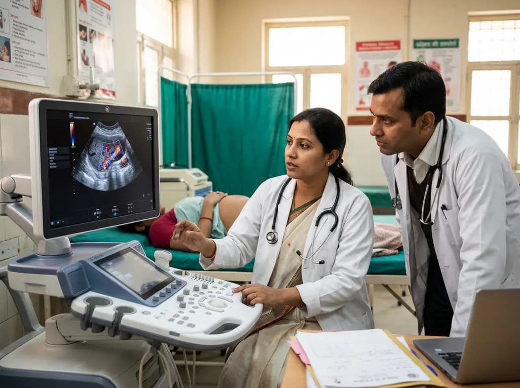
A study in Ultrasound in Obstetrics & Gynecology shows that quantitative vascularization of the anterior fornix/cervix with transvaginal ultrasound reliably identifies placenta accreta spectrum and predicts severe intraoperative hemorrhage, offering vital objective risk stratification.
Today
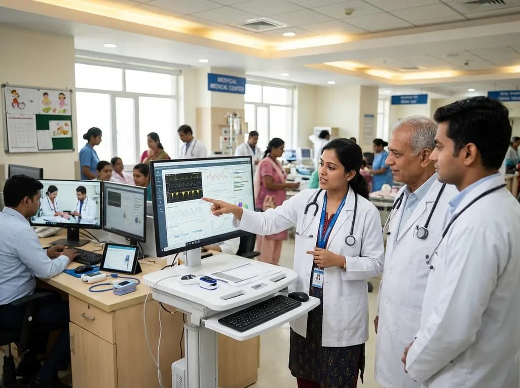
A landmark study details how an enterprise pediatric remote patient management model expanded from a single cardiology pilot to 16 diverse specialty programs by combining centralized digital operations with specialty clinical leadership.
Yesterday
A breakthrough study shows that 3D-planned corrective osteotomy successfully resolves distal radioulnar joint instability in pediatric diaphyseal forearm malunions. The precise osseous realignment eliminates chronic wrist pain and restores joint function without requiring secondary soft-tissue reconstruction.
Today

Protein O-GlcNAcylation couples cellular nutrient status to vital neuronal functions. This review explores how disrupted O-GlcNAc cycling drives both neurodevelopmental delays and neurodegenerative cascades, positioning metabolic post-translational modifications as therapeutic targets in clinical neurology.
Today
