.webp&w=384&q=75)

Loading, please wait...
Loading, please wait...
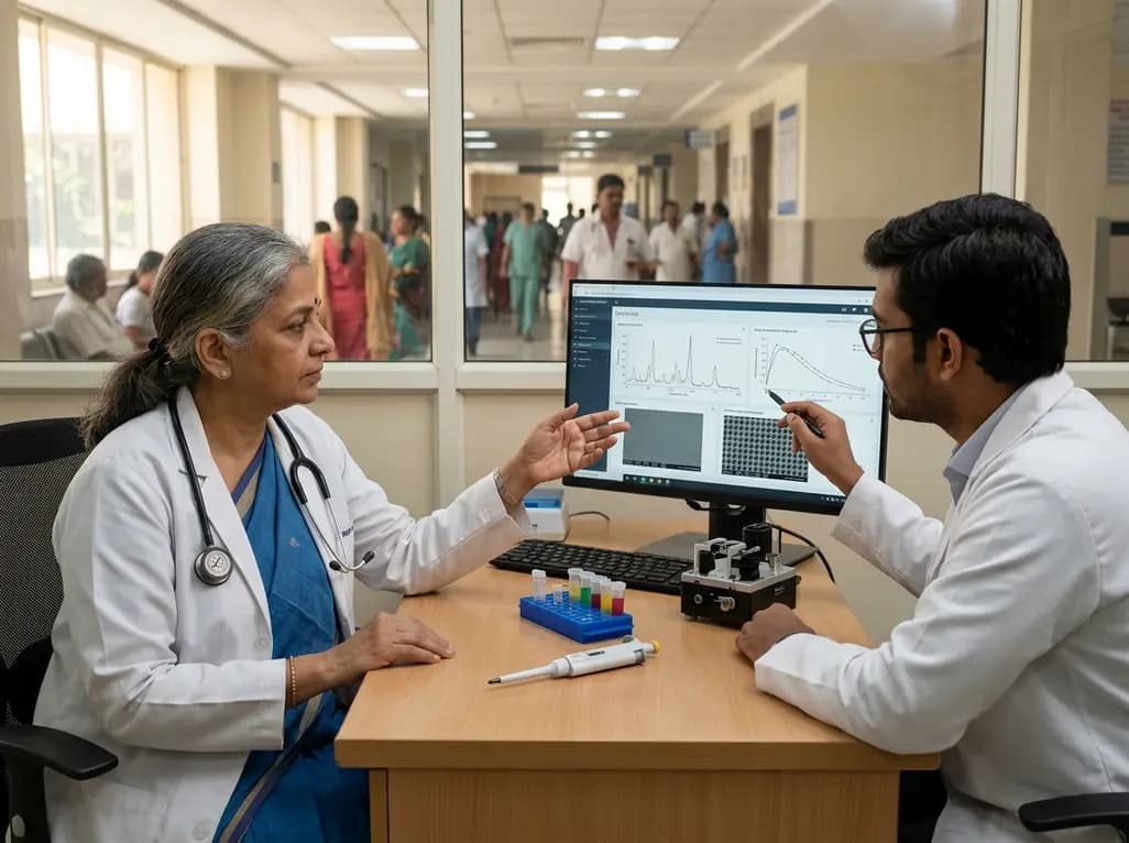
Personalized cancer therapy demands accurate therapeutic drug monitoring to balance maximum anti-tumor efficacy against life-threatening cytotoxic side effects. Implementing quantitative SERS drug monitoring provides clinicians with an advanced tool to quantify circulating antineoplastic agents with rapid turnaround times. Conventional cytotoxic regimens often rely on body surface area calculations, yet interindividual pharmacokinetic variability frequently leads to unpredictable systemic toxicity or underdosing. By stabilizing molecular interactions at deterministic plasmonic hotspots, this innovative nanoscale platform transforms surface-enhanced Raman spectroscopy from a qualitative laboratory method into an actionable diagnostic platform for real-time therapeutic decisions.
Traditional cytotoxic dosing protocols in oncology frequently produce unpredictable pharmacological exposure across diverse patient cohorts. Consequently, clinicians encounter severe dose-limiting toxicities such as myelosuppression, cardiotoxicity, and peripheral neuropathy in substantial subsets of patients. Although therapeutic drug monitoring could mitigate these adverse outcomes, conventional diagnostic tools remain poorly suited for point-of-care adaptation. Liquid chromatography-tandem mass spectrometry offers high chemical fidelity, but its procedural complexity and high operational costs restrict routine use. Therefore, developing rapid optical platforms like quantitative SERS drug monitoring addresses an urgent gap in acute oncological management.
Surface-enhanced Raman spectroscopy inherently delivers ultra-sensitive molecular fingerprinting through localized surface plasmon resonance. However, historical attempts to deploy standard SERS for quantitative pharmacological tracking encountered insurmountable technical barriers. Stochastic nanoparticle aggregation typically produces random electromagnetic hotspots, resulting in substantial signal variability across measurement replicates. Furthermore, photosensitive chemotherapeutic molecules often undergo photothermal degradation during continuous laser excitation. Consequently, clinicians could not rely on early plasmonic sensors for critical dose adjustments. To resolve these long-standing structural deficits, researchers designed a mesoporous-shell monolayer plasmonic architecture that delivers reproducible spectral quantification.
The newly engineered architecture stabilizes plasmonic fields by organizing metallic nanostructures into uniform monolayer assemblies beneath ordered mesoporous dielectric shells. Consequently, this geometry creates deterministic, highly reproducible electromagnetic hotspots rather than accidental aggregation zones. Furthermore, the mesoporous coating functions as a size-selective molecular sieve, filtering out large serum proteins while permitting rapid diffusion of targeted antineoplastic molecules. As a result, the platform prevents biofouling in untreated patient biofluids, preserving pristine signal clarity during rapid clinical analyses.
In addition, the dielectric mesoporous shell provides vital thermal dissipation and photoprotection for unstable small-molecule analytes. Photosensitive chemotherapy drugs maintain structural integrity throughout laser acquisition because the shell moderates local photothermal energy transfer. Therefore, the monolayer architecture generates consistent vibrational spectra with minimal background noise. Multiplexed detection of distinct antineoplastic agents achieves exceptional sensitivity across broad concentration gradients. By replacing random nanoparticle collisions with predictable nanofabricated geometries, this design ensures that spectral intensity directly correlates with absolute drug concentrations in complex biological matrices.
Accurate spectral deconvolution in whole blood or plasma requires sophisticated algorithmic interpretation to isolate subtle pharmaceutical signatures from complex endogenous backgrounds. Accordingly, investigators coupled the plasmonic platform with advanced machine learning regression models trained on extensive vibrational datasets. These algorithms rapidly extract multidimensional spectral features, correcting for baseline shifts and minor matrix interferences. Consequently, the combined computational and optical diagnostic system delivers precise concentration predictions within minutes of sample acquisition.
To establish clinical validity, researchers benchmarked the platform against gold-standard liquid chromatography-mass spectrometry using patient-derived blood samples. Notably, the machine learning-assisted SERS measurements demonstrated near-perfect correlation with mass spectrometric references across all evaluated cohorts. Unlike standard chromatography, this optical method requires negligible sample preparation and eliminates bulky chemical solvents. Therefore, hospital clinical biochemistry laboratories and intensive care units can rapidly determine circulating drug levels without relying on off-site reference laboratories, facilitating timely treatment personalization.
The ultimate test of any monitoring technology lies in its capacity to guide dynamic clinical decisions and improve therapeutic indices. To demonstrate this capability, researchers evaluated SERS-guided dosing protocols in orthotopic breast cancer animal models undergoing active chemotherapy. Serial micro-volume blood draws allowed investigators to track drug clearance kinetics and calculate real-time area-under-the-curve exposure for individual subjects. Consequently, treatment teams adjusted subsequent chemotherapy doses according to personal clearance rates rather than static baseline protocols.
Importantly, animals managed through SERS-guided dose titration achieved equivalent primary tumor regression compared to cohorts receiving conventional fixed-dose regimens. Furthermore, the individualized dosing cohort exhibited markedly lower organ toxicity, preserving hepatic, renal, and bone marrow function. Systemic weight loss and therapy-related morbidity decreased significantly in the guided intervention arm. Therefore, this direct link between plasmonic quantification and algorithmic dose adjustment proves that real-time therapeutic monitoring effectively widens the therapeutic window of toxic antineoplastic compounds.
Deploying rapid optical monitoring platforms presents transformative advantages for cancer care ecosystems across India and similar emerging healthcare environments. In high-volume tertiary cancer centers, central laboratory backlogs often delay therapeutic drug monitoring results by several days, rendering retrospective adjustments ineffective for acute infusion cycles. Because plasmonic SERS hardware can be miniaturized into portable Raman spectrometers, district-level cancer centers and day-care oncology units could perform decentralized point-of-care drug quantification prior to each infusion.
Moreover, personalized dose titration offers profound economic benefits by reducing emergency admissions secondary to chemotherapy-induced febrile neutropenia and severe organ failure. Malnourished or pharmacogenomically vulnerable oncology patients frequently exhibit altered drug clearance, making empirical dosing particularly hazardous. By adopting rapid point-of-care monitoring, Indian oncologists can individualize expensive or narrow-therapeutic-index therapies with high confidence. Ultimately, democratizing real-time pharmacological monitoring will elevate standard-of-care safety profiles across socioeconomically diverse patient populations.
The successful translation of mesoporous monolayer SERS architectures heralds a new era of feedback-controlled pharmacotherapy across multiple clinical disciplines. Beyond traditional cytotoxic chemotherapy, this platform can be readily adapted to monitor narrow-index targeted therapies, such as small-molecule tyrosine kinase inhibitors, immunosuppressants, and critical anti-infective agents. Furthermore, integrating microfluidic sampling cartridges with automated plasmonic readers could enable continuous bedside monitoring in intensive care settings.
As clinical trials progress toward human validation, regulatory standardization of plasmonic substrates and artificial intelligence algorithms will remain essential. Developing uniform quality control standards ensures cross-platform reproducibility across multi-center oncology networks. Additionally, combining real-time pharmacokinetic monitoring with pharmacogenomic profiling will empower oncologists to deliver truly customized therapeutic regimens. Consequently, this nanotechnological breakthrough establishes an indispensable foundation for safer, highly adaptive, and decision-guided precision medicine.
Historical SERS platforms relied on random nanoparticle aggregation, creating inconsistent electromagnetic hotspots that hindered reproducible quantification. The new platform uses a uniform monolayer of plasmonic structures coated with an engineered mesoporous dielectric shell. This structure establishes deterministic, predictable hotspots while shielding photosensitive drugs from photothermal degradation. Consequently, it achieves reproducible, highly accurate concentration measurements directly within complex patient biological fluids.
Machine learning-assisted SERS analysis demonstrates strong concordance with gold-standard liquid chromatography-tandem mass spectrometry across complex patient blood samples. The integrated computational models effectively filter background biological noise and isolate target spectral signatures. Unlike mass spectrometry, which requires labor-intensive extraction and lengthy instrument runs, this plasmonic method delivers rapid quantification with minimal sample processing, making it highly practical for point-of-care oncological testing.
In orthotopic breast cancer animal models, real-time SERS-guided dose adjustments maintained robust anti-tumor efficacy while significantly decreasing systemic toxicity. By titrating drug doses according to individual clearance kinetics, investigators avoided excessive drug accumulation. This adaptive approach prevented severe chemotherapy-induced organ damage and reduced overall morbidity, demonstrating that rapid feedback-controlled monitoring can substantially improve therapeutic indices during cancer management.
Disclaimer: This content is for informational and educational purposes only and does not constitute medical advice, diagnosis, or treatment recommendations. Clinical decisions should always be made by qualified healthcare professionals based on individual patient assessment and prevailing clinical guidelines. The drug dosages, analytical methods, and therapeutic strategies mentioned reflect emerging experimental data and may not represent standard of care. Refer to the latest local and national guidelines for clinical practice.
References
Huang G et al. Mesoporous-Shell Monolayer Plasmonic Architecture Enables Quantitative and Decision-Guided SERS. Adv Sci (Weinh). 2026 Aug 24. doi: 10.1002/advs.77299. PMID: 42635633.
Schilsky RL et al. Optimizing Dosing of Oncology Drugs. Friends of Cancer Research White Paper. 2023; 1-18.
Alnaim L. Therapeutic drug monitoring of cancer chemotherapy. J Oncol Pharm Pract. 2007;13(4):207-221. doi:10.1177/1078155207081133.

Read summarized clinical updates, watch expert medical content, and earn CME certifications right from your smartphone.
Open your smartphone camera to scan and install.





A breakthrough mesoporous-shell monolayer plasmonic architecture enables quantitative SERS drug monitoring, bridging the gap between nanoscale sensing and real-time chemotherapy dose adjustment to reduce toxicity while maintaining anti-tumor efficacy.
last month

Advances in pediatric cardiology have allowed most patients with congenital heart defects to reach adulthood. However, acquired cardiovascular risk factors like hypertension and diabetes now significantly drive late mortality in adult congenital heart disease, requiring proactive cardiometabolic intervention.
Today

A ten-year cohort study of ABU case logs reveals that URPS fellowship training significantly drives higher annual case volumes, advanced surgical complexity, and increased female representation in pelvic floor reconstruction, underscoring the vital need for structured subspecialty training.
Today
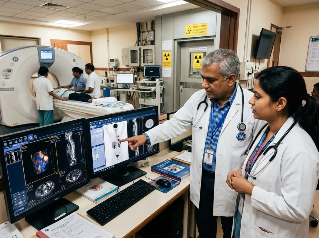
GE HealthCare has agreed to acquire Sofie Biosciences in a 945 million dollar all-cash transaction. The deal secures global rights to the investigational pan-cancer tracer FAPI-74 and expands cyclotron manufacturing infrastructure to accelerate precision theranostics and clinical molecular imaging workflows.
Today

Dual-nanofiber interpenetrating network scaffolds combining silica and PLLA/gelatin nanofibers with sodium alginate achieve complementary mechanical stability, immunomodulatory M2 macrophage polarization, and enhanced osteogenesis for advanced bone repair.
Today

New research reveals that amylin receptor signalling in the laterodorsal tegmental nucleus controls food intake through inhibitory projections to the ventral tegmental area. This study demonstrates how midbrain reward circuits regulate feeding, presenting crucial targets for next-generation obesity pharmacotherapy.
Today
