.webp&w=384&q=75)

Loading, please wait...
Loading, please wait...

Charcot-Marie-Tooth disease type 1A represents the most prevalent inherited peripheral neuropathy, presenting with progressive distal muscle weakness, sensory loss, and foot deformities. Clinicians face major challenges when tracking disease progression in Charcot-Marie-Tooth type 1A because symptoms evolve very slowly over decades. Standard functional scales frequently lack the sensitivity required to detect subtle neuromuscular changes during short clinical trials. Consequently, researchers increasingly rely on quantitative muscle MRI as an objective, non-invasive biomarker. Quantitative muscle MRI directly measures microstructural alterations, including muscle fat accumulation and edema, within affected limb muscles. By quantifying proton density fat fraction and muscle water relaxation times, clinicians establish precise baselines and detect subclinical disease progression before functional decline becomes obvious. Integrating quantitative imaging into routine clinical trials bridges a critical gap in neuromuscular medicine, enabling precise monitoring of disease dynamics and therapeutic response in hereditary demyelinating neuropathies.
To evaluate the longitudinal sensitivity of quantitative muscle MRI, investigators performed a prospective two-year study involving adult patients with genetically confirmed Charcot-Marie-Tooth type 1A and healthy controls. Participants underwent detailed assessments at baseline, twelve months, and twenty-four months. The imaging protocol focused on lower extremity musculature, utilizing automated three-dimensional whole-muscle segmentation across eighteen proximal thigh muscles and ten distal leg muscles. This automated segmentation approach ensured accurate evaluation of entire muscle volumes without relying on localized single-slice measurements. The primary magnetic resonance parameters quantified included proton density fat fraction, reflecting chronic muscle tissue fat replacement, and water T2 relaxation times, serving as a potential marker of active denervation and tissue edema. Healthy control data allowed researchers to distinguish disease-specific neuromuscular alterations from normal age-related changes. Furthermore, automated whole-muscle segmentation minimized operator bias, providing highly reproducible longitudinal measurements across consecutive clinical visits.
Alongside quantitative magnetic resonance imaging, the study evaluated a comprehensive battery of clinical and patient-reported outcome measures. Neurological impairment was assessed using the Charcot-Marie-Tooth Neuropathy Score version two and Overall Neuropathy Limitations Score. Physical function and motor performance were systematically evaluated through the thirty-two-item Motor Function Measure, six-minute walk distance, ten-meter walk test, thirty-second sit-to-stand test, and nine-hole peg test. In addition, isometric strength testing and Medical Research Council sum scores quantified lower limb muscle strength. Patient-reported assessments included the Individualized Neuromuscular Quality of Life questionnaire, ActivLim disability scale, Fatigue Severity Scale, and Brief Pain Inventory. Combining objective functional metrics with patient-reported outcomes allowed researchers to evaluate how structural muscle changes correlate with physical disability, fatigue, pain, and daily functional limitations over time. Consequently, this multidimensional framework provided a thorough characterization of disease burden in affected individuals.
At baseline evaluation, quantitative muscle MRI revealed significant structural differences between patients with Charcot-Marie-Tooth type 1A and healthy controls. Proton density fat fraction was markedly elevated across all lower limb muscle groups in affected individuals. In accordance with the typical distal-to-proximal gradient of disease involvement, fat accumulation was substantially higher in distal leg muscles than in proximal thigh musculature. Distal muscles, particularly in the anterior and lateral calf compartments, demonstrated extensive fibrofatty muscle replacement. Additionally, water T2 relaxation times were significantly increased in specific muscle groups, indicating active denervation, mild inflammation, or metabolic tissue alteration. Baseline fat fraction values correlated strongly with composite clinical impairment scores and reduced muscle strength. These baseline findings confirm that quantitative fat fraction accurately reflects cumulative neurogenic muscle damage, serving as a reliable surrogate marker for clinical severity in hereditary neuropathies.
Over the two-year observation period, quantitative muscle MRI demonstrated high sensitivity in detecting ongoing disease progression. Proton density fat fraction showed a statistically significant, steady increase across distal and proximal leg muscles in patients, whereas control values remained stable. Notably, fat fraction changes over twelve and twenty-four months were detectable even in patients whose functional clinical scores showed no measurable decline. Standard clinical functional tests demonstrated modest changes over time, requiring significantly larger sample sizes to detect statistical differences. Therefore, muscle fat fraction proved to be a highly responsive imaging biomarker capable of detecting disease progression within typical clinical trial timeframes. Water T2 values fluctuated in response to disease activity, suggesting utility in capturing active denervation processes. Ultimately, these longitudinal data demonstrate that quantitative imaging biomarkers reduce sample size requirements for prospective therapeutic trials.
The findings from this prospective two-year natural history study have significant implications for neuromuscular clinical practice and trial design. Identifying responsive, objective biomarkers has long been a primary hurdle in drug development for slowly progressive neuropathies. By proving that quantitative proton density fat fraction reliably tracks structural muscle loss over twelve to twenty-four months, this study supports using quantitative muscle MRI as a primary endpoint in upcoming clinical trials. Utilizing automated whole-muscle segmentation enhances trial feasibility by standardizing image analysis across multi-center studies. In routine clinical care, quantitative imaging helps clinicians monitor disease trajectories, evaluate rehabilitation strategies, and counsel patients regarding functional prognosis. Combining imaging markers with functional assessments ensures that novel disease-modifying therapies demonstrate both anatomical preservation and functional benefits. Overall, quantitative muscle imaging represents a transformative tool for accelerating clinical trial readiness and therapeutic development.
Quantitative muscle MRI provides an objective, highly sensitive method to track intramuscular fat accumulation and edema in Charcot-Marie-Tooth type 1A. Because CMT1A progresses slowly, standard functional clinical scales often fail to detect disease changes over short periods. Quantitative MRI captures subtle structural muscle alterations, making it an ideal surrogate endpoint for clinical trials.
Proton density fat fraction (PDFF) and water T2 relaxation times are the most effective magnetic resonance parameters. PDFF measures chronic fibrofatty muscle replacement, showing steady increases over time. Water T2 relaxation time reflects active denervation, metabolic changes, or low-grade muscle edema, offering vital insights into ongoing disease activity in peripheral neuropathies.
Quantitative muscle MRI significantly improves clinical trial design by providing high sensitivity to structural disease progression over 12 to 24 months. By using responsive imaging biomarkers like muscle fat fraction, investigators can reduce required sample sizes and shorten trial durations, accelerating the evaluation and regulatory approval of novel disease-modifying therapies.
Disclaimer: This content is for informational and educational purposes only. It does not constitute medical advice, diagnosis, or treatment. Healthcare professionals should rely on their clinical judgment and refer to the latest local and national guidelines for clinical practice.
References

Read summarized clinical updates, watch expert medical content, and earn CME certifications right from your smartphone.
Open your smartphone camera to scan and install.





A prospective 24-month study demonstrates that quantitative muscle MRI effectively tracks disease progression and muscle fat accumulation in Charcot-Marie-Tooth type 1A patients, offering a highly sensitive biomarker for upcoming clinical trials.
last month
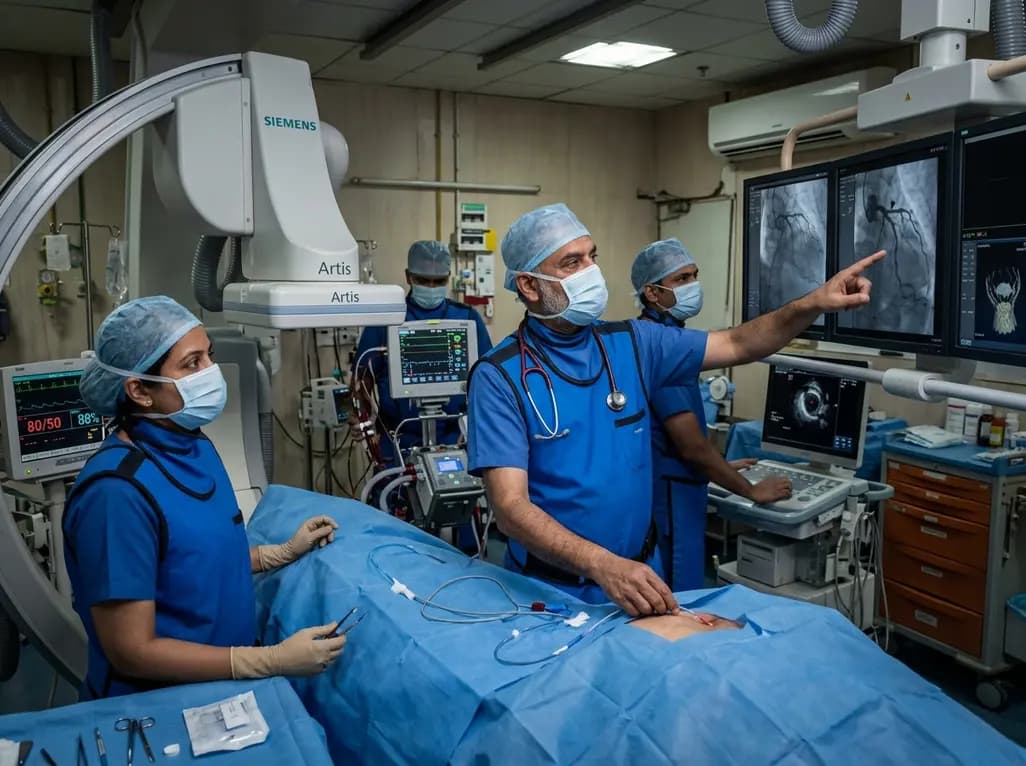
A rare case of acute coronary obstruction during TAVR caused by suspected calcified amorphous tumor embolization underscores the critical role of intracardiac imaging, emergency ECMO, and the paradox of enlarged-aperture valve geometry.
Today

Prelacteal feeding persists in South Asian communities despite known clinical risks. This review examines determinants from Chitral, pathophysiological hazards, and pediatric strategies to eliminate harmful feeds and support exclusive breastfeeding.
Today

Transcatheter mitral valve replacement in patients with degenerated bioprostheses carries a substantial risk of LVOT obstruction. Combining leaflet modification via the BATMAN technique with mechanical circulatory support offers a viable, life-saving solution for anatomically challenging surgical candidates.
Today
A comparative cohort study demonstrates that patients undergoing total hip arthroplasty after prior hip arthroscopy achieve comparable functional recovery, low complication rates, and equivalent implant survivorship when conversion occurs after six months.
Today

A cross-sectional study of 320 adults evaluated the link between habitual fasting duration and dietary quality using Nutrition Quotient scores, highlighting the clinical necessity of balancing time-restricted eating with nutrient density and meal quality in routine metabolic practice.
Today
