.webp&w=384&q=75)

Loading, please wait...
Loading, please wait...

A recent cadaveric study published in May 2026 introduces a standardized roadmap for **pudendal nerve surgery**. This guide aims to improve outcomes in complex pelvic procedures. Surgical precision remains paramount when navigating the female pelvis. Consequently, researchers detailed a three-stage, nine-step approach to safely expose the nerve. This guide provides a framework to avoid iatrogenic injury. It specifically supports nerve-sparing techniques for pudendal neuralgia and deep infiltrating endometriosis.
Specifically, the study analyzed 60 female hemipelvises to document key landmarks. The results emphasize the relationship between the nerve and the sacrospinous ligament. Moreover, identifying landmarks early can reduce neurovascular complications. Therefore, this framework serves as an essential tool for gynecologists. It helps those managing chronic pelvic pain and pelvic floor dysfunction. Furthermore, the findings clarify the branching patterns of the pudendal nerve across different subjects.
The dissection process begins with the exposure of the genitofemoral nerve and the obturator fossa. This first stage allows surgeons to clear the fossa and establish an initial baseline. Next, the procedure focuses on identifying the lumbosacral trunk and the sciatic nerve near the greater sciatic notch. This second stage is vital because it establishes the proximal boundaries of the operative field. Finally, the surgeon visualizes the pudendal nerve beneath the sacrospinous ligament. In addition to these steps, the study highlights branching variations that clinicians must anticipate during dissection.
Notably, the proximity of the nerve to the ischial spine makes it vulnerable during vaginal surgeries. Sacrospinous ligament fixations often occur near this site. Furthermore, a deep understanding of these anatomical relations supports the successful decompression of the pudendal canal. Consequently, adopting this nine-step protocol can enhance surgical precision across diverse specialties. Ultimately, this cadaveric roadmap offers a practical solution to the challenges of anatomical variations in pelvic neurovasculature.
The primary landmarks include the ischial spine, the sacrospinous ligament, and the greater sciatic notch. Identifying these structures helps surgeons locate the nerve and its branches safely.
By following a systematic nine-step process, surgeons can account for anatomical variations and establish clear visual boundaries. This structured method minimizes the risk of accidental nerve damage during complex dissections.
This approach is particularly beneficial for procedures involving pudendal neuralgia, deep infiltrating endometriosis, and complex gynecologic pelvic surgeries requiring nerve preservation.
Disclaimer: This content is for informational and educational purposes only. It does not constitute medical advice or establish a doctor-patient relationship. Always seek the advice of a qualified healthcare provider regarding a medical condition. Refer to the latest local and national guidelines for clinical practice.
References
Eşkin Tanrıverdi MD et al. Applied surgical anatomical approach to pudendal nerve: Step-by-step key neurovascular structures for pelvic nerve surgery. Int J Gynaecol Obstet. 2026 May 09. doi: 10.1002/ijgo.71065. PMID: 42104845.
OpenAnesthesia. Pudendal Nerve Blocks - Anatomy and Techniques. [Accessed May 2026].
StatPearls. Anatomy, Abdomen and Pelvis, Pudendal Nerve. [Accessed May 2026].

Read summarized clinical updates, watch expert medical content, and earn CME certifications right from your smartphone.
Open your smartphone camera to scan and install.





A cadaveric study defines a nine-step roadmap for pudendal nerve surgery, helping surgeons identify key landmarks and avoid nerve injury during pelvic proce...
5 months ago

A breakthrough ultrathin multimodal sensor decouples physical hemodynamic monitoring from disposable electrochemical calibration, achieving long-term wearable glucose prediction with 81.36% accuracy and a 9.12% MARD. This offers a robust, drift-free solution for outpatient metabolic surveillance.
Today

A clinical review of menopause and neurology highlights the neurobiological impacts of midlife hormonal shifts on cognition, epilepsy, migraine, multiple sclerosis, and stroke, emphasizing safe hormone therapy prescribing.
Today

Co-administering antiseizure medications and DOACs frequently causes pharmacokinetic interactions, subtherapeutic drug levels, and thromboembolic failures. This review highlights enzyme induction risks, unpredictable outcomes with levetiracetam, and the vital role of therapeutic drug monitoring.
Today
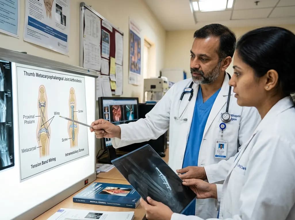
A comparative review of tension band wiring and intramedullary screw fixation in thumb metacarpophalangeal arthrodesis, evaluating union rates, hardware complications, and clinical outcomes.
Today

Following regulatory scrutiny from FSSAI regarding promotional representations and lean muscle preservation claims, Dr. Reddy's Laboratories and Nestlé Health Science are currently reviewing the branding and marketing strategy for their joint nutritional supplement Celevida GLP+.
Today
