.webp&w=384&q=75)

Loading, please wait...
Loading, please wait...

Gliomas represent the most common and aggressive primary malignant brain tumors in adult patients. Accurate histological grading guides surgical resection, radiation therapy planning, and chemotherapy regimens. In clinical neuro-oncology, the Ki-67 proliferation index serves as a fundamental immunohistochemical marker. Specifically, this nuclear protein reflects cellular proliferation rates, tumor invasiveness, and overall recurrence risk. Consequently, reliable preoperative Ki-67 prediction provides neurosurgeons with crucial biological information prior to entering the operating room. Traditional diagnostic methods depend upon stereotactic biopsy or open surgical resection. However, invasive biopsies carry inherent procedural risks, including intracranial hemorrhage, infection, and new focal neurological deficits. Furthermore, focal tissue sampling frequently misses spatial intratumoral heterogeneity, leading to inaccurate tumor grading. To resolve these diagnostic hurdles, clinicians actively seek noninvasive tools that estimate proliferation index preoperatively. Routine peripheral blood parameters offer an ideal diagnostic substrate. Standard blood tests are universally accessible, standardized, inexpensive, and fast. Therefore, integrating routine systemic biomarkers with modern artificial intelligence creates a practical clinical solution. By analyzing peripheral blood signals, surgical teams can characterize glioma aggressiveness well before surgical intervention, thereby improving preoperative planning and personalized patient counseling.
To construct a dependable predictive framework, researchers initiated a retrospective multicenter study across two medical institutions between January 2020 and December 2023. The final study cohort comprised 506 patients with pathologically confirmed glioma. The investigators partitioned these participants into three distinct groups: a training cohort of 352 patients, an internal validation cohort of 88 patients, and an external validation cohort of 66 patients. Clinicians collected all preoperative laboratory values within one week prior to neurosurgery from hospital laboratory information systems. In accordance with established pathological criteria, researchers stratified patients into a low Ki-67 group with an index below 10 percent and a high Ki-67 group with an index equal to or greater than 10 percent. Subsequently, investigators implemented three rigorous machine learning algorithms to screen candidate parameters and reduce dimensionality. Specifically, they utilized extreme gradient boosting, support vector machine algorithms, and least absolute shrinkage and selection operator regression. By requiring candidate markers to satisfy all three selection models simultaneously, the team eliminated uninformative noise and prevented statistical overfitting. Ultimately, this rigorous protocol narrowed dozens of routine blood parameters down to 15 core laboratory predictors, providing a robust dataset for model training.
Following feature extraction, the research team constructed and compared ten distinct machine learning models. The evaluated algorithms included support vector machines, extreme gradient boosting, and random forest classifiers. The team also examined adaptive boosting, neural networks, naive Bayes, and multivariable logistic regression. Investigators evaluated model performance across validation cohorts using receiver operating characteristic analysis, sensitivity, specificity, and accuracy metrics. In addition, decision curve analysis assessed clinical utility across varying threshold probabilities. Interestingly, while complex ensemble classifiers performed adequately, the logistic regression model demonstrated the highest overall diagnostic performance. Specifically, the logistic regression model achieved an area under the curve (AUC) of 0.838 in the primary training cohort. Furthermore, it achieved an AUC of 0.800 in the internal validation cohort, with an accuracy of 0.782 and an optimal sensitivity of 0.845. Most importantly, the model retained robust discriminative ability in the independent external validation set, recording an AUC of 0.757. To translate these results into clinical practice, the authors visualized the model as an intuitive nomogram. The nomogram incorporates six top laboratory features: patient age, anion gap, apolipoprotein A-1, apolipoprotein B, calcium, and creatinine. Therefore, this validated tool generates individualized probabilities at the bedside.
The statistical inclusion of these specific laboratory indicators reveals meaningful physiological links between intracranial tumors and host metabolism. Although the blood-brain barrier restricts large molecule exchange, invasive gliomas exert widespread systemic metabolic influences. For example, rapidly proliferating neoplastic cells heavily rely on aerobic glycolysis, famously termed the Warburg effect. Consequently, accelerated glycolysis releases excess lactic acid, which directly influences systemic serum anion gap measurements. In addition, aggressive brain tumors alter host lipid homeostasis to harvest cholesterol for cell membrane construction. Depleted apolipoprotein A-1 coupled with altered apolipoprotein B concentrations highlights progressive systemic inflammation and lipid depletion. Similarly, aberrant serum calcium dynamics mirror malignant signaling pathways that regulate glioma cell migration and angiogenesis. Meanwhile, fluctuations in serum creatinine reflect nutritional decline, muscle wasting, and cancer-associated systemic stress. Furthermore, patient age correlates with cumulative genetic mutations and diminished immunological surveillance against tumor growth. Therefore, routine blood biomarkers do not act as passive systemic bystanders. Instead, they serve as sensitive barometers of tumor-induced systemic disruption. Recognizing these physiological relationships reassures clinicians that the machine learning algorithm captures true biological tumor behavior rather than statistical artifacts.
This predictive nomogram offers substantial clinical relevance for neurosurgical and oncology centers throughout India. In many Indian healthcare settings, access to advanced neuroimaging techniques or rapid molecular pathology testing remains constrained. Advanced MRI sequences, such as MR spectroscopy and perfusion imaging, entail significant equipment costs and specialized radiological expertise. In contrast, standard complete blood counts, electrolyte panels, renal function tests, and lipid profiles are widely accessible across district and tertiary hospitals. Consequently, Indian neurosurgeons can implement this inexpensive nomogram during initial hospital admission without increasing financial burdens on patients. Identifying a high Ki-67 proliferation index preoperatively alerts surgical teams to plan maximal safe surgical resection. Teams can arrange advanced intraoperative tools, such as neuronavigation and fluorescence guidance, whenever high cellular proliferation is anticipated. In addition, surgical teams can schedule early postoperative chemoradiotherapy consultations, preventing dangerous treatment delays. Conversely, predicting low proliferative activity helps surgeons preserve functional neurological tissue in eloquent brain regions. Furthermore, early risk estimation guides communication with patients and families regarding expected treatment intensity. Ultimately, adopting blood-based machine learning tools supports equitable, data-driven neuro-oncological care across resource-variable healthcare environments.
Preoperative Ki-67 prediction helps neurosurgeons gauge tumor proliferation and biological aggressiveness before entering the operating room. High Ki-67 expression typically warrants maximal safe resection alongside accelerated adjuvant chemoradiotherapy planning. Conversely, predicting low proliferation helps clinicians avoid overtreatment and tailor individualized perioperative monitoring pathways.
The logistic regression model identified six primary clinical parameters: patient age, anion gap, apolipoprotein A-1, apolipoprotein B, serum calcium, and serum creatinine. These accessible laboratory markers reflect systemic metabolic alterations, inflammation, and cellular turnover driven by invasive intracranial glioma growth.
No, this machine learning nomogram is not an absolute replacement for definitive histopathological and molecular tissue testing. Instead, it serves as a noninvasive, complementary triage tool. It empowers surgical teams to anticipate glioma aggressiveness preoperatively when stereotactic biopsies are high risk, delayed, or anatomically challenging.
Disclaimer: This content is for informational and educational purposes only... Refer to the latest local and national guidelines for clinical practice.
References

Read summarized clinical updates, watch expert medical content, and earn CME certifications right from your smartphone.
Open your smartphone camera to scan and install.





Researchers developed a machine learning model utilizing standard routine blood parameters to achieve noninvasive preoperative Ki-67 prediction in patients with glioma. This accessible nomogram demonstrates high diagnostic accuracy, aiding neurosurgeons and oncologists in risk stratification and surgical planning.
Today

Digital health tools often perform unevenly across diverse populations, reinforcing systemic inequities. Addressing this challenge requires transparent reporting of subgroup performance, representative dataset curation, and rigorous postdeployment monitoring across commercial healthcare technologies.
Today

Prelacteal feeding persists in South Asian communities despite known clinical risks. This review examines determinants from Chitral, pathophysiological hazards, and pediatric strategies to eliminate harmful feeds and support exclusive breastfeeding.
Today
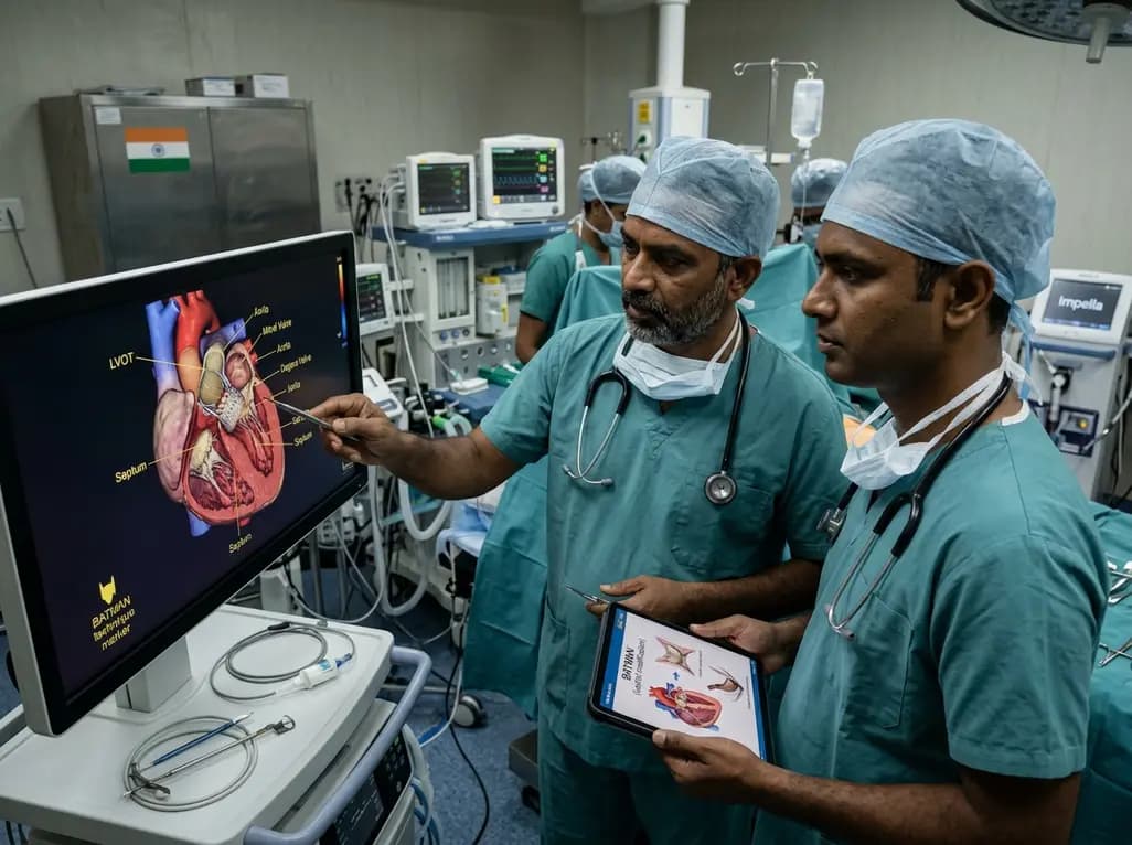
Transcatheter mitral valve replacement in patients with degenerated bioprostheses carries a substantial risk of LVOT obstruction. Combining leaflet modification via the BATMAN technique with mechanical circulatory support offers a viable, life-saving solution for anatomically challenging surgical candidates.
Today

A comparative cohort study demonstrates that patients undergoing total hip arthroplasty after prior hip arthroscopy achieve comparable functional recovery, low complication rates, and equivalent implant survivorship when conversion occurs after six months.
Today
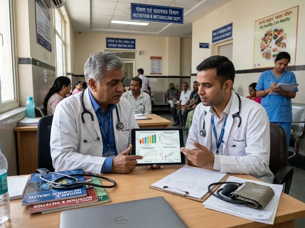
A cross-sectional study of 320 adults evaluated the link between habitual fasting duration and dietary quality using Nutrition Quotient scores, highlighting the clinical necessity of balancing time-restricted eating with nutrient density and meal quality in routine metabolic practice.
Today
