.webp&w=384&q=75)

Loading, please wait...
Loading, please wait...

Accurate oral cancer prediction remains one of the greatest clinical challenges in modern oncology. Clinicians frequently encounter oral potentially malignant disorders, such as leukoplakia, erythroplakia, and oral submucous fibrosis. However, predicting which precursor lesions will undergo malignant transformation presents substantial difficulty. Currently, malignant progression rates across these lesions vary widely, ranging from less than one percent to over ten percent annually. Consequently, clinicians must balance the hazard of underestimating aggressive lesions against the harm of overtreating indolent mucosal alterations. In high-prevalence countries like India, where betel quid and tobacco consumption drive significant disease burdens, this clinical dilemma escalates rapidly. Early detection undeniably improves survival rates, yet standard clinical examinations cannot reliably differentiate progressing lesions from stable mimics. Therefore, oncology teams urgently require objective risk stratification frameworks. Healthcare practitioners must detect neoplastic changes well before invasive malignancy develops. By advancing predictive surveillance methods, clinicians can timely intervene, preserve vital oral architecture, and notably minimize morbidity. Developing standardized prognostic workflows represents an imperative clinical goal for head and neck oncology teams worldwide.
For decades, surgical pathologists have relied primarily on routine hematoxylin and eosin staining to assess epithelial dysplasia. Nevertheless, light microscopic evaluation alone exhibits persistent diagnostic limitations. Inter-observer and intra-observer variability significantly plague conventional dysplasia grading schemes, creating substantial discordance among experienced pathologists. In addition, architectural atypia and cytological abnormalities do not always correlate directly with ultimate genomic instability. A lesion classified as mild dysplasia may unexpectedly harbor aggressive driver alterations, whereas moderate dysplasia might remain quiescent for years. Furthermore, conventional biopsy sampling captures only a localized tissue snapshot, overlooking spatial heterogeneity within extensive mucosal fields. Carcinogen exposure frequently generates widespread field cancerization throughout the upper aerodigestive tract. Hence, a single focal biopsy may completely miss adjacent clones undergoing active malignant transition. Consequently, histopathology alone delivers an incomplete assessment of biological risk. Clinicians cannot rely purely on static morphological features to guide definitive surgical or ablative interventions. These diagnostic uncertainties underscore an urgent imperative for multimodal biological characterization. Integrating high-throughput molecular markers provides the necessary precision to resolve these diagnostic ambiguities.
To overcome existing diagnostic hurdles, researchers propose an analytical framework aligned with the National Cancer Institute's Human Tissue Atlas Network. This sophisticated approach systematically evaluates multiple biological layers within oral mucosal tissue. First, whole exome sequencing identifies critical somatic mutations, copy number variations, and overarching genomic risk. Second, comprehensive methylation arrays measure epigenetic modifications that reflect environmental carcinogen exposure and tobacco-induced cellular reprogramming. Third, bulk RNA sequencing profiles transcriptional dynamics, capturing dysregulated signaling cascades, immune evasion pathways, and proliferative programs. Most importantly, spatial transcriptomics resolves gene expression within intact tissue architecture. By preserving topography, spatial technologies illuminate cellular interactions across the neoplastic boundary and reveal critical tumor-microenvironment dynamics. Standardizing these multimodal protocols across academic centers ensures high reproducibility and creates comparable multi-institutional registries. Moreover, mapping multiomic data across longitudinal time points elucidates the precise biological transitions driving malignant initiation. Ultimately, this multi-layered biological profiling replaces subjective grading with objective, quantifiable molecular metrics. Such comprehensive stratification enables clinicians to distinguish benign inflammatory conditions from high-risk, progressing premalignancies with unprecedented accuracy.
Although comprehensive multiomic profiling yields extraordinary biological insights, its substantial economic cost and technical complexity restrict widespread clinical adoption. To bridge this critical gap, investigators are deploying modern machine learning architectures. Specifically, researchers utilize deep learning algorithms to correlate microscopic morphology on routine hematoxylin and eosin slides with paired multiomic profiles. Once these models learn subtle image-molecular relationships, developers formulate an innovative Teacher-Student computational framework. In this paradigm, the sophisticated multiomic Teacher model transfers its diagnostic knowledge into a streamlined Student neural network. Consequently, the deployed Student model infers complex molecular alterations using only digital histopathology images and basic clinical covariates. Clinicians can derive profound prognostic predictions without requiring expensive genomic sequencing for every individual patient. Furthermore, this computational methodology democratizes access to advanced precision diagnostics across resource-constrained clinical settings. Pathologists can rapidly analyze routine biopsies, identify occult molecular drivers, and flag high-risk premalignant lesions during everyday diagnostic workflows. Therefore, integrating machine learning with traditional microscopy converts routine histology into an informative, molecularly annotated diagnostic asset.
The clinical translation of standardized multimodal prediction models promises to revolutionize head and neck surgical oncology. Currently, clinicians frequently face acute dilemmas regarding optimal surveillance intervals and surgical margins. By providing individualized risk scores, automated computational models empower surgeons to tailor interventions according to actual biological behavior. Low-risk patients can safely undergo conservative observational surveillance, avoiding unnecessary invasive surgical procedures and post-treatment morbidity. Conversely, individuals demonstrating high-risk molecular phenotypes can receive prompt operative excision, photodynamic therapy, or chemoprevention. Furthermore, this standardized framework holds immense promise for high-burden regions like India. Deploying scalable artificial intelligence tools in district clinics will rapidly enhance diagnostic triage and prioritize oncology referrals. Looking ahead, prospective multi-center validation remains essential to confirm algorithm accuracy across diverse patient cohorts. Developers must ensure that artificial intelligence models remain transparent and interpretable for practicing pathologists. In addition, future research should integrate liquid biopsy biomarkers, such as salivary microRNAs, into existing histological networks. Ultimately, combining non-invasive screening with image-based molecular inference will shift oral cancer management from reactive treatment toward proactive, personalized cancer prevention.
Spatial transcriptomics uniquely preserves the anatomical architecture of tissue while profiling gene expression across specific cellular niches. Conventional bulk sequencing homogenizes whole biopsies, obscuring crucial microenvironmental interactions. In contrast, spatial profiling localizes transcripts to distinct dysplastic cells, invasive margins, and infiltrating immune subsets. Consequently, clinicians gain deep biological clarity regarding early invasive changes. This detailed spatial context helps researchers identify malignant transformation long before overt morphological destruction becomes visible under standard microscopy.
The Teacher-Student model bridges complex multiomic science and practical patient care. First, a complex Teacher network learns multi-layered molecular and spatial associations from high-cost genomic assays and paired histology. Subsequently, this knowledge trains a lightweight Student model. Once trained, the Student algorithm infers underlying molecular signatures directly from standard hematoxylin and eosin slides and basic clinical covariates alone. Consequently, resource-limited clinics can deliver high-accuracy precision prognostication without requiring expensive laboratory genetic testing.
India bears a disproportionately high global burden of oral cancer due to rampant use of areca nut, gutkha, and tobacco. These habits trigger chronic inflammation and widespread field cancerization across the oral cavity. Conventional biopsies often miss progressing subclones in extensive mucosal lesions. Furthermore, standardized multiomic analysis decodes unique carcinogen-induced mutational signatures and epigenetic changes. Deploying trained predictive models into community centers allows early risk triage, guiding timely surgical care and conserving critical oncology resources.
Disclaimer: This content is for informational and educational purposes only... Refer to the latest local and national guidelines for clinical practice.
References

Read summarized clinical updates, watch expert medical content, and earn CME certifications right from your smartphone.
Open your smartphone camera to scan and install.





Standardized multiomic analysis and machine learning models enable accurate risk prediction for oral precursor lesions, transforming malignant transformation assessment.
Today
A landmark cross-sectional study in Algeria and Türkiye reveals distinct age-related molecular IgE sensitization profiles between children and adults, highlighting higher house dust mite reactivity in youth and providing vital insights for precision allergen immunotherapy.
Today
A recent case-control study identified key protein changes in peritoneal fluid-derived small extracellular vesicles, highlighting candidates like S100-A10 and ALDOA as potential biomarkers to overcome diagnostic delays in endometriosis.
Today
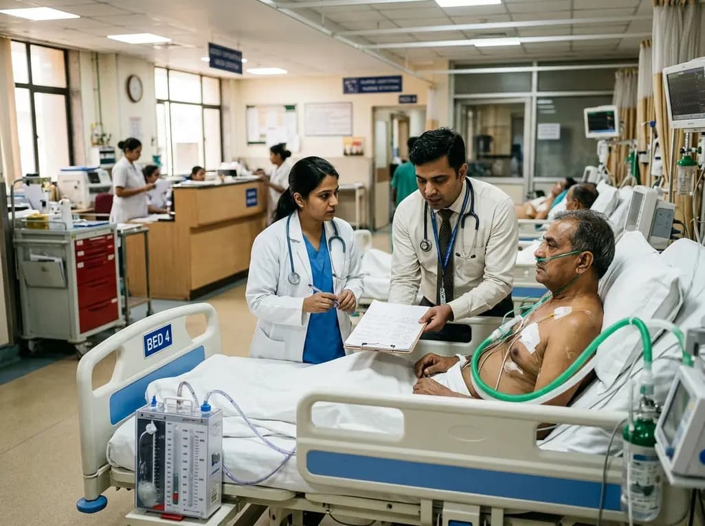
Managing refractory hypoxemia after cardiothoracic surgery becomes perilous when prolonged air leak limits positive-pressure ventilation. This case-based review details how high-flow nasal cannula, paired with awake rehabilitation, enabled successful extubation and avoided invasive re-intubation.
Today
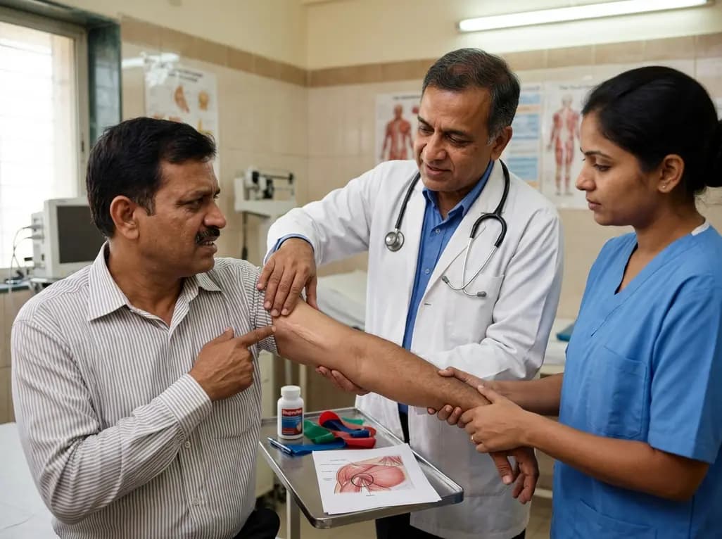
Idiopathic axillary web syndrome (IAWS) is a rare cause of shoulder pain and mobility restriction characterized by palpable axillary cording without prior surgery or trauma. Early diagnosis, nonsteroidal anti-inflammatory therapy, and physical rehabilitation lead to complete resolution of symptoms.
Today

SWEDEPAD-1 trial insights show paclitaxel-coated devices do not improve long-term limb salvage in patients with chronic limb-threatening ischemia and tissue loss. While one-year reinterventions decreased, the devices were linked to a higher risk of major amputation at three months.
Today
