.webp&w=384&q=75)

Loading, please wait...
Loading, please wait...

Managing post-cardiotomy respiratory failure presents significant clinical hurdles, particularly when alveolar-pleural disruptions complicate conventional management. The application of a high-flow nasal cannula offers a viable non-invasive avenue to support gas exchange when conventional mechanical strategies pose risks. Invasive positive-pressure ventilation often perpetuates pleural fistula flow and delays healing. Therefore, clinicians require adaptable weaning strategies that secure systemic oxygen delivery while safeguarding healing pulmonary tissue.
Prolonged air leak represents one of the most frustrating thoracic complications following complex cardiothoracic procedures. When patients undergo extensive surgical interventions, pleural disruptions can fail to seal spontaneously. Mechanical ventilation with high positive end-expiratory pressure delivers continuous distending force across the injured visceral pleura. Consequently, high airway pressures prevent parenchymal closure, worsen subcutaneous emphysema, and increase chest drain duration. Prolonged pleural drainage escalates systemic infection risks and dramatically lengthens critical care stays. Furthermore, persistent air leakage impairs normal diaphragmatic mechanics and predisposes lung tissue to progressive collapse. Physicians frequently face an agonizing compromise between providing adequate ventilatory support and minimizing fistula progression. Traditional management heavily relies on reducing airway driving pressures, yet this adjustment often produces hypercapnia and critical arterial desaturation.
Patients undergoing simultaneous coronary artery bypass grafting and valve replacements inherently endure prolonged cardiopulmonary bypass durations. Systemic inflammation and pulmonary endothelial permeability regularly peak within the initial seventy-two hours postoperatively. When extensive bilateral air leaks arise during this vulnerable window, traditional alveolar recruitment maneuvers become exceedingly hazardous. Clinicians cannot simply increase positive airway pressures to counteract progressive atelectasis, because alveolar overdistension immediately accelerates transpleural air transit. Moreover, bilateral thoracostomy tubes compromise thoracic wall compliance and induce substantial chest wall pain. Splinting impairs deep breathing and suppresses the native cough reflex. Consequently, severe arterial hypoxemia may persist despite maximal tolerable ventilator settings. Escalating invasive ventilation under these circumstances risks catastrophic tension physiology and barotrauma. Therefore, the multidisciplinary intensive care team must promptly recognize the futility of prolonged invasive support and orchestrate safe extubation.
Liberating the patient from endotracheal intubation removes constant positive-pressure duress from the pulmonary parenchyma. In this complex setting, high-flow nasal cannula serves as a versatile bridge to restore physiological respiratory dynamics. Following planned extubation at hour seventy-two, initiating heated and humidified high-flow oxygen delivers continuous anatomical dead space clearance. Clinical observations indicate substantial gas exchange enhancement within twenty-four hours of initiation, demonstrating marked PaO2 gains alongside significant PaCO2 reductions. Furthermore, delivering warm, conditioned gases preserves ciliary activity and liquefies retained bronchial secretions. Because the circuit functions as an open system, it avoids the deleterious peak alveolar pressures seen during invasive mechanical breaths. The device stabilizes resting respiratory frequency and substantially decreases dyspnea sensations. Consequently, patients tolerate early liberation from mechanical ventilation without experiencing post-extubation respiratory collapse.
The therapeutic superiority of high-flow nasal therapy relies on multiple distinct physiological mechanisms. Firstly, administering high gas flow rates matches or exceeds the peak inspiratory demand of dyspneic post-surgical patients. This dynamic flow rate eliminates ambient air dilution and guarantees consistent fraction of inspired oxygen delivery. Secondly, continuous pharyngeal flushing washes out expired carbon dioxide from the upper airway dead space. This washout optimizes alveolar ventilation efficiency, directly explaining rapid hypercapnia clearance. Additionally, the device generates modest, flow-dependent positive airway pressure throughout the respiratory cycle. This low-level pressure provides gentle end-expiratory stenting to open microatelectatic alveolar units without perpetuating pleural disruption. Importantly, the open circuit configuration prevents excessive intrathoracic pressure spikes during uncoordinated coughing episodes. Thus, the system enhances baseline alveolar recruitment while allowing pleural fistulas to seal undisturbed.
Successful management requires an integrated bedside protocol combining advanced oxygen delivery with proactive physical therapy. Because high-flow interfaces leave the mouth unobstructed, patients communicate effortlessly and participate actively in targeted pulmonary rehabilitation. Bedside therapists can execute awake mobilization, thoracic expansion maneuvers, and incentive spirometry earlier in the postoperative timeline. Furthermore, clinicians can safely introduce intermittent sessions of noninvasive positive-pressure support as a complementary recruitment adjunct. Careful scheduling ensures that brief positive-pressure intervals recruit dependent basal segments without reopening fragile alveolar-pleural defects. Adequate regional analgesia mitigates sternotomy discomfort and enhances voluntary patient effort. Over successive days, clinicians progressively titrate down gas flow rates and inspired oxygen concentrations as pleural drainage ceases. Consequently, this collaborative multimodal pathway prevents secondary atelectrauma and successfully circumvents hazardous tracheal re-intubation.
Intensive care teams managing cardiothoracic air leaks must maintain rigorous objective monitoring. Clinicians should track arterial blood gas kinetics, dynamic chest tube air bubbling, and clinical breathing workload around the clock. If persistent hypercapnia or deteriorating hypoxemic ratios develop, teams must promptly exclude tension pneumothorax or tube malposition. Moreover, selecting appropriately sized nasal prongs ensures accurate dead space washout while avoiding unintentional occlusion of the nares. Early enteral nutrition and meticulous fluid balance further accelerate visceral pleural remodeling. By establishing a cooperative multidisciplinary environment, cardiac surgeons, intensivists, and respiratory therapists can deploy non-invasive options confidently. Transforming traditional weaning paradigms minimizes invasive mechanical exposure and improves recovery trajectories for fragile cardiothoracic patients.
The modality operates via an open circuit that generates only modest, dynamic end-expiratory pressure. Because peak inspiratory airway pressures remain significantly lower than those delivered during invasive mechanical ventilation, the transpulmonary pressure gradient across the damaged visceral pleura decreases. Consequently, the alveolar tear experiences minimal shear stress, which allows the persistent pleural fistula to close naturally while maintaining adequate functional residual capacity.
Extubation becomes appropriate once the patient displays stable hemodynamics, adequate airway reflexes, acceptable mental status, and reasonable metabolic stability. If persistent air leak prevents effective invasive alveolar recruitment, transitioning to spontaneous unassisted breathing reduces intrathoracic distending pressure. Clinicians can then deploy high-flow support immediately after tube removal to sustain adequate oxygenation while expediting spontaneous visceral pleural healing.
Yes, teams can safely employ intermittent noninvasive positive-pressure ventilation alongside continuous high-flow nasal support under vigilant supervision. Short, targeted noninvasive ventilation intervals provide transient alveolar recruitment for refractory basal atelectasis. Meanwhile, high-flow oxygenation maintains physiological comfort, dead space flushing, and continuous secretion clearance between positive-pressure sessions, thereby preventing respiratory muscle exhaustion without over-pressurizing healing pleural tissue.
Disclaimer: This content is for informational and educational purposes only. It is not intended to be a substitute for professional medical advice, diagnosis, or treatment. Always seek the advice of your physician or other qualified health provider with any questions you may have regarding a medical condition or treatment procedure. Never disregard professional medical advice or delay in seeking it because of something you have read here. Refer to the latest local and national guidelines for clinical practice.
References

Read summarized clinical updates, watch expert medical content, and earn CME certifications right from your smartphone.
Open your smartphone camera to scan and install.





Managing refractory hypoxemia after cardiothoracic surgery becomes perilous when prolonged air leak limits positive-pressure ventilation. This case-based review details how high-flow nasal cannula, paired with awake rehabilitation, enabled successful extubation and avoided invasive re-intubation.
Today
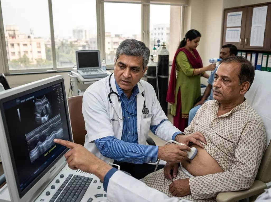
Diaphragmatic dysfunction affects over a third of chronic stroke patients, worsening sleep, mood, and functional mobility. Ultrasound assessment and inspiratory training improve outcomes.
Today

The ClinGen Prenatal Gene Curation Expert Panel evaluated 63 disease relationships across 61 genes, establishing clinical validity for severe fetal phenotypes like hydrops and stillbirth to enhance prenatal genomic interpretation and clinical care.
Yesterday
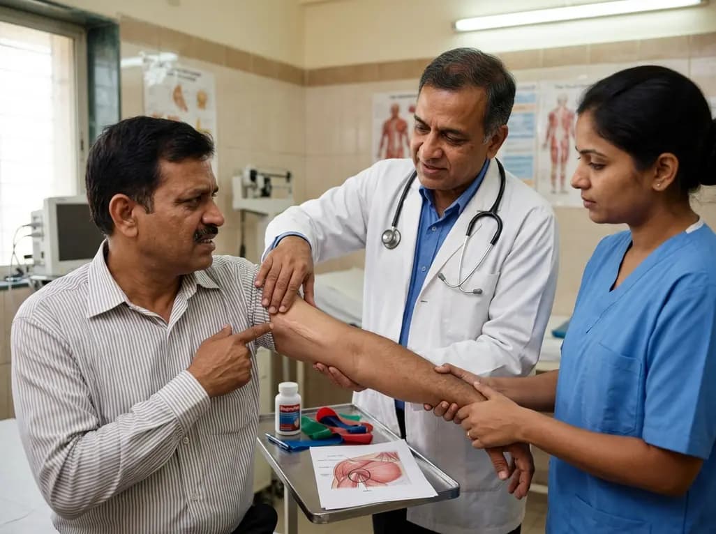
Idiopathic axillary web syndrome (IAWS) is a rare cause of shoulder pain and mobility restriction characterized by palpable axillary cording without prior surgery or trauma. Early diagnosis, nonsteroidal anti-inflammatory therapy, and physical rehabilitation lead to complete resolution of symptoms.
Today

SWEDEPAD-1 trial insights show paclitaxel-coated devices do not improve long-term limb salvage in patients with chronic limb-threatening ischemia and tissue loss. While one-year reinterventions decreased, the devices were linked to a higher risk of major amputation at three months.
Today
