.webp&w=384&q=75)

Loading, please wait...
Loading, please wait...

Cervical degenerative disc disease frequently causes radiculopathy and myelopathy, requiring surgical decompression when nonoperative therapy fails. Anterior cervical discectomy and fusion remains the established gold standard for resolving nerve root and spinal cord compression. Historically, spine surgeons utilized autografts, allografts, or polyetheretherketone implants combined with anterior plating systems. However, anterior plates often increase surgical operative duration, elevate soft tissue retraction risks, and cause persistent postoperative dysphagia. Consequently, the adoption of stand-alone interbody devices has expanded globally to mitigate anterior approach complications. In recent years, additive manufacturing has revolutionized spinal implant architecture through three-dimensional printing technology. Modern porous titanium cages provide an innovative alternative that addresses the mechanical and biological shortcomings of earlier designs. These advanced porous structures aim to enhance primary mechanical stability while actively encouraging osseous tissue ingrowth across endplate interfaces. Additionally, their engineered elastic modulus closely approximates native trabecular bone, theoretically lowering graft subsidence rates and preserving cervical sagittal alignment. A recent multicenter investigation highlights compelling real-world evidence regarding the clinical safety and efficacy of these stand-alone devices. Spine surgeons must critically evaluate these radiographic and clinical outcomes to optimize patient selection and surgical protocols.
For decades, polyetheretherketone dominated anterior cervical discectomy procedures due to its radiolucency and favourable elastic properties. Nevertheless, smooth polymer materials remain inherently hydrophobic and biologically inert, frequently provoking fibrous tissue encapsulation rather than direct osseous integration. Standard solid titanium implants offer high biocompatibility, but their excessive stiffness creates an elastic modulus mismatch that precipitates endplate subsidence. In contrast, 3D-printed porous titanium cages effectively overcome these persistent material limitations. Additive manufacturing creates interconnected macroporous and microporous architectures that mimic the natural trabecular matrix of human cancellous bone. Consequently, this interconnected network facilitates cellular adhesion, osteoblast proliferation, and vascularization throughout the implant volume. Furthermore, the porous construct significantly reduces overall cage stiffness, matching the modulus of human subchondral bone far better than solid metals. As a result, physiological load transfers uniformly across the interbody space according to Wolff's law of bone remodeling. Surgeons also gain superior friction-based frictional stability against vertebral endplates, which drastically reduces early implant migration without supplemental screws. Therefore, porous titanium represents a major biomaterial advancement for cervical spine reconstruction.
A notable multicenter study led by Panico and colleagues provides robust clinical evidence regarding stand-alone porous titanium spacers. The researchers evaluated 120 patients who underwent non-plated anterior cervical procedures across three tertiary neurosurgical units. At 12 months postoperatively, computed tomography and dynamic flexion-extension radiographs confirmed successful solid interbody fusion in 88.3 percent of patients. Furthermore, patient-reported outcome measures revealed dramatic, statistically significant improvements in subjective pain scores. The mean Numerical Rating Scale score dropped by 4.2 points, demonstrating excellent clinical relief of radicular and axial neck symptoms. Importantly, operative times were noticeably shorter because the technique eliminated anterior cervical plating and screw insertion steps. Patients mobilized rapidly following surgery without rigid external cervical orthoses in most instances. Although general physical and mental health subscores on the SF-12 questionnaire showed modest changes, patient satisfaction remained uniformly high. The absence of persistent swallowing difficulties was particularly striking compared to historical cohorts receiving anterior hardware. Thus, stand-alone porous titanium cages reliably achieve solid structural fusion and clinically meaningful symptom resolution within 12 months of surgery.
Implant subsidence represents one of the primary historical concerns when surgeons omit anterior plate fixation during cervical arthrodesis. When a cage collapses into the adjacent vertebral endplates, loss of intervertebral height can cause recurrent neuroforaminal stenosis. In the multicenter cohort, however, radiographic cage subsidence occurred in only 5.8 percent of patients over the 12-month observation period. Moreover, zero patients experienced anterior or posterior cage migration, confirming exceptional primary implant stability under physiological cervical loading. Detailed radiographic measurements demonstrated that cervical sagittal alignment parameters remained well preserved across long-term follow-up intervals. The segmental lordosis achieved intraoperatively did not deteriorate significantly during healing. Similarly, investigators detected no significant statistical correlations between baseline patient factors, such as age or baseline bone quality, and adverse radiographic settling. The roughened surface micro-topography and open porous geometry effectively grip cortical bone while distributing axial compressive forces across wide contact surfaces. Consequently, the porous structure minimizes focal stress peaks that typically cause endplate breakdown. These encouraging radiographic findings indicate that stand-alone 3D-printed porous implants mitigate subsidence risks effectively without supplemental anterior plates.
Spine surgeons continually weigh the comparative advantages of stand-alone cages against traditional plate-augmented anterior constructs. Anterior cervical plates provide rigid internal fixation, but their physical profile often irritates the anterior esophagus and sympathetic trunk. As a result, chronic dysphagia remains a frequent, frustrating complication in up to twenty percent of plated patients. In contrast, stand-alone implants reside completely within the intervertebral disc boundary, completely eliminating direct esophageal compression and friction. Furthermore, avoiding plate installation decreases surgical exposure requirements, reduces intraoperative blood loss, and shortens operative duration. Recent comparative trials confirm that stand-alone porous titanium spacers achieve equivalent fusion rates and lordotic maintenance compared to anchored or plated systems. Additionally, stand-alone cages reduce adjacent segment stress concentrations by avoiding plate impingement near un-fused disc spaces. Nevertheless, surgeons must carefully identify appropriate candidates for stand-alone interventions. Cases exhibiting severe multi-level instability, severe traumatic disruption, or significant osteoporosis may still necessitate supplemental anterior or posterior instrumentation. Overall, the stand-alone porous approach offers a refined balance between minimal invasiveness and reliable structural stability for single-level and select two-level degenerative cervical pathology.
In the Indian healthcare landscape, degenerative cervical spine disorders are rising rapidly due to aging demographics and sedentary modern lifestyles. Indian spine specialists encounter a diverse patient cohort ranging from young manual laborers to osteopenic geriatric patients. Introducing stand-alone porous titanium cages into Indian surgical centers offers distinct clinical and logistical benefits. By eliminating the necessity for cervical plates and ancillary screws, operating room turnover increases and overall implant inventory simplifies. Shorter operating times also decrease general anesthesia exposure and reduce surgical site infection risks in high-volume public and private hospitals. Moreover, patients typically experience smoother recoveries, reporting less early swallowing discomfort and requiring shorter postoperative hospital admissions. However, meticulous surgical technique remains paramount for ensuring optimal outcomes with stand-alone cages. Surgeons must perform comprehensive endplate decortication without violating the underlying cortical bone integrity to prevent catastrophic subsidence. In addition, accurate interbody footprint sizing is essential to distribute axial cervical loads across the dense peripheral ring apophysis. When applied with rigorous surgical discipline, stand-alone porous titanium cages deliver outstanding clinical durability and cost-effective value in routine spinal reconstructive practice.
Stand-alone porous titanium cages enhance osteointegration through an interconnected 3D-printed biomimetic lattice while eliminating the need for anterior plating. Consequently, this design reduces operative duration, decreases blood loss, minimizes soft tissue retraction, and significantly prevents chronic postoperative dysphagia while providing strong mechanical stability and low subsidence rates.
Porous titanium implants exhibit low subsidence rates, reported around 5.8 percent, which is comparable to or lower than conventional PEEK cages. The 3D-printed porous structure lowers the elastic modulus closer to cancellous bone, distributing compressive forces evenly and preventing focal endplate breakdown without sacrificing intervertebral height or cervical alignment.
Ideal candidates include patients suffering from single-level or select two-level cervical disc herniations or spondylotic radiculopathy with preserved vertebral bone density. Conversely, patients with severe traumatic cervical disruption, profound segment instability, significant osteoporosis, or multi-level kyphotic deformity often require supplemental anterior plate fixation or posterior pedicle screw instrumentation to ensure fusion.
Disclaimer: This content is for informational and educational purposes only and is not intended as a substitute for professional medical advice, diagnosis, or treatment. Always seek the advice of your physician or other qualified health provider with any questions you may have regarding a medical condition. Never disregard professional medical advice or delay in seeking it because of something you have read here. Refer to the latest local and national guidelines for clinical practice.
References

Read summarized clinical updates, watch expert medical content, and earn CME certifications right from your smartphone.
Open your smartphone camera to scan and install.





A multicenter study evaluates stand-alone 3D-printed porous titanium cages in ACDF, demonstrating an 88.3% fusion rate, significant pain reduction, and a low 5.8% subsidence rate without anterior plating.
Today

A mixed-methods within-subject study reveals that patients report significantly higher overall satisfaction, clearer communication, and greater staff courtesy in an acute palliative care unit compared to prior non-palliative hospital stays, despite persistent logistical challenges like nursing response times.
Today

A recent cohort study reveals that reproductive-age women with cerebral venous thrombosis have nearly double the odds of developing intracranial hypertension compared to older women. Superior sagittal and lateral sinus occlusions heighten this risk, predisposing patients to persistent visual impairment.
Today
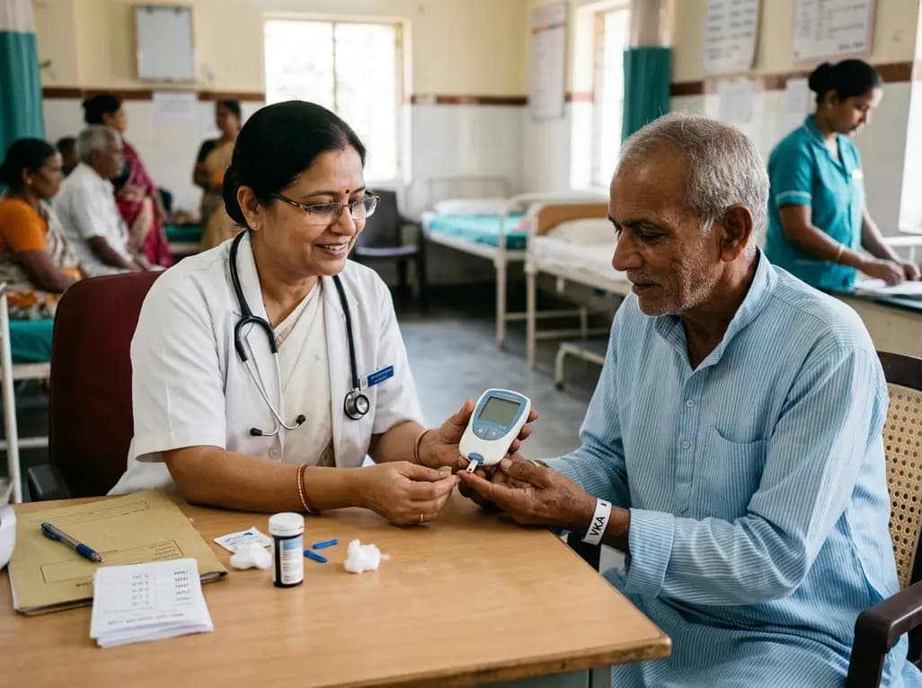
A new implementation study demonstrates that an integrated home INR monitoring network for vitamin K antagonist therapy cuts test turnaround times from three hours to 20 minutes, enhancing clinical oversight, diagnostic accuracy, and patient quality of life.
Today
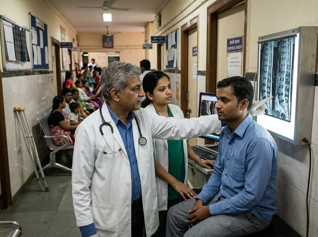
Diagnostic data shows that nearly one in four spine investigations now involve patients younger than 40 years. This comprehensive overview explores the drivers behind this demographic trend, evaluates ergonomic hazards, clarifies red-flag signs, and highlights clinical indications for judicious diagnostic imaging.
Today

A study reveals how adrenal hormones maintain glucose homeostasis during prolonged fasting when glucagon is absent. In glucagon-deficient mice, corticosterone-driven lipolysis prevents severe hypoglycemia, offering key insights for managing counterregulatory failure and fasting in diabetes.
Today
