.webp&w=384&q=75)

Loading, please wait...
Loading, please wait...
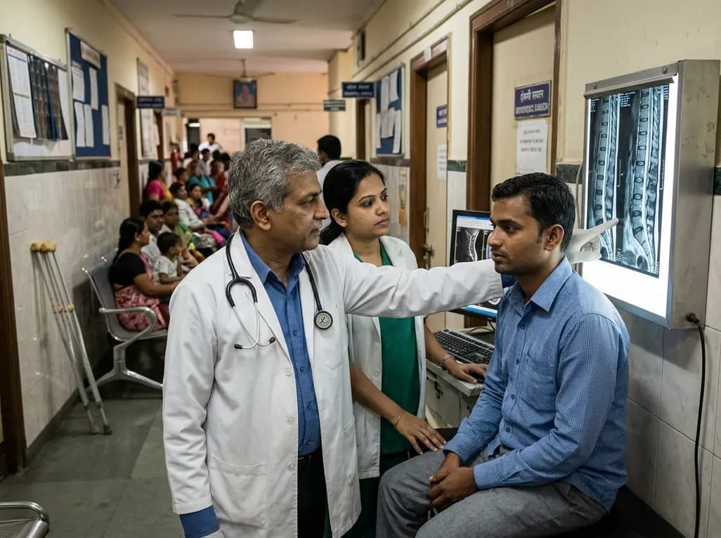
Recent clinical diagnostic reports indicate an unexpected shift in musculoskeletal disease burden. Specifically, data reveals that spine investigations are increasing rapidly among younger populations who traditionally experienced minimal degenerative complaints. A thirteen-month assessment demonstrated that individuals under forty years old accounted for nearly one in four diagnostic requests. In addition, overall imaging volume surged by twenty-four percent year-over-year. Consequently, healthcare providers must recognize that chronic back pain is no longer confined to older demographics. This shift demands a refreshed approach to physical assessments, early ergonomic counseling, and evidence-based diagnostic protocols in modern clinical practice.
Recent diagnostic analytics from premier imaging centers offer revealing perspectives on community spine health. Between September 2025 and September 2026, an analysis of over 29,000 cases highlighted surprising demographic trends. Adults aged sixty and above still comprised the largest single demographic cohort, representing approximately thirty-nine percent of requests. Furthermore, middle-aged adults between forty and sixty years constituted thirty-seven percent of the evaluated volume. Nevertheless, patients younger than forty years accounted for nearly twenty-four percent of all diagnostic procedures. This cohort represented 6,925 individual studies within thirteen months. Moreover, the absolute monthly volume of spinal studies climbed by twenty-four percent across all age groups during this interval. The diagnostic data also uncovered an intriguing gender disparity across the patient population. Specifically, female patients represented fifty-seven percent of total diagnostic scans, while male patients comprised forty-three percent. However, healthcare specialists emphasize that investigation volumes do not automatically reflect confirmed spinal pathologies or true disease prevalence. Instead, elevated referral rates reflect growing symptom awareness, easier access to diagnostic centers, and escalating lifestyle stressors. Therefore, clinicians must interpret these imaging numbers thoughtfully rather than assuming rampant premature anatomical degeneration.
Modern occupational realities have profoundly altered physical biomechanics across urban populations. Most young professionals now maintain prolonged sitting postures that exceed eight to ten hours every day. In addition, poor ergonomic workstation configurations place persistent mechanical load on the lumbar and cervical spine. Consequently, paraspinal muscular fatigue develops rapidly, which destabilizes normal lumbo-pelvic rhythm. Inadequate daily physical movement also deprives intervertebral discs of regular nutrient exchange through hydrostatic imbibition. Over time, physical deconditioning reduces core abdominal strength and diminishes pelvic stabilization. Furthermore, excessive handheld screen usage fosters cervical hyperflexion, commonly described as forward head posture or text neck. This sustained alignment significantly increases biomechanical strain on upper thoracic vertebrae and cervical facets. In contrast, regular postural breaks and targeted postural exercises help maintain healthy spinal biomechanics. Dr. Harsh Mahajan observed that prolonged inactivity and poor physical conditioning directly fuel musculoskeletal discomfort. However, transient muscular fatigue rarely indicates structural destruction or irreversible disk injury. Therefore, clinicians should educate young adults about posture ergonomics rather than quickly ordering expensive advanced imaging tests. Promoting structured activity breaks and active core conditioning provides durable clinical benefits for non-specific axial discomfort.
When young adults seek medical consultation for persistent spinal discomfort, differential diagnosis requires thorough clinical acumen. Most primary care encounters involve benign mechanical low back pain secondary to soft tissue strain. However, clinicians must distinguish simple postural pain from early axial spondyloarthritis and ankylosing spondylitis. Typically, mechanical back pain worsens with strenuous physical activity and improves during rest. In contrast, inflammatory spinal diseases present with prominent morning stiffness lasting over thirty minutes. Furthermore, inflammatory symptoms generally improve with physical exercise rather than complete recumbency. Patients with spondyloarthritis often experience nocturnal awakenings during the early morning hours due to sacroiliac irritation. Additionally, alternating buttock pain and elevated systemic inflammatory markers provide critical diagnostic clues for physicians. Clinicians must also remain vigilant for red-flag symptoms indicating urgent neurological or oncological conditions. For example, progressive bilateral lower extremity weakness or saddle paresthesia necessitates immediate emergency decompression evaluation. Similarly, unexplained constitutional weight loss or systemic fever raises suspicion for malignancy or spinal infections like tuberculosis. Therefore, careful history taking and focused physical examinations remain paramount before selecting diagnostic imaging pathways.
Major international medical organizations, including the American College of Radiology, emphasize conservative diagnostic stewardship. Clinical guidelines strongly advise against routine early radiographs or magnetic resonance scans for uncomplicated back discomfort. In fact, most acute spinal episodes resolve spontaneously within four to six weeks under conservative management. Furthermore, early unindicated imaging frequently identifies incidental anatomical variations that do not correlate with clinical symptoms. For example, asymptomatic disc bulges, facet arthropathy, and annular fissures occur commonly in healthy young individuals. Consequently, premature imaging reports can induce substantial patient distress, leading to kinesiophobia and unnecessary clinical interventions. Guidelines recommend diagnostic imaging primarily when patients present with objective neurological deficits or red-flag warning signs. In addition, imaging is appropriate when persistent symptoms fail to respond after six weeks of supervised conservative therapy. When clinicians genuinely suspect serious structural disease or progressive radiculopathy, magnetic resonance imaging serves as the premier diagnostic modality. Conversely, clinicians should avoid repetitive lumbar radiographs to minimize unnecessary cumulative ionizing radiation exposure. Therefore, diagnostic restraint aligns with evidence-based practice and protects patients from excessive financial and psychological burdens.
Effective management of mechanical spinal discomfort centers on proactive rehabilitation rather than passive medical interventions. Clinicians should reassure young patients that acute discomfort rarely signals irreversible structural damage. Moreover, healthcare providers must actively discourage prolonged bed rest, which accelerates muscle atrophy and prolongs functional disability. Instead, clinicians should encourage early resumption of comfortable daily activities and low-impact cardiovascular conditioning. For example, regular walking, swimming, and stationary cycling safely preserve spinal mobility without overloading fatigued tissues. Additionally, targeted physical therapy programs that strengthen transverse abdominis and multifidus muscles significantly reduce recurrent symptoms. Clinicians can also prescribe short courses of oral non-steroidal anti-inflammatory agents to facilitate early exercise participation. Furthermore, ergonomic modifications in office environments yield remarkable long-term improvements for sedentary desk workers. Ergonomic chairs, adjustable monitor heights, and sit-to-stand desks reduce sustained static loading on spinal ligaments. In addition, routine dynamic stretching breaks interrupt prolonged sitting and promote tissue perfusion. Finally, clinicians must foster realistic expectations regarding recovery timelines through supportive patient education. Consequently, empowering patients with active self-management tools builds lasting confidence, restores functional mobility, and prevents superfluous diagnostic testing.
Q1: Why are spine investigations becoming more frequent among younger individuals?
Spine investigations are rising among young adults primarily due to prolonged sedentary work habits, poor physical conditioning, and inadequate daily movement. Additionally, modern desk environments and extended screen usage impose sustained mechanical strain on spinal structures. Growing health awareness and accessible diagnostic imaging facilities also prompt earlier clinical consultations. However, clinicians recognize that increased imaging requests reflect investigative volume rather than confirmed severe anatomical pathologies.
Q2: When should a physician recommend urgent spinal imaging for acute back pain?
Physicians should order urgent spinal imaging when clear red-flag symptoms suggest serious underlying systemic or neurological disorders. Specifically, indications include progressive motor weakness, bowel or bladder incontinence, saddle anesthesia, or suspected cauda equina syndrome. Furthermore, imaging is warranted when patients present with unexplained constitutional weight loss, persistent fever, significant trauma, or active cancer histories. In contrast, uncomplicated acute back pain rarely requires immediate diagnostic scanning.
Q3: How does unnecessary early spinal imaging affect patient outcomes and recovery?
Unnecessary early spinal imaging often reveals incidental, age-related structural changes that bear no correlation to the patient's actual symptoms. Consequently, these benign findings can trigger heightened health anxiety, illness behaviors, and unhelpful fear of physical movement. Moreover, early imaging does not improve clinical pain relief or functional recovery. Instead, unwarranted scanning frequently leads to unnecessary specialist referrals, excessive healthcare expenses, and unneeded invasive interventional procedures.
Disclaimer: This content is for informational and educational purposes only. It does not constitute medical advice or replace professional judgment. Refer to the latest local and national guidelines for clinical practice.
References

Read summarized clinical updates, watch expert medical content, and earn CME certifications right from your smartphone.
Open your smartphone camera to scan and install.





Diagnostic data shows that nearly one in four spine investigations now involve patients younger than 40 years. This comprehensive overview explores the drivers behind this demographic trend, evaluates ergonomic hazards, clarifies red-flag signs, and highlights clinical indications for judicious diagnostic imaging.
Today

A retrospective study shows that symptom-monitored loading with lorazepam, chlordiazepoxide, or gabapentin significantly reduces hospital length of stay compared to CIWA-Ar-only therapy in severe alcohol withdrawal, providing a safer, more proactive management strategy.
Today

A retrospective cohort study of 5,481 children aged 3–6 years evaluated whether surgical sperm retrieval for ICSI affects offspring cognitive and physical growth compared with ejaculated sperm and IVF. Results demonstrate comparable IQ scores and growth parameters, providing reassuring safety evidence for clinical practice.
Today
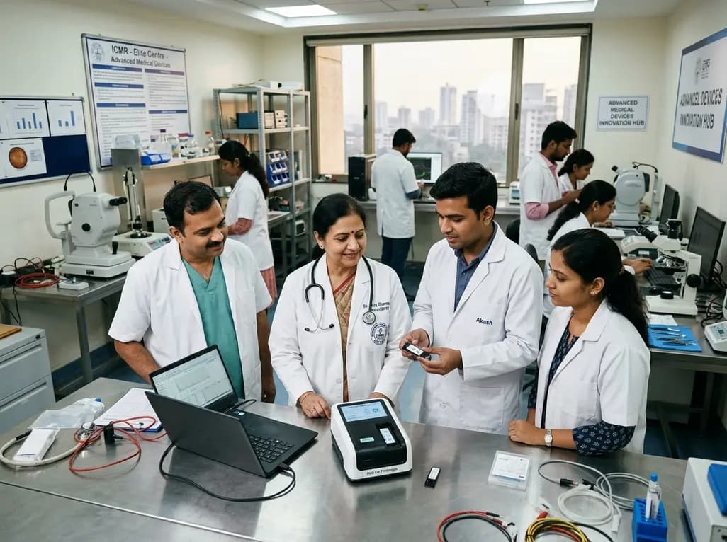
The Indian Council of Medical Research has funded seven top academic hubs to build indigenous healthcare technologies. Targeting point-of-care diagnostics, biomaterials, and ophthalmology, these multidisciplinary centres aim to tackle unmet clinical needs and make medical care affordable across India.
Today
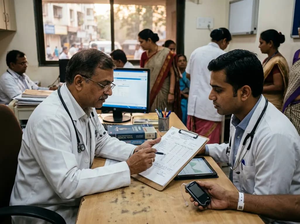
A multicenter cross-sectional study of 1,315 adults with type 1 diabetes reveals that the GMI/HbA1c ratio does not independently predict microvascular complications. Clinicians should prioritize sustained time in range and blood pressure management over glycation discordance metrics.
Today
