.webp&w=384&q=75)

Loading, please wait...
Loading, please wait...
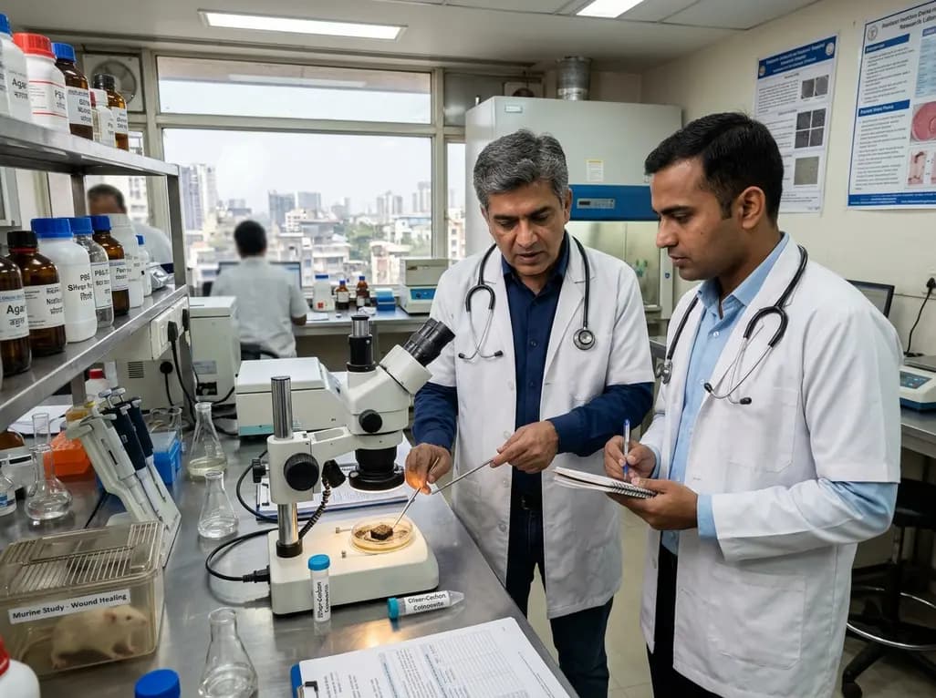
Chronic cutaneous ulcers and complex surgical wounds present significant therapeutic challenges for healthcare providers worldwide. Clinicians frequently encounter recalcitrant infections because bacterial pathogens form dense, structured communities known as biofilms. Within this protective matrix, organisms such as Staphylococcus aureus and Escherichia coli resist standard topical antimicrobials and host immune defenses. Conventional therapies often deploy high concentrations of silver to penetrate these barriers. However, excessive silver ion exposure causes localized cytotoxicity, delays re-epithelialization, and induces heavy metal accumulation. Consequently, wound care specialists require innovative biomaterials that eliminate sessile pathogens without damaging healthy granulation tissue. A newly engineered porous silver-carbon composite offers a promising solution to this persistent clinical paradox. In situ glucose foaming and carbonization produce this sponge-like matrix, balancing robust antibacterial potency with minimal cellular toxicity. Therefore, modern wound management can transition away from indiscriminate silver flooding toward calibrated delivery. As wound infections prolong hospital stays, discovering safe biomaterials remains an urgent priority for surgical teams.
To overcome the physical trade-offs of existing dressings, investigators synthesized a sponge-like porous silver-carbon composite using glucose foaming followed by thermal carbonization. This specialized technique creates an interconnected hierarchical pore network spanning macroporous, mesoporous, and microporous domains. Nitrogen adsorption analysis demonstrated an extraordinary Brunauer-Emmett-Teller surface area of 1067.8 square meters per gram. Furthermore, complete acid digestion confirmed that the carbonaceous scaffold contained a total silver content of 9.22% by weight. This vast surface area enables massive bacterial entrapment while providing active sites for metal interaction. In addition, interconnected porous pathways facilitate effortless exudate fluid exchange across the wound interface. Traditional silver dressings often shed loose nanoparticles or rapidly dissolve, causing rapid metal depletion and host tissue irritation. In contrast, this carbon scaffold securely anchors metallic silver domains within its structural framework. Consequently, the material retains mechanical integrity when hydrated by biological fluids. By combining light carbon architecture with well-distributed metallic centers, the composite functions as a durable barrier against microbial invasion. Thus, material architecture directly dictates biological performance in infected tissue microenvironments.
The therapeutic advantage of this composite stems from its tightly controlled ion-release kinetics. During dialysis experiments against phosphate-buffered saline containing 1% bovine serum albumin, the composite released approximately 0.27% of its total silver content over 72 hours. This sustained, minute release profile prevents toxic systemic exposure while supplying lethal concentrations of silver ions to adjacent pathogens. Standard silver formulations typically exhibit burst release kinetics, which exhaust the dressing and damage delicate keratinocytes. Conversely, the dialyzable silver release from this composite establishes a stable, long-lasting bactericidal gradient. Laboratory testing revealed remarkable antimicrobial activity against Gram-negative and Gram-positive pathogens. Specifically, the material achieved minimum inhibitory concentrations of 4 micrograms per milliliter for Escherichia coli and 8 micrograms per milliliter for Staphylococcus aureus. Furthermore, the corresponding minimum bactericidal concentrations were 8 micrograms per milliliter and 16 micrograms per milliliter, respectively. Therefore, clinicians can appreciate how low concentrations of dialyzable silver achieve dependable microbial suppression. Because the carbon matrix regulates ion detachment, the composite maintains consistent local efficacy without requiring frequent, painful dressing changes.
Understanding how antimicrobial dressings eradicate protective biofilms is crucial for clinical adoption. Comprehensive mechanistic evaluations confirmed that the composite exerts rapid, multi-target bactericidal actions against colonizing microorganisms. Propidium iodide fluorescence assays demonstrated profound increases in bacterial membrane permeability shortly after material contact. Furthermore, scanning electron microscopy revealed severe morphological collapse, cell wall corrugated folds, and extensive membrane lysis in treated bacterial populations. In addition to physical structural destruction, molecular assays highlighted significant oxidative damage. Fluorescent probes utilizing 2',7'-dichlorodihydrofluorescein diacetate along with electron spin resonance measurements confirmed robust intracellular reactive oxygen species generation. These reactive radicals disrupt bacterial respiratory enzymes, oxidize structural lipids, and fragment genomic DNA. Most importantly, plate-counting assays confirmed concentration-dependent suppression of newly forming bacterial colonies. The composite also significantly reduced the recovery of viable bacteria from established biofilms. Because extracellular matrix barriers shield embedded bacteria from conventional antibiotic penetration, this multimodal destructive mechanism is vital. Consequently, the composite neutralizes recalcitrant bacterial populations before systemic bacteremia or deep tissue destruction can occur.
A major clinical limitation of existing silver therapeutics is cellular toxicity toward mammalian repair cells. Fortunately, extensive biocompatibility profiling revealed that this composite preserves healthy tissue viability. Murine fibroblast cultures maintained over 95% viability following exposure, demonstrating excellent host cytocompatibility. Furthermore, red blood cell hemolysis remained well below 5%, satisfying international standards for blood-contacting medical devices. In a validated full-thickness mouse wound model infected with Escherichia coli, researchers compared the composite directly against traditional silver nitrate at an identical elemental silver dose of 35 micrograms. On post-infection day 4, wound beds treated with the composite showed significantly lower bacterial colony counts than wounds receiving silver nitrate. Moreover, composite-treated wounds attained approximately 95% total re-epithelialization and closure by day 14. Histological examination of vital organs, including liver, kidneys, spleen, and lungs, revealed no pathological abnormalities or structural lesions at the study endpoint. These findings confirm that controlled dialyzable release eliminates bacterial burdens without triggering organ toxicity. Therefore, the composite demonstrates superior therapeutic index and regenerative acceleration compared to older ionic silver standards.
The material prevents cytotoxicity by controlling silver ion release through its interconnected carbon scaffold. During physiological exposure, the composite releases only 0.27% of its silver content over 72 hours. This minute dialyzable rate provides sufficient antimicrobial concentrations to kill bacteria without overwhelming mammalian cells. In laboratory testing, murine fibroblasts maintained over 95% viability and hemolysis remained under 5%, proving that low, sustained silver kinetics protect delicate repair tissues effectively during wound healing.
Biofilm communities produce a protective extracellular polymeric matrix that acts as a physical and electrostatic shield. This matrix impedes antimicrobial penetration, while bacteria within the biofilm enter a dormant metabolic state with down-regulated targets. Standard topical silver dressings frequently generate rapid burst release that binds superficial surface proteins without infiltrating deep biofilm clusters. In contrast, hierarchically porous composites adsorb matrix components and maintain sustained dialyzable silver release, achieving deeper penetration and superior eradication of persistent pathogens.
Conventional silver nitrate delivers an uncontrolled, rapid bolus of ionic silver that precipitates rapidly with wound fluid anions. This process leads to chemical irritation, localized tissue tattooing, and cellular necrosis that stalls re-epithelialization. Conversely, dialyzable silver release provides a steady, regulated antimicrobial gradient without chemical burn risks. In animal models, this controlled mechanism lowered bacterial colony counts much faster than silver nitrate while achieving 95% wound closure within fourteen days without damaging underlying internal organs.
Disclaimer: This content is for informational and educational purposes only... Refer to the latest local and national guidelines for clinical practice.
References

Read summarized clinical updates, watch expert medical content, and earn CME certifications right from your smartphone.
Open your smartphone camera to scan and install.





A novel hierarchically porous silver-carbon composite provides sustained dialyzable silver release, achieving potent biofilm eradication, high cytocompatibility, and 95% closure in infected murine wounds without organ toxicity.
Today

Bacteroides thetaiotaomicron utilizes an intricate multiprotein extracellular relay and outer membrane vesicles for cobamide scavenging in the human gut. This review examines its transport mechanics, riboswitch regulation, and potential clinical implications for host-microbe vitamin competition and nutrient absorption.
Today

An independent clinical evaluation from Assam confirms the excellent safety profile of CERVAVAC, India's indigenous HPV vaccine. Evaluating 3,800 doses administered to 2,000 young girls, researchers documented zero serious adverse events, providing critical real-world evidence to resolve vaccine hesitancy.
Today

This clinical analysis examines heparin-free versus heparin-containing Impella purge solutions in patients receiving concomitant ECMO support (ECPELLA). Evidence suggests anticoagulant-free purges maintain device patency while reducing bleeding risks, offering a viable strategy alongside systemic anticoagulation.
Today

A 10-year Markov state-transition model shows minimally invasive tubular decompression dominates open laminectomy for pure lumbar spinal stenosis, yielding 5.95 vs 5.91 QALYs and saving $4,167 per patient through reduced complications and lower index costs.
Today

A preclinical study demonstrates that naringin-loaded phytosomes significantly enhance oral bioavailability and provide neuroprotective efficacy in diabetic neuropathy by dampening inflammation, reducing oxidative stress, and relieving neuropathic pain.
Today
