.webp&w=384&q=75)

Loading, please wait...
Loading, please wait...

Traumatic brain injury represents a devastating global health crisis that causes profound, lifelong neurological disability across millions of patients annually. While the primary mechanical impact inflicts immediate vascular shearing and irreversible tissue contusion, secondary injury cascades propagate neurological damage over subsequent days and weeks. In particular, aberrant microglial polarization in TBI acts as a primary catalyst of progressive tissue degeneration and cell death. Following acute biomechanical trauma, resident microglia rapidly transition toward an aggressive, pro-inflammatory M1 phenotype. Consequently, these reactive immune cells release massive quantities of neurotoxic mediators, including tumor necrosis factor-alpha, interleukin-1beta, and interleukin-6. This self-amplifying neuroinflammatory cascade degrades extracellular matrix architecture, destabilizes the blood-brain barrier, and drives secondary neuronal apoptosis across adjacent peri-lesional brain tissue. Conversely, driving microglial polarization toward an anti-inflammatory M2 phenotype fosters tissue repair, suppresses cytotoxic mediators, and supports axonal preservation. Unfortunately, conventional systemic neuroprotective drugs routinely fail in clinical translation because compromised cerebral microcirculation severely impairs drug delivery to damaged areas. Furthermore, high systemic drug concentrations provoke intolerable off-target toxicities. Therefore, neurosurgeons and bioengineers urgently require targeted physical modalities that remodel local neuroimmune responses directly at the injury site without causing systemic complications.
Mitochondrial impairment sits at the direct crossroads of traumatic metabolic collapse, cellular energetic depletion, and sustained neuroinflammation following brain trauma. Under physiological conditions, continuous fusion and fission cycles sustain mitochondrial dynamic homeostasis, supporting efficient oxidative phosphorylation and vital cell viability. However, severe traumatic biomechanical strain abruptly disrupts this fine equilibrium. Mechanical trauma induces uncontrolled intracellular calcium influx that accelerates aberrant mitochondrial fission and impairs endogenous quality control mechanisms. Consequently, fragmented mitochondria undergo rapid mitochondrial membrane potential collapse and electron transport chain breakdown. These destabilized organelles generate toxic surges of mitochondrial superoxide and cellular reactive oxygen species. In addition, the trauma drastically depletes endogenous superoxide dismutase activity, paralyzing native cellular antioxidant defense systems. Importantly, this pervasive oxidative stress dictates microglial inflammatory fate. Excessive intracellular reactive oxygen species permanently trap microglia in the cytotoxic M1 state, preventing phenotypic resolution and tissue regeneration. Thus, preserving mitochondrial dynamic homeostasis serves as an essential gatekeeper for neural survival. Stabilizing mitochondrial membrane potential prevents reactive oxygen species surges, protects vulnerable adjacent neurons, and terminates persistent inflammatory signaling cascades. Accordingly, direct mitochondrial intervention offers a compelling molecular strategy to attenuate secondary post-traumatic damage.
To overcome traditional biochemical delivery hurdles, investigators engineered an innovative ultrasound-responsive piezoelectric fibrous membrane for implantation at the traumatic brain lesion site. Piezoelectric biomaterials possess the unique capability to generate surface electrical polarization in direct response to applied mechanical deformation. In this therapeutic paradigm, low-intensity pulsed ultrasound acts as an external, non-invasive mechanical trigger. Low-intensity pulsed ultrasound waves easily penetrate overlying soft tissues and cranial defects to reach the underlying fibrous implant safely. Upon acoustic irradiation, the scaffold undergoes microscale physical vibrations that produce precise, localized electric fields across the injured cortex. Furthermore, this wireless electromechanical transduction operates reliably without invasive wires, transcutaneous cables, or bulky internal battery packs. The fibrous architecture also mirrors the natural extracellular matrix, ensuring excellent tissue integration and minimizing mechanical shear against soft cerebral cortex. Moreover, neurosurgeons can adjust ultrasound intensity, duty cycles, and pulse frequencies to modulate bioelectric output dynamically based on patient requirements. Consequently, this versatile platform provides on-demand biophysical stimulation directly into the damaged microenvironment. Therefore, ultrasound-responsive piezoelectric scaffolds represent an innovative synthesis of materials engineering and wireless bioelectronics for acute neurosurgical care.
The ultrasound-driven piezoelectric membrane exerts therapeutic neuroprotection primarily through precise neuroimmunometabolic remodeling within the injured microenvironment. During ultrasonic activation, the generated electrical microcurrents interact directly with microglial membrane structures and voltage-sensitive ion channels. Mechanistically, this biophysical stimulation restores mitochondrial dynamic homeostasis by rebalancing mitochondrial fission and fusion machinery. Consequently, the therapy stabilizes mitochondrial membrane potential and curtails excessive mitochondrial superoxide generation within stressed cells. Concurrently, cellular reactive oxygen species levels drop markedly, while endogenous superoxide dismutase activity increases substantially. This dual reduction in oxidative stress directly reprograms intracellular metabolic pathways toward restorative oxidative phosphorylation. Relieved of oxidative pressure, microglia suppress nuclear factor-kappa B signaling, significantly attenuating the synthesis of pro-inflammatory cytokines. Specifically, the membrane treatment dramatically downregulates tumor necrosis factor-alpha, interleukin-1beta, and interleukin-6 expression. In addition, this bioelectric intervention actively drives microglial polarization toward the neuroprotective M2 phenotype within the pericontusional parenchyma. These M2 microglia release beneficial neurotrophic factors and clear cellular debris efficiently, preventing secondary neurotoxicity. Thus, by targeting mitochondrial stability, the platform shifts the inflammatory equilibrium from destructive neurotoxicity to proactive tissue repair.
Preclinical evaluation in animal models of traumatic brain injury provides compelling evidence of meaningful functional, histological, and neurobehavioral recovery. Experimental subjects treated with the ultrasound-activated piezoelectric membrane demonstrated striking structural preservation across the cerebral cortex. Histological evaluations revealed markedly smaller contusion volumes and significantly reduced pericontusional edema compared to sham-treated cohorts. Furthermore, the therapeutic regimen significantly elevated neuronal survival rates within vulnerable pericontusional cortex and CA1 hippocampal subfields. Most importantly, these histological improvements translated into tangible behavioral recovery across multiple domains. Standardized sensorimotor assessments showed rapid resolution of motor deficits and enhanced limb coordination. In cognitive testing using the Morris water maze, treated subjects demonstrated superior spatial learning and spatial memory retention compared to controls. From a clinical neurosurgical perspective, these observations carry tremendous implications for patient management. Surgeons frequently perform decompressive craniectomies or duraplasties in severe trauma cases. Utilizing bioactive piezoelectric dural grafts could provide continuous neuroprotection without altering the standard operative workflow. However, researchers must verify long-term material biodegradation, dielectric longevity, and thermal safety profiles before human trials commence. Nonetheless, this electromechanical strategy represents an extraordinary leap toward targeted neurorehabilitation.
Low-intensity pulsed ultrasound delivers non-invasive acoustic pressure waves across the cranial defect directly to the implanted piezoelectric fibrous membrane. Consequently, the mechanical deformations generate controllable local electrical potentials without requiring internal batteries or invasive transcutaneous wires. These dynamic bioelectric microcurrents directly stimulate damaged neural and glial cells. Therefore, this electroacoustic coupling provides targeted physical cues that modulate cellular pathways and restore microglial homeostasis at the focal lesion site.
Mitochondrial dynamics balance continuous fusion and fission processes to support cellular bioenergetics and survival. In traumatic brain injury, severe mitochondrial fragmentation triggers excessive reactive oxygen species production and membrane depolarization, locking microglia into a destructive M1 phenotype. Consequently, restoring dynamic equilibrium through piezoelectric stimulation stabilizes mitochondrial membrane potential and boosts endogenous antioxidants like superoxide dismutase. As a result, microglia successfully reprogram toward the neuroprotective M2 phenotype, dampening persistent neuroinflammation.
Traumatic brain injury remains notoriously difficult to treat because conventional pharmacotherapies fail to penetrate damaged parenchyma effectively. Furthermore, systemic drug administration often causes dose-limiting toxicities. Ultrasound-responsive piezoelectric biomaterials offer an innovative, minimally invasive theranostic platform that clinicians can activate on demand. While clinical implementation requires comprehensive human trials and long-term biocompatibility evaluation, this electromechanical strategy provides neurosurgeons with an adaptable scaffold to mitigate secondary neurodegenerative cascades effectively.
Disclaimer: This content is for informational and educational purposes only... Refer to the latest local and national guidelines for clinical practice.
References

Read summarized clinical updates, watch expert medical content, and earn CME certifications right from your smartphone.
Open your smartphone camera to scan and install.





A novel ultrasound-responsive piezoelectric fibrous membrane restores mitochondrial dynamic homeostasis and drives neuroprotective microglial polarization in traumatic brain injury, suppressing oxidative damage and neuroinflammation to accelerate functional recovery.
Yesterday

FDA staff reviewers have raised no major safety or analytical concerns regarding Grail's Galleri blood test ahead of an advisory panel meeting. This milestone advances liquid biopsy technologies designed to identify tumor methylation signals across dozens of malignancies before clinical symptoms emerge.
Today

Meghalaya recorded a historic 55% reduction in maternal deaths, dropping from 244 to 107 over five years. Through the MOTHER app, dedicated transit homes, and expanded infrastructure like the 150-bed Tura Maternity Hospital with its first public NICU, the state demonstrates an effective public health model.
Today

Explore modern perioperative management protocols for clinical xenotransplantation. Learn how costimulation blockade, anti-inflammatory perfusion, and coagulopathy rescue overcome interspecies hurdles.
Today
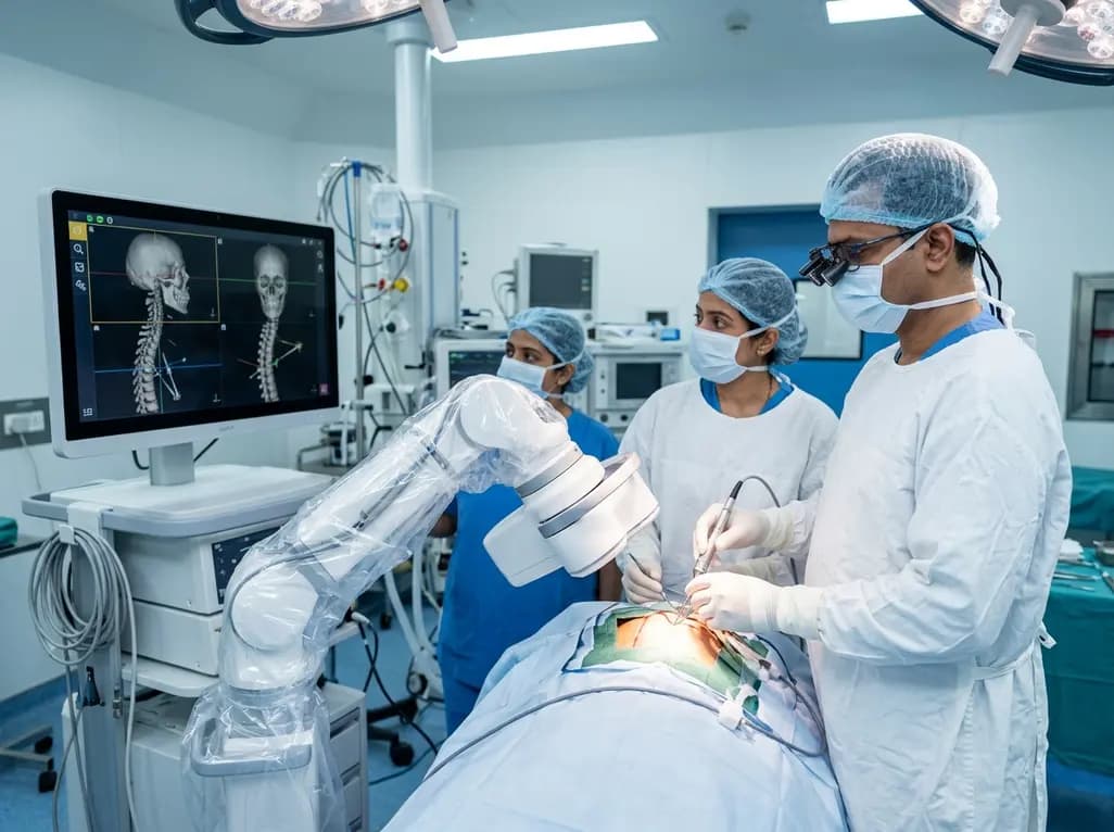
North India's first Centre of Excellence for Brain and Spine at Amrita Hospital Faridabad integrates Brainlab's Loop-X mobile robot. This landmark facility combines cone-beam computed tomography with surgical navigation to elevate precision, optimize operative workflows, and enhance specialized clinician training.
Today

At Homa CME 2026 at Hyderabad's T-Hub, healthcare leaders emphasized moving beyond routine metrics through digital calculators and precision stratification. Discover why characterizing individual insulin secretion, resistance patterns, and vascular risks transforms chronic metabolic care in India.
Today
