.webp&w=384&q=75)

Loading, please wait...
Loading, please wait...
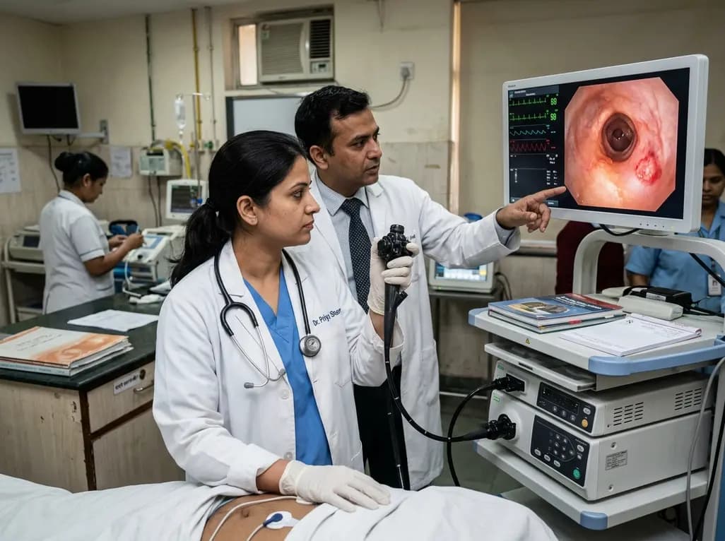
Managing central airway malignancies requires balancing effective local tumor ablation with the preservation of critical lung function. Clinicians frequently encounter central-type early-stage lung cancer in patients who present with underlying chronic obstructive pulmonary disease or field cancerization. In such scenarios, aggressive surgical resection might severely compromise respiratory reserve or carry prohibitive surgical risk. Therefore, photodynamic therapy for lung cancer has emerged as an essential, lung-sparing intervention. This non-thermal ablative technique utilizes a tumor-localizing photosensitizing drug that becomes activated upon exposure to specific light wavelengths delivered via flexible bronchoscopy. Consequently, light activation generates cytotoxic reactive oxygen species that selectively induce localized microvascular occlusion and tumor cell necrosis. Because the underlying connective tissue architecture remains largely spared, photodynamic therapy for lung cancer preserves airway integrity and pulmonary function. Furthermore, the non-ionizing nature of light activation permits repeated applications when local recurrences or new endobronchial lesions arise. Recent clinical evaluations demonstrate that this strategy achieves durable local disease control while maintaining functional capacity. Consequently, understanding its long-term efficacy, procedural repeatability, and morphological response patterns is crucial for refining multidisciplinary thoracic oncology care.
Bronchoscopic evaluation plays a pivotal role in selecting appropriate candidates for endobronchial light activation. Lesion morphology significantly influences treatment outcomes and dictates the necessity for adjunctive therapeutic modalities. Endoscopists routinely categorize central airway lesions into superficial, thickened, or nodular phenotypes based on white-light bronchoscopy, autofluorescence imaging, and endobronchial ultrasonography. Superficial lesions, which remain strictly confined to the mucosal and submucosal layers, consistently demonstrate high rates of complete response following photodynamic ablation alone. Because light penetration depth is naturally limited to a few millimeters, superficial mucosal changes respond exceptionally well without requiring aggressive physical debulking. Furthermore, when localized mucosal recurrences develop during longitudinal follow-up, repeated light application successfully restores complete disease eradication without causing cumulative pulmonary toxicity. Long-term cohort studies confirm that repeating treatment sessions for superficial lesions achieves sustained local control over extended observation periods. Thus, accurate morphological classification enables interventionalists to tailor light delivery parameters and predict clinical responses reliably. Additionally, high-resolution endobronchial imaging helps clinicians establish precise tumor boundaries, ensuring complete light coverage across the affected bronchial segment and minimizing residual disease.
Although photodynamic therapy achieves excellent local control in superficial lesions, management of thickened or nodular airway lesions presents distinct therapeutic challenges. Thickened and nodular endobronchial tumors frequently extend deeper into the cartilage layer or project into the bronchial lumen. As a result, primary light penetration alone may prove insufficient to achieve complete microscopic eradication across the entire tumor mass. To overcome these limitations, clinicians increasingly adopt hybrid, multimodal therapeutic strategies. Combining photodynamic ablation with electrosurgical snaring, thermal ablation, or cryotherapy effectively reduces bulk endobronchial tumor volume prior to light delivery. Alternatively, integrating external beam radiotherapy or brachytherapy addresses deep-seated invasive components while light activation targets mucosal surface extension. Clinical outcomes show that structured multimodal interventions can convert initial partial responses into durable long-term local control. Moreover, timely integration of complementary interventional tools prevents acute airway obstruction and mitigates treatment-related inflammatory stenosis. Consequently, interventional pulmonologists and radiation oncologists must collaborate closely to formulate individualized multimodal plans. By combining initial debulking with targeted light activation, multidisciplinary teams successfully manage complex airway lesions that would otherwise resist monotherapy.
Long-term survival data highlight the clinical viability of photo-ablative techniques for patients with early central airway malignancies. Clinical follow-up spanning more than a decade demonstrates favorable long-term overall survival rates. Specifically, overall survival estimates reach approximately 93.8% at three years and 84.4% at five years following initial treatment, with median overall survival remaining unreached in key cohorts. Indeed, some long-term survivors achieve disease-free survival exceeding 4,000 days. These impressive outcomes validate photodynamic ablation as a definitive, lung-preserving option for medically inoperable individuals or those seeking alternatives to parenchymal resection. However, extended follow-up also highlights the profound impact of competing mortality risks in this patient population. Because diffuse field cancerization affects the entire respiratory epithelium, long-term mortality is frequently driven by second primary lung cancers rather than local failure of the treated index lesion. Metachronous malignancies represent a primary challenge during extended survivorship. Consequently, achieving high initial complete response rates does not eliminate future oncologic threats. Clinicians must recognize that long-term survival depends not only on effective local ablation, but also on aggressive management of emerging second primary neoplasms.
Given the significant risk of metachronous airway tumors and local recurrence, establishing rigorous surveillance protocols remains essential after photodynamic therapy. Lifelong endoscopic and radiologic follow-up is imperative for early detection of second primary malignancies across the aerodigestive tract. Interventionalists should schedule regular surveillance bronchoscopy, incorporating high-definition white-light and autofluorescence techniques, alongside low-dose computed tomography imaging. Early identification of superficial metachronous lesions allows for prompt, repeat photodynamic interventions before invasive progression occurs. Furthermore, clinicians must optimize secondary prevention strategies, including rigorous smoking cessation counseling and chemoprevention evaluation where applicable. In clinical practice, patient selection should balance tumor morphology, functional cardiopulmonary status, and anatomical location. For centers establishing endobronchial therapy programs, standardized training in photosensitizer administration, light dosimetry, and post-procedure airway care is necessary to ensure safety and efficacy. Additionally, multidisciplinary tumor boards should routinely evaluate patients to determine whether monotherapy or hybrid multimodal therapy offers the highest probability of local cure. Through diligent lifelong surveillance and adaptive re-intervention, clinicians can maximize functional preservation and long-term survival for patients facing complex central airway neoplasms.
Photodynamic therapy is ideal for early central lung cancer because it selectively destroys malignant tissue while preserving underlying bronchial cartilage and parenchymal tissue. This lung-sparing mechanism is especially beneficial for patients with compromised pulmonary function or those with multiple airway lesions. Additionally, because the non-ionizing light delivery does not cause cumulative radiation toxicity, clinicians can safely repeat the procedure if local recurrences or metachronous malignancies occur during follow-up.
Thickened and nodular lesions present greater depth of invasion, which limits the effective penetration of light activation alone. Therefore, clinical guidelines recommend a multimodal strategy combining initial mechanical or thermal debulking, such as electrosurgical snaring or cryotherapy, with photodynamic light activation. In addition, external beam radiation or brachytherapy may be integrated to target deeper parenchymal extension, thereby achieving complete disease eradication and preventing obstructive airway complications.
Lifelong surveillance is essential because field cancerization exposes the entire respiratory epithelium to malignant transformation. Although photodynamic ablation achieves high rates of local disease control for the primary lesion, patients remain at significant risk for developing second primary lung cancers or metachronous malignancies over time. Routine bronchoscopic and radiologic follow-up ensures early detection of emerging superficial lesions, enabling timely, repeatable endobronchial light treatments before advanced invasive disease develops.
Disclaimer: This content is for informational and educational purposes only and does not constitute medical advice, diagnosis, or treatment. Healthcare professionals should rely on their clinical judgment and refer to the latest local and national guidelines for clinical practice.
References

Read summarized clinical updates, watch expert medical content, and earn CME certifications right from your smartphone.
Open your smartphone camera to scan and install.





Photodynamic therapy (PDT) offers durable local airway control for early-stage central lung cancer. This review explores long-term survival, repeatable ablation, morphological classification, hybrid treatments, and the critical role of lifelong surveillance for metachronous malignancies.
last month
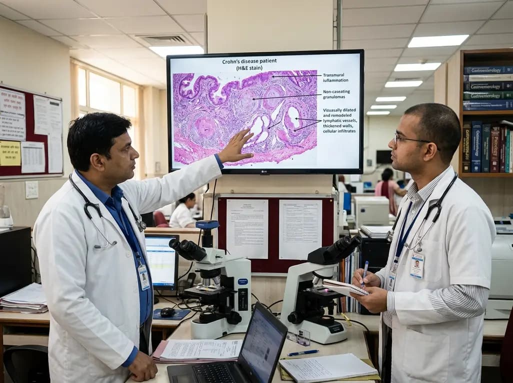
Intestinal lymphatic dysfunction is re-emerging as a fundamental driver of Crohn's disease pathogenesis. Explore how structural remodeling, lymphangitis, and defective clearance shape transmural inflammation and offer novel therapeutic targets.
Today
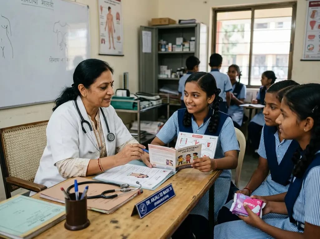
A school-based intervention using the Health Action Process Approach (HAPA) significantly enhanced adolescents' perceived benefits of consulting healthcare providers and promoted proactive menstrual health behaviors, offering key clinical insights for managing adolescent dysmenorrhea and endometriosis.
Today

A comprehensive analysis of the Translational Maturity Framework for nanomedicine-device combination products, evaluating regulatory complexity, manufacturing hurdles, and clinical readiness across oncology, cardiology, and orthopedics.
Today

A cross-sectional study reveals that sarcopenic obesity independently increases functional impairment and severe disc degeneration in patients with degenerative lumbar spinal stenosis, highlighting the need for body composition assessment.
Today
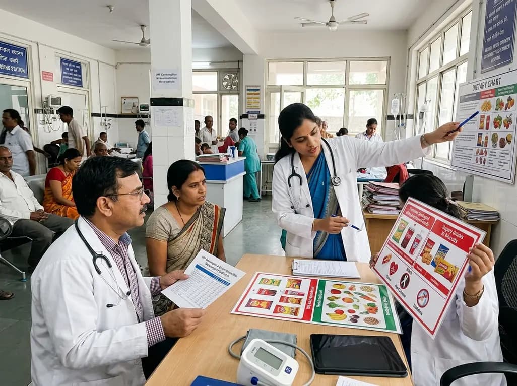
A landmark dose-response meta-analysis of 8.8 million adults reveals that high ultra-processed food intake significantly elevates risks for cardiovascular disease, cancer, diabetes, depression, and all-cause mortality, highlighting urgent needs for clinical lifestyle interventions.
Today
