.webp&w=384&q=75)

Loading, please wait...
Loading, please wait...

The convergence of rapid population aging and the obesity epidemic has created a critical health crisis, highlighting the role of sarcopenic obesity in older adults. Sarcopenic obesity represents a detrimental phenotype where low skeletal muscle mass and strength coexist with excessive body fat. In spinal care, degenerative lumbar spinal stenosis is a leading reason for chronic back pain, neurogenic claudication, and walking disability. However, clinicians often overlook how muscle deterioration and excess adiposity simultaneously aggravate spinal degeneration. Historically, practitioners relied primarily on body mass index to evaluate weight. Unfortunately, this crude measure masks severe skeletal muscle depletion in overweight individuals. Consequently, patients with hidden muscle wasting endure progressive functional decline without proper identification. Furthermore, poor musculature impairs axial spinal stabilization while excess weight intensifies biomechanical loading. When paraspinal muscles fail to maintain alignment, shear forces damage delicate neural structures. Therefore, recognizing sarcopenic obesity is vital for orthopedic surgeons, neurosurgeons, and geriatricians managing spinal pathologies. Understanding this interaction allows clinicians to address underlying metabolic risks and personalize treatment strategies effectively.
To evaluate these clinical relationships, researchers executed a retrospective cross-sectional analysis enrolling 408 patients aged 50 years and older who had degenerative lumbar spinal stenosis. Specifically, investigators applied the consensus criteria defined by the European Society for Clinical Nutrition and Metabolism and the European Association for the Study of Obesity. This diagnostic protocol combines handgrip strength for muscle performance, dual-energy X-ray absorptiometry for appendicular lean mass, and total body fat percentage. Among the study population, 66 participants met the diagnostic criteria for this combined disorder. In addition, researchers gathered validated patient-reported outcome measures, specifically the Visual Analogue Scale for pain and the Oswestry Disability Index for functional impairment. Meanwhile, certified radiologists scored spinal magnetic resonance images across multiple degenerative parameters. These radiological evaluations assessed disc degeneration via Pfirrmann grading, facet arthrosis, central canal stenosis, and paraspinal muscle fatty infiltration. Additionally, researchers performed multivariable regression and moderation analyses to isolate confounding baseline variables. This structured design offered comprehensive objective evidence regarding spinal deterioration.
Statistical analyses revealed striking correlations between poor body composition and exacerbated spinal disease severity. After adjusting for confounding factors, the concurrent presence of sarcopenia and obesity remained independently associated with worse physical disability. Specifically, affected patients scored significantly higher on the Oswestry Disability Index, displaying an adjusted increase exceeding seven points compared to counterparts. This heightened disability severely restricts standing tolerance, walking distance, and daily physical independence. Furthermore, structural imaging evaluations demonstrated that this condition substantially accelerates tissue breakdown. Multivariable logistic regression revealed an adjusted odds ratio exceeding 5.0 for advanced intervertebral disc degeneration using Pfirrmann classification. Patients also suffered significantly greater facet joint arthrosis, severe central spinal canal narrowing, and pronounced paraspinal muscle fatty atrophy. Moreover, moderation analyses demonstrated that higher body fat percentage actively magnified the detrimental impact of sarcopenia on physical mobility. Thus, excess adipose tissue acts not as an inert load, but as a potent modifier that compounds musculoskeletal collapse and impairs recovery.
The pronounced spinal deterioration observed in these patients stems from synergistic mechanical and biochemical mechanisms. Mechanically, deep paraspinal muscles, particularly the multifidus and longissimus, maintain dynamic segmental stability and absorb compressive axial forces. When sarcopenia depletes functional muscle fibers and replaces them with fatty infiltration, paraspinal stiffness declines markedly. Consequently, uncontrolled mechanical stress shifts directly onto passive structural elements, including intervertebral discs, facet cartilage, and supporting ligaments. Meanwhile, expanded visceral and subcutaneous fat depots function as active endocrine organs. Adipose tissue continuously releases circulating pro-inflammatory mediators, including tumor necrosis factor-alpha and interleukin-6. These inflammatory cytokines diffuse into poorly vascularized intervertebral discs, upregulating matrix metalloproteinases and accelerating proteoglycan loss. In addition, systemic inflammation promotes facet synovitis, osteophyte formation, and ligamentum flavum hypertrophy. These microstructural modifications progressively narrow the central canal and neuroforamina, exacerbating radicular symptoms. Therefore, heightened mechanical instability combines with persistent biochemical degradation to accelerate degenerative lumbar disease. Because both processes continually reinforce one another, structural damage advances much faster than observed in patients with isolated obesity or sarcopenia alone.
These clinical findings demand that clinicians shift from traditional weight management toward targeted body composition optimization. Because body mass index fails to capture underlying sarcopenia, spine specialists should integrate functional screenings, such as handgrip dynamometry and chair rise tests, into regular practice. Furthermore, radiologists and surgeons can utilize routine lumbar computed tomography or magnetic resonance imaging to quantify paraspinal muscular cross-sectional area and myosteatosis. Early risk stratification identifies vulnerable surgical candidates who face heightened complication rates, prolonged rehabilitation, or persistent postoperative pain. Therapeutically, simple caloric restriction often worsens outcomes by accelerating muscle loss. Instead, multidisciplinary teams must implement structured nutritional support, providing 1.2 to 1.5 grams of daily protein per kilogram of body weight. Simultaneously, physical therapists should prescribe progressive resistance training and core stabilization exercises to rebuild spinal musculature without provoking neurogenic symptoms. In addition, medical teams must address metabolic comorbidities, optimize glycemic control, and correct vitamin D deficiency. Ultimately, combining targeted nutrition with customized exercise preserves lean muscle mass, reduces adiposity, and enhances physical recovery in vulnerable spinal stenosis patients.
Clinicians diagnose this condition using a standardized two-step framework established by international nutrition and obesity societies. First, teams assess muscle function using handgrip dynamometry or chair stand tests. Next, they evaluate body composition through dual-energy X-ray absorptiometry or bioelectrical impedance analysis. A confirmed diagnosis requires documented low skeletal muscle mass relative to body weight alongside an elevated fat mass percentage. Routine imaging scans can also evaluate paraspinal muscle myosteatosis.
Excessive adipose tissue accelerates disc breakdown through two complementary mechanisms. Mechanically, increased upper body weight exerts excessive axial compressive loads across the lumbar intervertebral discs and facet joints, accelerating structural wear. Biologically, visceral adipose tissue functions as an active endocrine organ that secretes destructive pro-inflammatory adipokines and cytokines. These circulating inflammatory chemicals enter disc tissues, promoting chondrocyte death and enzymatic matrix breakdown, which ultimately speeds up intervertebral degeneration.
Patients benefit most from progressive resistance training combined with non-impact aerobic conditioning under qualified supervision. Aquatic exercise and stationary cycling minimize axial compressive loading on stenotic spinal canals while building cardiovascular fitness. Concurrently, clinicians prescribe targeted isometric trunk exercises to strengthen the transversus abdominis, multifidus, and gluteal musculature. High-impact exercises, uncontrolled lumbar hyperextension, and aggressive flexion should be avoided to prevent nerve root compression and acute symptom flare-ups.
Disclaimer: This content is for informational and educational purposes only... Refer to the latest local and national guidelines for clinical practice.
References
Feng T et al. Sarcopenic obesity is associated with more severe functional impairment and lumbar degeneration in patients with degenerative lumbar spinal stenosis. Eur Spine J. 2026 Sep 22. doi: 10.1007/s00586-026-10381-w. PMID: 42771025.
Donini LM, Busetto L, Bischoff SC, Cederholm T, Ballesteros-Pomar MD, Batsis JA, et al. Definition and diagnostic criteria for sarcopenic obesity: ESPEN and EASO consensus statement. Clin Nutr. 2022;41(4):990-1000.
Batsis JA, Villareal DT. Sarcopenic obesity in older adults: aetiology, epidemiology and treatment strategies. Nat Rev Endocrinol. 2018;14(9):513-537.

Read summarized clinical updates, watch expert medical content, and earn CME certifications right from your smartphone.
Open your smartphone camera to scan and install.





A cross-sectional study reveals that sarcopenic obesity independently increases functional impairment and severe disc degeneration in patients with degenerative lumbar spinal stenosis, highlighting the need for body composition assessment.
Today
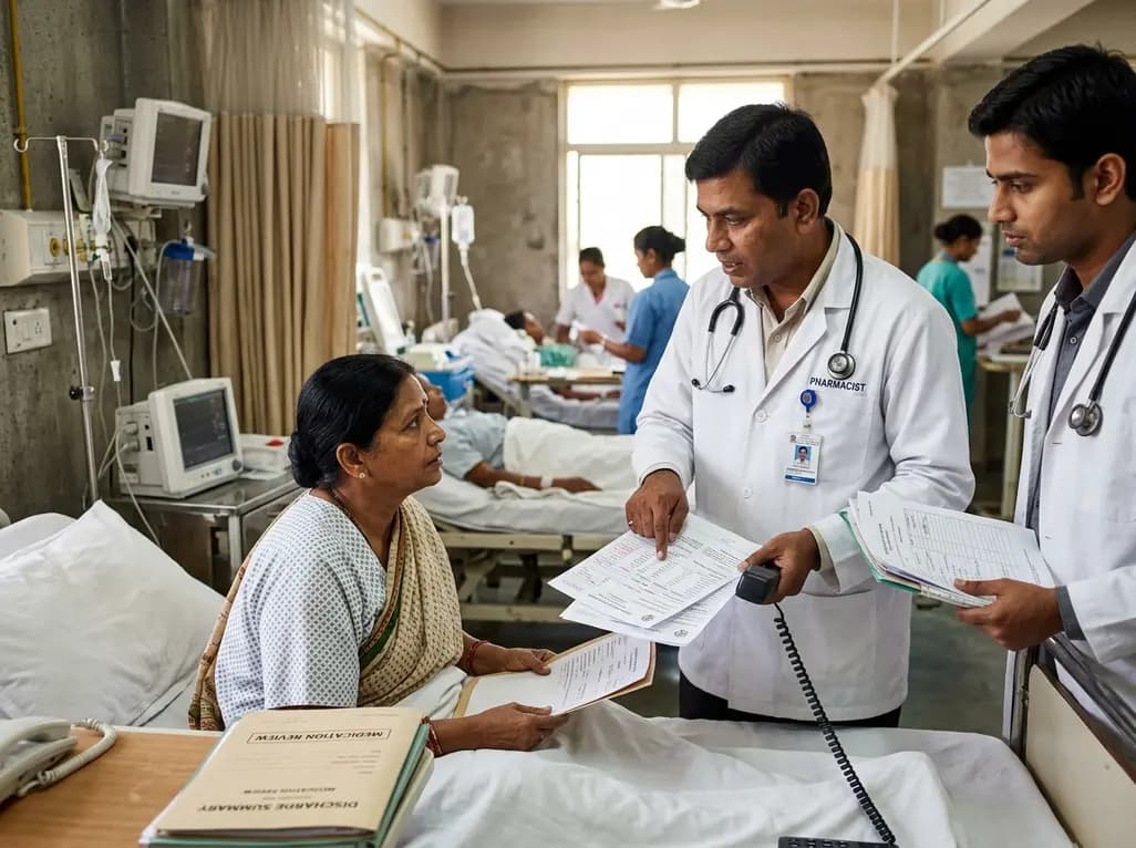
A retrospective cohort study demonstrates that comprehensive pharmacy transitions of care—including admission medication history, discharge review, and 72-hour telephone follow-up—significantly reduced 30-day readmission rates in acute care patients, highlighting the value of pharmacist-led clinical interventions.
Today

Meghalaya recorded a historic 55% reduction in maternal deaths, dropping from 244 to 107 over five years. Through the MOTHER app, dedicated transit homes, and expanded infrastructure like the 150-bed Tura Maternity Hospital with its first public NICU, the state demonstrates an effective public health model.
Today

Explore modern perioperative management protocols for clinical xenotransplantation. Learn how costimulation blockade, anti-inflammatory perfusion, and coagulopathy rescue overcome interspecies hurdles.
Today

At Homa CME 2026 at Hyderabad's T-Hub, healthcare leaders emphasized moving beyond routine metrics through digital calculators and precision stratification. Discover why characterizing individual insulin secretion, resistance patterns, and vascular risks transforms chronic metabolic care in India.
Today
