.webp&w=384&q=75)

Loading, please wait...
Loading, please wait...
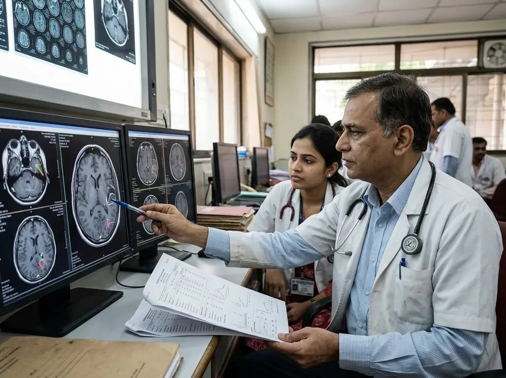
Glioblastoma multiforme remains one of the most aggressive primary central nervous system malignancies in adult oncology. Despite comprehensive multimodal therapeutic regimens comprising maximal safe surgical resection, concurrent radiotherapy, and alkylating temozolomide chemotherapy, tumor recurrence remains practically inevitable. Clinicians continuously seek reliable, cost-effective, and non-invasive diagnostic tools to track postoperative disease dynamics. Recent clinical investigations indicate that systemic inflammatory indices derived from routine complete blood counts provide critical diagnostic insights into tumor behavior. In particular, multivariable risk scoring models utilizing these peripheral parameters help clinicians detect early glioblastoma disease progression, thereby refining post-surgical patient surveillance, prognosis assessment, and individualized treatment planning.
Monitoring patients following surgical resection and adjuvant chemoradiotherapy presents substantial diagnostic hurdles for neuro-oncology teams. Currently, serial contrast-enhanced magnetic resonance imaging serves as the primary cornerstone for postoperative surveillance. However, clinicians frequently encounter complex radiological dilemmas during routine follow-up, most notably pseudoprogression and radiation necrosis. These treatment-induced reactive changes often mimic true neoplastic recurrence on conventional neuroimaging sequences, creating severe diagnostic uncertainty. Consequently, neuro-oncologists may inadvertently delay necessary salvage interventions or inappropriately discontinue effective therapeutic regimens based solely on ambiguous radiographic findings.
Furthermore, advanced physiological neuroimaging modalities, such as magnetic resonance spectroscopy, perfusion imaging, and positron emission tomography, require substantial financial investment, specialized equipment, and dedicated technical expertise. These logistical limitations severely restrict their widespread availability across routine community clinics and resource-constrained healthcare settings. Therefore, researchers actively explore peripheral circulating biomarkers that can reliably complement standard neuroimaging protocols. Integrating readily accessible systemic markers into existing clinical workflows promises to bridge these critical diagnostic gaps and support timely, evidence-based management decisions.
Glioblastoma tumors profoundly alter both the localized intracranial microenvironment and the broader systemic host immunity. High-grade malignant gliomas continuously secrete a wide array of pro-inflammatory cytokines, chemokines, and angiogenic signaling factors into the circulation. These signaling molecules actively mobilize bone marrow myeloid precursors, alter leukocyte differentiation pathways, and disrupt normal systemic homeostasis. Consequently, active tumor growth and impending recurrence frequently manifest as distinct, quantifiable shifts in routine peripheral blood parameters.
Elevated total white blood cell counts, increased absolute neutrophil counts, and higher platelet levels frequently reflect an aggressive, pro-tumorigenic, and immunosuppressive systemic environment. Neutrophils actively promote glioblastoma cell invasion, extracellular matrix remodeling, and neo-angiogenesis by releasing matrix metalloproteinases and vascular endothelial growth factors. Conversely, circulating lymphocyte counts often decline due to tumor-mediated immune exhaustion and cytotoxic chemotherapy regimens. Therefore, derived systemic inflammatory ratios, particularly the neutrophil-to-lymphocyte ratio and platelet-to-lymphocyte ratio, function as potent surrogates for systemic tumor activity. These dynamic hematological landscapes offer a real-time reflection of host-tumor interactions evolving throughout the postoperative disease course.
A recent pivotal study evaluated the clinical utility of routine peripheral hematological parameters for detecting glioblastoma disease progression at six months following surgical resection. Investigators analyzed clinical and laboratory profiles from a cohort of newly diagnosed glioblastoma patients, establishing a dedicated multivariable risk score model alongside an internal validation group. In univariate analyses, patients experiencing progressive disease demonstrated significantly higher white blood cell counts, absolute neutrophil counts, platelet counts, and neutrophil-to-lymphocyte ratios compared to stable patients.
Subsequently, multivariable statistical regression identified independent hematological predictors of early tumor progression, enabling the development of an objective, point-based risk scoring system. This multivariable approach demonstrated high diagnostic accuracy and discriminatory performance across both derivation and validation cohorts. By synthesizing multiple continuous peripheral parameters into a single quantitative risk metric, the model provides clear, standardized prognostic stratification. Clinicians can readily compute these scores using standard, low-cost complete blood count panels obtained during routine postoperative visits, ensuring seamless translation into daily clinical oncology workflows.
The clinical distinction between true progressive disease and pseudoprogression remains a notorious diagnostic bottleneck in modern neuro-oncology. Pseudoprogression typically occurs within the first three to six months following the completion of concurrent chemoradiotherapy, presenting as transiently increased contrast enhancement and peritumoral edema without genuine tumor growth. Relying exclusively on standard radiographic response criteria frequently leads to diagnostic misclassification and premature changes in oncological therapy.
Systemic inflammatory biomarkers offer indispensable assistance in resolving this persistent clinical conundrum. While localized radiation-induced tissue inflammation generates focal intracranial enhancement, true glioblastoma recurrence typically incites a marked, systemic pro-inflammatory state characterized by peripheral neutrophilia and elevated inflammatory ratios. Therefore, tracking serial peripheral hematological trends alongside neuroimaging provides clinicians with essential biological corroboration. When surveillance imaging reveals questionable lesion expansion, a concurrently elevated multivariable hematological risk score strongly indicates active tumor progression. This multimodal diagnostic synthesis empowers clinicians to make confident decisions regarding re-resection, second-line pharmacotherapy, or safe continued observation.
Integrating hematological risk scoring models into routine neuro-oncological practice offers immediate, tangible clinical advantages. Complete blood count tests represent an essential, standardized, and globally accessible laboratory investigation already performed at nearly every patient encounter. Consequently, implementing hematological risk assessment requires no additional healthcare expenditure, specialized laboratory platforms, or burdensome diagnostic procedures for patients. Clinicians can effortlessly monitor longitudinal biomarker trajectories over time, identifying subtle upward trends that precede overt clinical deterioration or radiological progression.
Nevertheless, clinicians must interpret these hematological landscapes within a comprehensive clinical framework. Concomitant systemic infections, high-dose dexamethasone administration, and bone marrow suppression secondary to cytotoxic chemotherapy can significantly alter circulating leukocyte and platelet counts. Therefore, ongoing multi-center prospective studies seek to further refine these risk models by integrating molecular markers, such as IDH mutation and MGMT promoter methylation status. Combining peripheral blood analytics, advanced neuroimaging, and molecular genetics will ultimately establish robust, multimodal surveillance frameworks that optimize individualized patient outcomes.
Glioblastoma cells release pro-inflammatory cytokines and growth factors that stimulate systemic myelopoiesis and suppress adaptive immunity. Consequently, active tumor progression causes elevated neutrophil and platelet counts alongside decreased lymphocytes. These peripheral alterations mirror systemic immunosuppression, making derived inflammatory ratios valuable indicators of advancing disease activity.
No, hematological risk scoring models cannot replace magnetic resonance imaging. Instead, they serve as non-invasive, complementary surveillance tools. When neuroimaging displays ambiguous findings, such as pseudoprogression or radiation necrosis, peripheral blood risk scores provide valuable biological context to help clinicians distinguish true tumor recurrence from treatment-related radiographic artifacts.
Dexamethasone therapy, systemic infections, and chemotherapy-induced myelosuppression represent common confounding factors that significantly alter leukocyte and platelet counts. Therefore, clinicians must carefully account for corticosteroid dosing, recent infections, and cytotoxic treatment cycles when calculating and interpreting peripheral hematological risk scores in postoperative glioblastoma management.
Disclaimer: This content is for informational and educational purposes only. It is not intended to provide medical advice, diagnosis, or treatment and should not be used as a substitute for professional healthcare guidance. While we endeavor to provide accurate and up-to-date information, medical knowledge is constantly evolving. Always consult a qualified physician or healthcare provider with any questions you may have regarding a medical condition. Do not disregard professional medical advice or delay in seeking it because of something you have read here. Refer to the latest local and national guidelines for clinical practice.
References

Read summarized clinical updates, watch expert medical content, and earn CME certifications right from your smartphone.
Open your smartphone camera to scan and install.





A study highlights how peripheral blood markers, including neutrophil and platelet counts, combined into a multivariable risk score, improve the early detection of postoperative glioblastoma disease progression alongside standard neuroimaging.
4 weeks back
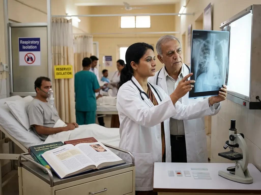
A laboratory technician's death in Siberia from an unverified respiratory pathogen has renewed global vigilance for pneumonic plague. Clinicians must recognize early presentations of Yersinia pestis, enforce immediate respiratory isolation, and start early bactericidal therapy to prevent severe secondary spread.
Today

Tripura has launched a comprehensive statewide oncology campaign to address late-stage breast cancer presentations. By strengthening community screening for women aged 30 and older and expanding District Day Care Cancer Centres, the initiative improves early clinical detection and decentralized chemotherapy access.
Today
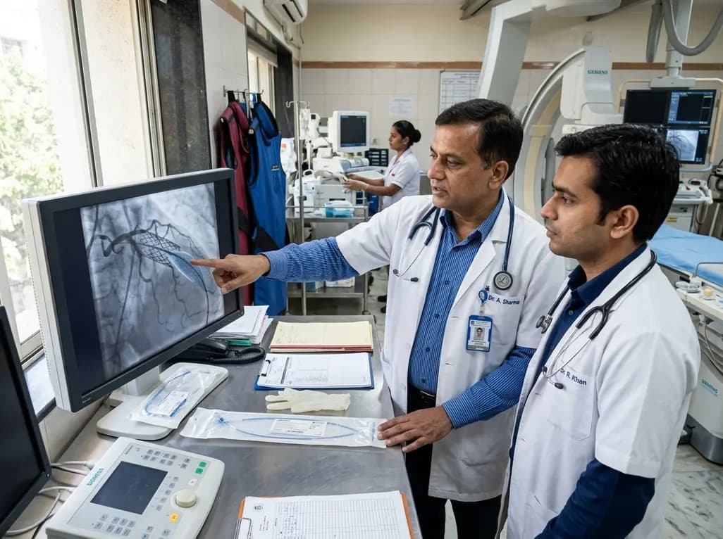
The US FDA has granted approval to the SELUTION SLR drug-eluting balloon for treating coronary in-stent restenosis. This milestone introduces a sustained-release sirolimus platform that eliminates the need for additional permanent metallic stents while maintaining robust vascular healing and luminal patency.
Today

The Thumbs-Out Classification (TOC) is a reliable screening tool for detecting thumb-in-palm deformity in children with cerebral palsy. Utilizing traffic-light tiers for radial abduction, TOC enables clinicians to monitor upper limb function and initiate timely therapeutic or surgical interventions.
Today

A comprehensive review analyzes periconceptional GLP-1 receptor agonist exposure, revealing reassuring data on congenital anomalies and adverse perinatal outcomes while emphasizing the need for cautious patient counseling.
Today
