

Loading, please wait...
Loading, please wait...

Pediatric multiple sclerosis represents a distinct clinical entity within acquired central nervous system demyelinating syndromes, accounting for approximately three to ten percent of all demyelinating presentations. Clinicians frequently encounter diagnostic and prognostic dilemmas because the disease course in children diverges noticeably from adult patterns. While adult disease involves both robust neuroinflammation and progressive neurodegeneration, young patients often exhibit aggressive acute relapse activity paired with a remarkable capacity for clinical recovery. Understanding the precise anatomical substrates of this condition remains paramount for pediatric neurologists, radiologists, and general physicians managing early demyelination.
Conventional magnetic resonance imaging continues to serve as the cornerstone for diagnosing demyelinating syndromes across all age brackets. In adult cohorts, spinal cord pathology represents an established surrogate marker of overall disease severity. Cervical cord lesions and progressive tissue atrophy correlate directly with chronic physical disability and secondary progressive conversion. However, pediatric multiple sclerosis exhibits distinctive radiological behavior that does not simply mirror adult neurodegenerative patterns. Advanced neuroimaging protocols now enable precise quantification of localized parenchymal changes, moving beyond qualitative lesion identification to volumetric assessments. Voxel-wise analytical techniques and three-dimensional high-resolution sequences allow researchers to examine microstructural variations throughout the upper cervical spine. Consequently, clinicians gain deeper insight into how acute myelin injury interacts with developmental neurobiology in youth. These sophisticated imaging modalities reveal that childhood central nervous system compartments experience distinct biological stresses. By examining cross-sectional spinal cord metrics early in the clinical course, clinicians can differentiate between transient inflammatory edema and irreversible axonal loss, ensuring tailored clinical expectations and accurate baseline evaluations.
To determine whether cervical cord atrophy develops early in pediatric disease, clinical investigators conducted a controlled study assessing thirty-eight young patients alongside thirteen healthy controls. The participants underwent comprehensive neurological examinations, disability grading, and volumetric spinal cord magnetic resonance imaging. Researchers paid particular attention to the upper cervical cord area, which classically demonstrates significant thinning in long-standing adult multiple sclerosis. Surprisingly, the quantitative analyses demonstrated no significant reduction in upper cervical cord area when comparing pediatric patients to healthy peer controls. Moreover, cord dimensions did not differ significantly between pediatric patients who harbored focal spinal lesions and those who exhibited normal-appearing spinal scans. Rather than displaying diffuse thinning or structural parenchymal loss, the spinal cord in these young individuals preserved its overall regional volume. These observations suggest that diffuse axonal degeneration and widespread cord atrophy do not represent prominent initial hallmarks of childhood demyelination. Instead, the macroscopic structural integrity of the spinal cord remains remarkably preserved during the early phases of disease manifestation.
Although overall cervical cord area showed stability, detailed voxel-wise evaluations uncovered compelling regional volumetric differences tied to acute inflammation. Specifically, pediatric patients presenting with focal lesions exhibited localized increases in regional cord volume directly co-localizing with those inflammatory plaques. This focal expansion reflects acute edema, hypercellular immune cell infiltration, and reactive glial swelling within active demyelinating plaques. Therefore, in pediatric multiple sclerosis, active lesions expand cord volume locally rather than contributing immediately to tissue thinning. In contrast to adult phenotypes where destructive lesions frequently induce rapid focal tissue cavitation and subsequent atrophy, childhood spinal lesions display predominantly inflammatory characteristics. The surrounding neurovascular unit reacts with marked vasogenic permeability and localized tissue expansion. Furthermore, this dynamic swelling demonstrates that the immature spinal cord reacts aggressively with cellular and fluid accumulation during autoimmune attacks. Recognizing this inflammatory tumefaction prevents clinicians from mistaking focal cord expansion for intramedullary neoplasia or extensive myelopathy. It confirms that the primary pathophysiological driver at disease onset is acute, reversible inflammatory swelling.
These neuroimaging discoveries carry profound implications for the therapeutic management of pediatric demyelinating conditions. Because early damage stems predominantly from acute inflammatory activity rather than irreversible neuroaxonal demise, clinicians possess a critical therapeutic window of opportunity. Highly effective disease-modifying therapies can suppress active immune cell trafficking across the blood-brain barrier, effectively terminating focal lesion formation. Consequently, timely immunomodulatory intervention halts recurrent tissue injury before destructive neurodegenerative cascades take hold. Because children maintain substantial neuroplastic reserve, their nervous systems can repair myelin sheaths and preserve functional integrity when neuroinflammation is successfully controlled. Furthermore, monitoring spinal cord dimensions may serve as an objective safety and efficacy marker in clinical trials evaluating emerging pediatric treatments. Clinicians must recognize that persistent inflammatory activity, if left unsuppressed over decades, will ultimately overwhelm local repair mechanisms and initiate secondary atrophy. Therefore, early escalation to robust disease-modifying regimens prevents cumulative disability, protects neural architecture, and ensures sustained neurological health into adulthood.
In developing regions such as India, evaluating spinal cord swelling in children demands careful exclusion of infectious and alternate inflammatory etiologies. Clinicians must differentiate demyelinating lesions from tuberculosis of the central nervous system, viral transverse myelitis, and acute disseminated encephalomyelitis. In addition, neuromyelitis optica spectrum disorder and myelin oligodendrocyte glycoprotein antibody-associated disease occur with notable frequency in Asian populations, often producing longitudinally extensive spinal cord lesions. High-resolution magnetic resonance imaging protocols must be interpreted alongside serological autoantibody panels and cerebrospinal fluid examinations. Because focal spinal swelling can mimic infectious intramedullary granulomas or neoplastic processes, identifying typical demyelinating features protects young patients from unnecessary surgical biopsies or delayed therapy. Radiologists should look for short-segment, peripherally located cervical plaques rather than central, longitudinally extensive abnormalities. Establishing an accurate diagnosis early enables clinicians to initiate pulse corticosteroid therapy promptly, followed by targeted immunotherapy. Comprehensive multidisciplinary collaboration between pediatricians, neurologists, and diagnostic neuroradiologists ensures that Indian children receive timely interventions tailored to their exact inflammatory profile.
Spinal cord atrophy is not a prominent early feature in pediatric cases. Quantitative magnetic resonance imaging demonstrates preserved upper cervical cord dimensions in young patients compared to healthy controls, indicating that diffuse neurodegenerative thinning develops much later in disease progression than in adult-onset presentations.
Active cervical cord lesions cause localized cord enlargement due to robust acute inflammation. Intense perivascular immune cell infiltration, vasogenic edema, and local glial activation induce tissue swelling at the lesion site, temporarily increasing regional cord volume rather than producing immediate structural loss or focal cavitation.
Because early disease pathology is predominantly inflammatory rather than degenerative, prompt administration of high-efficacy disease-modifying therapies can arrest inflammatory relapses. Intervening during this therapeutic window protects the high intrinsic repair capacity of children, preventing permanent neuroaxonal loss and long-term secondary neurodegenerative progression.
Disclaimer: This content is for informational and educational purposes only and should not be construed as medical advice or replace clinical judgment. Healthcare professionals must exercise independent clinical discretion and verify information against official sources. Treatment protocols and diagnostic pathways should be individualized to patient needs and institutional standards. Refer to the latest local and national guidelines for clinical practice.
References

Read summarized clinical updates, watch expert medical content, and earn CME certifications right from your smartphone.
Open your smartphone camera to scan and install.





A landmark MRI evaluation reveals that early pediatric multiple sclerosis presents with focal cord inflammation and localized swelling rather than significant cervical cord atrophy, highlighting distinctive pathophysiological mechanisms compared to adult-onset disease.
Today

Review how tailored exercise programs benefit frail older patients with heart failure. Explore pathophysiological mechanisms, individualized training prescriptions, and strategies to overcome adherence barriers in clinical practice.
Today
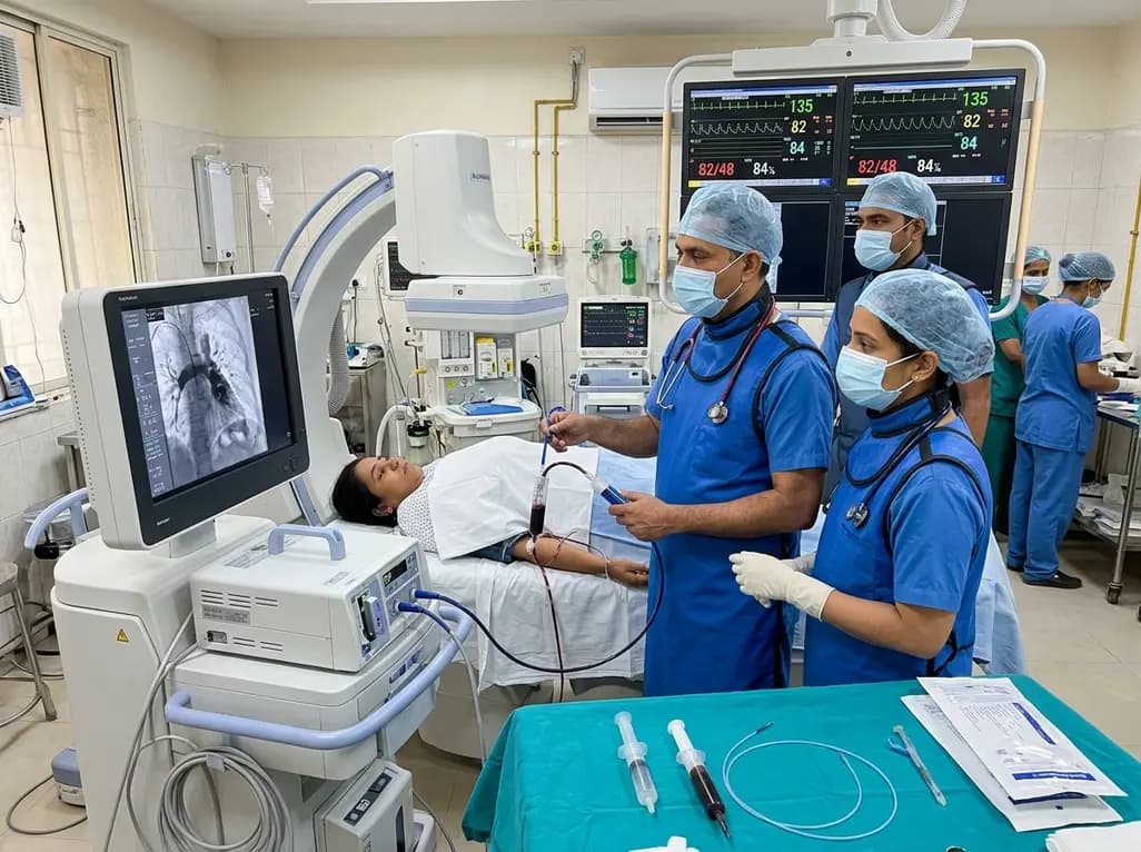
In high-risk pulmonary embolism complicated by severe anemia and active uterine bleeding, systemic thrombolysis is contraindicated. Large-bore aspiration thrombectomy provides rapid hemodynamic rescue without fibrinolysis, supported by inferior vena cava filter placement and multidisciplinary critical care.
Today

A cross-sectional study in Zanzibar reveals that 46.5% of adults with type 2 diabetes experience multidomain autonomic dysfunction on the COMPASS-31 tool, primarily orthostatic intolerance, while 45.9% exhibit cardiovascular autonomic neuropathy, highlighting the urgent need for structured clinical screening.
Today

This comparative study evaluates posterior labral tears with and without glenohumeral instability, highlighting distinct demographic patterns, MRI underestimation in stable shoulders, and successful arthroscopic repair outcomes.
Today

A recent case-control study reveals that urinary clusterin provides superior diagnostic performance for early diabetic kidney disease detection in pediatric type 1 diabetes mellitus, identifying tubular damage before conventional microalbuminuria occurs.
Today
