.webp&w=384&q=75)

Loading, please wait...
Loading, please wait...
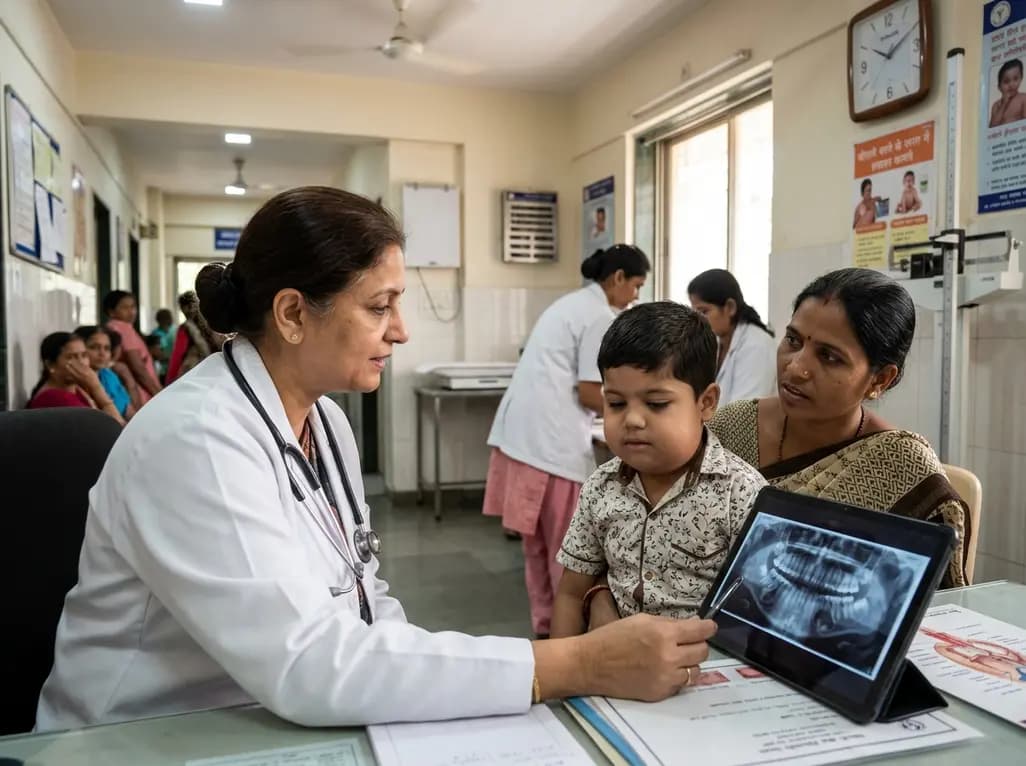
Pediatric cherubism represents a rare, benign, autosomal dominant bone disorder characterized by the symmetric expansion of the jaws. The condition primarily affects children between two and seven years of age. Clinicians frequently trace the underlying genetic etiology of cherubism to gain-of-function point mutations in the SH3BP2 gene located on chromosome 4p16.3. Consequently, these genetic alterations cause excessive osteoclastogenesis and hyperactive bone resorption within the maxillomandibular complex.
Furthermore, the mutated protein disrupts intracellular signaling pathways in myeloid and osteoclast precursor cells. This abnormal signaling promotes intense osteoclast recruitment and chronic inflammatory bone destruction. As a result, dense fibrovascular tissue containing numerous multinucleated giant cells progressively replaces normal medullary trabeculae. Although most patients inherit the condition in an autosomal dominant pattern with variable penetrance, spontaneous de novo mutations also occur in sporadic cases. Therefore, clinicians must recognize that the absence of a positive family history does not exclude the diagnosis of this disorder.
Clinically, pediatric cherubism typically presents as painless, bilateral swelling localized to the posterior mandible and maxilla. The expansive lesions create a characteristic round, cherub-like facial contour that gives the disorder its historic name. In severe cases, maxillary expansion elevates the orbital floors and stretches the lower eyelid skin downward. Consequently, this anatomical displacement exposes the inferior sclera, causing a classic upward-gazing ocular appearance.
Moreover, the progressive expansion of the alveolar processes significantly impacts dental development and oral function. Affected children frequently present with severe tooth displacement, delayed eruption of permanent dentition, premature exfoliation of primary teeth, and marked dental agenesis. In addition, extensive bone expansion can compromise airway patency, resulting in obstructive sleep apnea or difficulty swallowing. Nevertheless, patients typically maintain normal general physical health, intellectual development, and routine biochemical parameters, including serum calcium, phosphorus, and parathyroid hormone levels. Because of this localized nature, systematic clinical examinations help differentiate the disease from generalized systemic metabolic bone disorders.
Radiographic assessment plays a central role in confirming the diagnosis and evaluating the structural extent of the condition. Standard panoramic radiographs typically demonstrate bilateral, multilocular, expansile radiolucent lesions within the mandible and maxilla. Furthermore, these lesions impart a classic soap-bubble or honeycomb appearance to the jawbones. The pathological processes commonly begin near the mandibular angles and ascend through the ascending rami, characteristically sparing the mandibular condyles.
Additionally, modern cross-sectional imaging modalities provide invaluable anatomical detail for complex cases. Computed tomography (CT) and cone-beam computed tomography (CBCT) clearly depict severe cortical thinning, expansive osteolytic defects, and displaced tooth follicles. In contrast, magnetic resonance imaging (MRI) reveals soft-tissue extension and demonstrates non-specific intermediate T1 and T2 signal intensities with homogeneous post-contrast enhancement. Thus, longitudinal imaging protocols allow clinicians to track dynamic bone remodeling and detect spontaneous re-ossification as affected children mature toward adulthood.
Histopathological examination provides vital diagnostic confirmation when clinical and radiographic features present ambiguities. Under light microscopy, the lesions consist of abundant multinucleated giant cells uniformly dispersed throughout a highly cellular, vascularized fibrous stroma. Furthermore, pathognomonic eosinophilic perivascular cuffing of collagen around small blood vessels represents a distinctive histological hallmark in cherubism cases.
However, clinicians must carefully distinguish cherubism from several other giant cell-rich and fibro-osseous lesions of the maxillofacial skeleton. For example, central giant cell granuloma (CGCG) exhibits identical histological features but typically presents as a solitary, unilateral lesion. Similarly, brown tumors associated with primary or secondary hyperparathyroidism show abundant osteoclastic giant cells; nevertheless, abnormal serum parathyroid hormone and calcium levels readily identify them. In addition, craniofacial fibrous dysplasia produces characteristic ground-glass bony trabeculae rather than prominent multinucleated giant cells. Therefore, a comprehensive diagnostic approach that integrates histopathology, molecular testing, endocrine laboratory profiles, and advanced imaging remains mandatory.
The natural clinical trajectory of pediatric cherubism strongly guides therapeutic decision-making. In the vast majority of cases, the osteolytic process exhibits an indolent, self-limiting behavior that naturally stabilizes around puberty. Subsequently, spontaneous osteogenesis and progressive bone remodeling fill the lytic cavities during the second and third decades of life. Therefore, watchful waiting and conservative clinical surveillance represent the primary standard of care for asymptomatic patients.
Conversely, clinicians reserve aggressive surgical intervention exclusively for patients experiencing severe functional impairment or disfiguring facial deformity. When airway compromise, significant masticatory dysfunction, or vision-threatening orbital compression develops, surgical options include conservative curettage, bone re-contouring, or surgical decompression. Furthermore, surgeons generally avoid extensive early resection during the active inflammatory phase because aggressive interventions can paradoxically accelerate lesion growth. In recent years, researchers have investigated targeted medical therapies, including calcitonin, anti-TNF agents, and denosumab, to suppress osteoclastogenesis; however, watchful surveillance combined with supportive dental care remains the cornerstone of pediatric management.
Optimal long-term care for patients with cherubism requires a coordinated multidisciplinary approach. The treatment team should include pediatricians, maxillofacial surgeons, pediatric dentists, orthodontists, radiologists, and clinical geneticists. Because dental disruption occurs frequently, early orthodontic planning and prosthetic rehabilitation help preserve chewing efficiency, maintain arch symmetry, and protect developing permanent tooth buds.
Moreover, clinicians must prioritize the psychosocial well-being of affected children. Symmetrical facial swelling and dental deformities can cause substantial emotional distress, peer anxiety, and reduced quality of life during critical school years. Consequently, regular supportive psychological counseling and family education significantly improve treatment compliance and self-esteem. In addition, genetic counseling remains indispensable for affected families to explain autosomal dominant transmission risks, anticipate de novo occurrences, and guide family planning. Through structured longitudinal follow-up, medical teams ensure prompt detection of complications while minimizing unnecessary surgical morbidity.
Pediatric cherubism primarily arises from heterozygous gain-of-function mutations in the SH3BP2 gene located on chromosome 4p16.3. This genetic defect disrupts normal osteoclast signaling cascades, leading to hyperactive osteoclastogenesis and localized autoinflammatory bone resorption. Consequently, proliferative fibrovascular tissue rich in multinucleated giant cells replaces normal jawbone architecture. While patients usually inherit the condition in an autosomal dominant manner, spontaneous de novo mutations also occur frequently in children without family history.
Clinicians strongly favor conservative observation because pediatric cherubism follows a predictable, self-limiting clinical course. The expansive lesions typically halt their growth around puberty and progressively undergo spontaneous re-ossification and remodeling during early adulthood. Furthermore, aggressive surgical resection during the active childhood phase can stimulate accelerated lesion recurrence. Therefore, surgeons reserve operative procedures, such as curettage or aesthetic re-contouring, strictly for patients experiencing airway compromise, visual deficits, or severe functional impairments.
Physicians distinguish cherubism from central giant cell granulomas through an integrative approach combining clinical distribution, imaging, and genetic testing. Cherubism typically presents as bilateral, symmetrical multilocular expansile lesions in early childhood, whereas central giant cell granulomas usually appear as solitary, unilateral lesions in older individuals. Additionally, cherubism demonstrates characteristic perivascular collagen cuffing on histopathology and gain-of-function SH3BP2 mutations, which are absent in conventional central giant cell granulomas.
Disclaimer: This content is for informational and educational purposes only and does not constitute professional medical advice, diagnosis, or treatment. Always seek the advice of your physician or other qualified health provider with any questions you may have regarding a medical condition. Never disregard professional medical advice or delay in seeking it because of something you have read here. The authors and publishers are not liable for any losses, injuries, or damages arising from the use of this information. Refer to the latest local and national guidelines for clinical practice.
References

Read summarized clinical updates, watch expert medical content, and earn CME certifications right from your smartphone.
Open your smartphone camera to scan and install.





Cherubism is a rare, benign, autosomal dominant disorder causing bilateral jaw expansion in children. This review examines SH3BP2 mutations, diagnostic imaging, histopathology, differential diagnosis, and why conservative surveillance remains the preferred management approach for pediatric patients.
last month
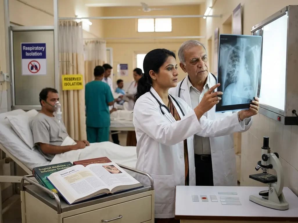
A laboratory technician's death in Siberia from an unverified respiratory pathogen has renewed global vigilance for pneumonic plague. Clinicians must recognize early presentations of Yersinia pestis, enforce immediate respiratory isolation, and start early bactericidal therapy to prevent severe secondary spread.
Today
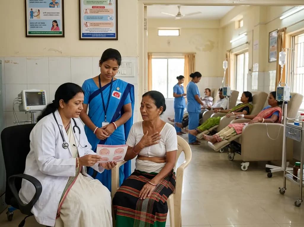
Tripura has launched a comprehensive statewide oncology campaign to address late-stage breast cancer presentations. By strengthening community screening for women aged 30 and older and expanding District Day Care Cancer Centres, the initiative improves early clinical detection and decentralized chemotherapy access.
Today
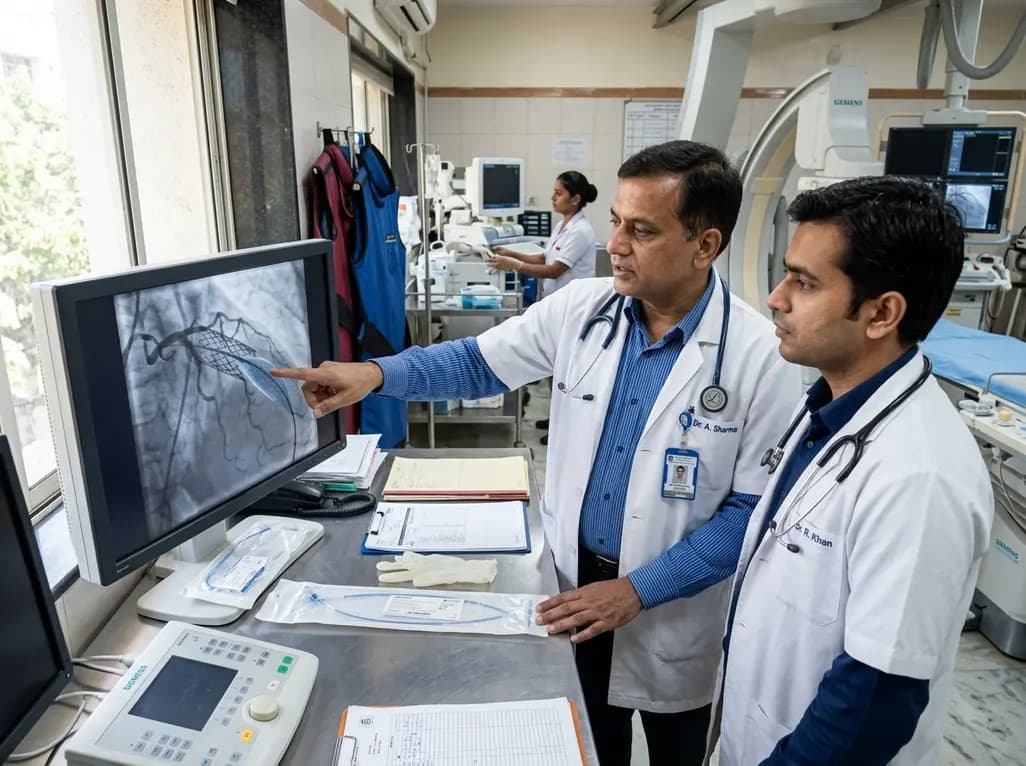
The US FDA has granted approval to the SELUTION SLR drug-eluting balloon for treating coronary in-stent restenosis. This milestone introduces a sustained-release sirolimus platform that eliminates the need for additional permanent metallic stents while maintaining robust vascular healing and luminal patency.
Today

The Thumbs-Out Classification (TOC) is a reliable screening tool for detecting thumb-in-palm deformity in children with cerebral palsy. Utilizing traffic-light tiers for radial abduction, TOC enables clinicians to monitor upper limb function and initiate timely therapeutic or surgical interventions.
Today

A comprehensive review analyzes periconceptional GLP-1 receptor agonist exposure, revealing reassuring data on congenital anomalies and adverse perinatal outcomes while emphasizing the need for cautious patient counseling.
Today
