.webp&w=384&q=75)

Loading, please wait...
Loading, please wait...

Primary breast neuroendocrine neoplasms represent an exceedingly rare diagnostic entity in clinical oncology. However, pathologists and clinicians often encounter considerable diagnostic ambiguity when identifying these lesions. Consequently, recent updates in classification systems establish clearer criteria that facilitate personalized treatment.
Pathologists have vigorously debated the diagnostic framework of breast neuroendocrine neoplasms for decades. Initially, historical World Health Organization criteria required neuroendocrine differentiation in over fifty percent of tumour cells. However, clinical studies demonstrated that this arbitrary threshold caused high interobserver variability among pathologists. Therefore, the fifth edition of the World Health Organization classification reorganized these tumours into well-differentiated neuroendocrine tumours and poorly differentiated neuroendocrine carcinomas. Furthermore, experts created a distinct category for invasive carcinomas of no special type that display focal neuroendocrine differentiation. Poorly differentiated carcinomas comprise small cell and large cell variants that follow aggressive clinical courses. In addition, recent guidelines discourage diagnosing pure neuroendocrine tumours when lesions lack diffuse organoid morphology. Meanwhile, consensus panels emphasize integrating cytological characteristics with quantitative immunohistochemistry. Thus, the revised hierarchical framework provides much-needed diagnostic clarity for pathologists globally.
Microscopic evaluation reveals characteristic morphological patterns that suggest neuroendocrine lineage. Well-differentiated tumours typically display solid nests, trabeculae, ribbons, or insular arrangements. Furthermore, delicate fibrovascular networks consistently surround these uniform cellular aggregates. Cytologically, these tumour cells exhibit plasmacytoid or polygonal features with finely stippled salt-and-pepper nuclear chromatin. In contrast, poorly differentiated neuroendocrine carcinomas demonstrate diffuse sheets, extensive geographic necrosis, and abundant apoptotic debris. Specifically, small cell variants display hyperchromatic nuclei, scant cytoplasm, nuclear moulding, and frequent crush artefacts. Similarly, large cell neuroendocrine carcinomas feature vesicular nuclei, prominent nucleoli, and elevated mitotic rates. Moreover, pathologists frequently encounter diagnostic dilemmas with solid papillary carcinomas and mucinous carcinomas because both entities show neuroendocrine differentiation. Therefore, pathologists must distinguish pure neoplasms from these morphologically overlapping tumours. In addition, identifying extracellular mucin or delicate fibrovascular cores helps exclude secondary variants. When neuroendocrine features comprise less than ninety percent of the mass, specialists diagnose invasive carcinoma with neuroendocrine differentiation.
Immunohistochemical staining remains indispensable for validating neuroendocrine differentiation in mammary tissue. Synaptophysin offers the highest sensitivity among classic neuroendocrine markers in surgical specimens. However, synaptophysin can show non-specific cytoplasmic background staining in conventional ductal carcinomas. Consequently, pathologists evaluate chromogranin A because it exhibits superior diagnostic specificity despite lower sensitivity. Recently, insulinoma-associated protein 1 emerged as an exceptional novel nuclear biomarker for neuroendocrine neoplasms. Additionally, insulinoma-associated protein 1 provides clean nuclear staining that significantly reduces diagnostic ambiguity in core needle biopsies. Pathologists strongly discourage using neuron-specific enolase due to unacceptably high rates of false-positive results. Furthermore, complete diagnostic assessment requires testing for estrogen receptor, progesterone receptor, and human epidermal growth factor receptor 2. Most well-differentiated tumours demonstrate strong hormone receptor positivity and lack HER2 expression. In contrast, poorly differentiated carcinomas usually display triple-negative profiles and extremely high Ki-67 proliferation indices. As a result, comprehensive immunohistochemical panels prevent costly interpretive errors.
Distinguishing primary mammary neuroendocrine tumours from metastatic lesions represents a critical clinical objective. Metastatic neuroendocrine neoplasms originating in the lungs, pancreas, or gastrointestinal tract frequently mimic primary breast malignancies. Therefore, pathologists meticulously search for associated ductal carcinoma in situ to confirm mammary origin. However, small core biopsies often lack in situ components due to limited tissue sampling. Consequently, clinicians must rely on organ-specific immunohistochemical markers to resolve the primary site. Mammary-specific markers including GATA3, mammaglobin, and gross cystic disease fluid protein 15 strongly support a primary breast neoplasm. Conversely, thyroid transcription factor 1 expression confirms a pulmonary origin. Similarly, CDX2 positivity indicates intestinal origin, while PAX8 and islet 1 expression indicate pancreatic origin. In addition, oncologists should perform cross-sectional imaging and somatostatin receptor scintigraphy whenever biomarker results appear ambiguous. Thus, thorough clinical correlation prevents misdiagnosis and avoids unnecessary surgical interventions.
Recent genomic sequencing has illuminated the divergent pathogenesis of breast neuroendocrine neoplasms. Well-differentiated neuroendocrine tumours demonstrate molecular profiles that closely resemble conventional luminal breast cancers. Specifically, genomic assays frequently detect mutations in PIK3CA, GATA3, and MAP3K1 within well-differentiated lesions. Furthermore, copy number analyses show recurrent gains on chromosome 1q and losses on chromosome 16q. These molecular findings indicate that low-grade tumours arise from luminal mammary precursors rather than specialized neuroendocrine cells. In contrast, poorly differentiated neuroendocrine carcinomas display aggressive genomic alterations comparable to pulmonary small cell carcinomas. Researchers consistently identify concurrent TP53 and RB1 inactivation in high-grade mammary carcinomas. Additionally, these aggressive tumours harbour prominent chromosomal instability and frequent MYC amplifications. Therefore, targeted next-generation sequencing provides valuable prognostic information for ambiguous cases. Ultimately, genomic profiling assists multidisciplinary teams in distinguishing indolent disease from highly aggressive malignancies.
Accurate pathological diagnosis directly guides systemic and surgical therapy in modern breast oncology. Because prospective randomized clinical trials are sparse, clinicians tailor treatment strategies according to histological grade and receptor expression. Patients with well-differentiated neuroendocrine tumours generally receive standard breast cancer protocols. Consequently, surgical oncologists perform lumpectomy or mastectomy with sentinel lymph node biopsy. Furthermore, medical oncologists prescribe adjuvant endocrine therapy for hormone-receptor-positive tumours. In contrast, poorly differentiated neuroendocrine carcinomas require aggressive multimodal systemic therapy. Oncology teams typically administer platinum agents combined with etoposide, mirroring established small cell lung cancer regimens. Additionally, clinicians may consider somatostatin receptor-targeted therapy or peptide receptor radionuclide therapy for advanced somatostatin-receptor-positive cases. Routine multidisciplinary case discussions ensure that surgeons, pathologists, and medical oncologists collaborate effectively. Therefore, standardizing pathological evaluation directly enhances patient survival and clinical outcomes.
Primary breast neuroendocrine neoplasms are exceptionally rare tumours, representing less than one percent of all primary breast malignancies. Their true incidence remains difficult to measure because past classification systems utilized differing histological thresholds. Most patients are postmenopausal women presenting with palpable, solitary breast nodules. Because these neoplasms are uncommon, clinicians and pathologists must always rule out metastatic neuroendocrine disease from gastrointestinal or pulmonary origins before confirming a primary breast tumour diagnosis.
Pathologists primarily evaluate synaptophysin, chromogranin A, and insulinoma-associated protein 1 to identify neuroendocrine differentiation in breast specimens. While synaptophysin demonstrates high sensitivity, chromogranin A offers superior diagnostic specificity. Furthermore, insulinoma-associated protein 1 serves as an emerging nuclear biomarker that resolves equivocal cases effectively. Conversely, pathologists avoid neuron-specific enolase due to high non-specific background reactivity. In addition, assessing estrogen receptor, progesterone receptor, and HER2 remains mandatory for treatment planning.
Pathologists and oncologists distinguish primary breast tumours from metastatic lesions using histopathology, immunohistochemistry, and clinical imaging. Identifying an associated ductal carcinoma in situ definitively confirms primary mammary origin. Furthermore, positive staining for mammary biomarkers like GATA3 and mammaglobin supports a breast primary. Conversely, expression of TTF-1, CDX2, or PAX8 indicates extra-mammary origins. Additionally, clinicians use whole-body computed tomography or somatostatin receptor scintigraphy to detect hidden primary tumours elsewhere.
Disclaimer: This content is for informational and educational purposes only and does not constitute medical advice or establish a doctor-patient relationship. Healthcare professionals must exercise independent clinical judgement when applying this information. While based on peer-reviewed research, it is not an exhaustive clinical manual. Refer to the latest local and national guidelines for clinical practice.
References

Read summarized clinical updates, watch expert medical content, and earn CME certifications right from your smartphone.
Open your smartphone camera to scan and install.





A comprehensive clinical review of breast neuroendocrine neoplasms, detailing the evolution of WHO diagnostic criteria, morphological patterns, novel immunohistochemical markers like INSM1, differential diagnoses against metastases, genomic landscapes, and grade-adapted treatment protocols.
Today

India's antidepressant market crossed Rs 2,842 crore in 2026 even as unit sales declined. Clinicians analyze the shift toward newer drugs, widespread clonazepam combination use, social media-driven self-diagnosis, and persistent treatment gaps to assess genuine therapeutic recovery across diverse patient groups.
Today

Integrating adult genetic services into reproductive and pregnancy care substantially alters clinical management. A study reveals that diagnostic genetic evaluations directly inform maternal, fetal, and neonatal care in nearly 57% of tested patients, highlighting the value of preconception and antenatal genomics.
Today
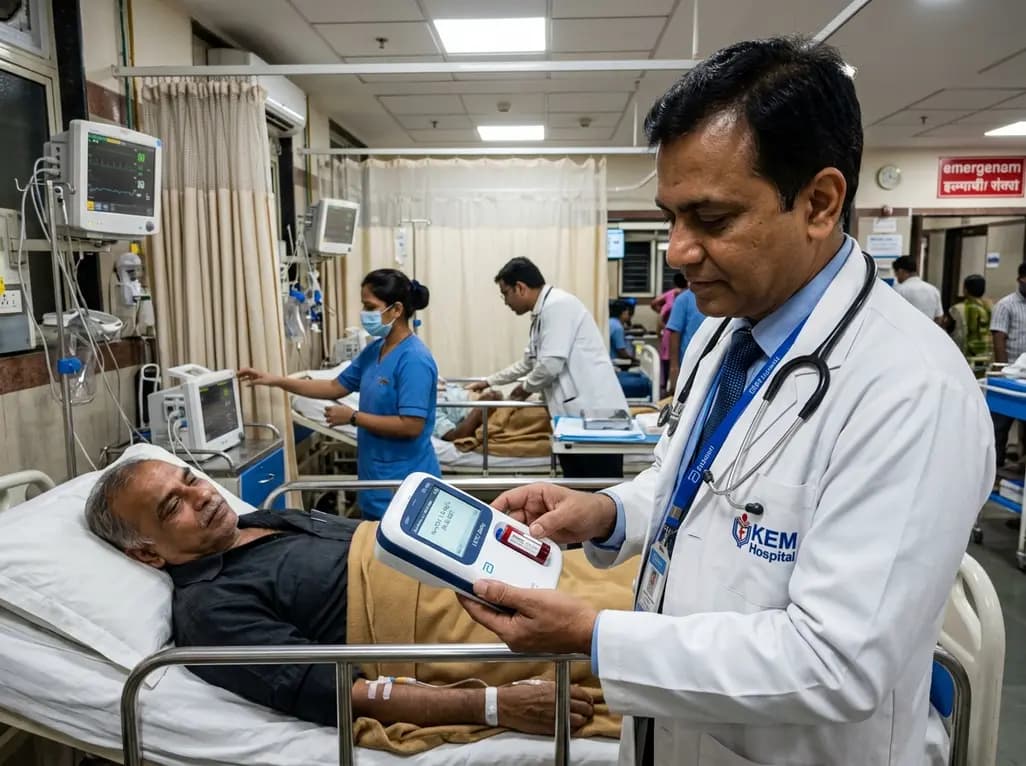
A real-world emergency department evaluation demonstrates that the Abbott i-STAT Alinity point-of-care high-sensitivity troponin I assay achieves excellent analytical precision and safely rules out myocardial infarction without false negatives, offering rapid bedside results to decongest acute care pathways.
Today

A recent scoping review highlights the efficacy, usability, and patient satisfaction of virtual reality pain management for chronic musculoskeletal pain, emphasizing psychotherapy-based VR protocols.
Today

A cross-sectional study in Nepal evaluates cardiovascular risk factors among adults working above 4,000 meters. The findings reveal unexpected rates of diabetes, hypertension, and mountain sickness, underscoring the urgent need for targeted cardiometabolic screening in high-altitude occupational cohorts.
Today
