

Loading, please wait...
Loading, please wait...

Epiphora and medial canthal swelling represent routine clinical presentations in ophthalmology clinics, yet unusual pathogens can complicate diagnosis. Clinicians frequently encounter persistent inflammatory conditions that fail routine conservative care. Indeed, parasitic lacrimal infections represent rare but clinically impactful conditions that closely mimic bacterial dacryocystitis or lacrimal neoplasms. Because these entities occur infrequently, practitioners often delay accurate diagnoses, subjecting patients to ineffective antibiotic regimens or inappropriate probing. However, recent systematic evidence sheds vital light on these insidious ocular pathologies. By recognizing unusual clinical features early, clinicians can avoid treatment delays and choose targeted, organ-sparing surgical therapies with confidence.
A recent comprehensive systematic review examined forty-four international studies encompassing 172 individual patient cases. Interestingly, the geographical distribution reflects distinct tropical and subtropical environments where individuals encounter contaminated water or endemic soil. The analysis revealed that Rhinosporidium seeberi accounts for 85.5 percent of all reported infections worldwide. Consequently, aquatic protistan exposure represents the primary driver of lacrimal parasitic disease. Furthermore, protozoal pathogens such as Leishmania species contribute 4.7 percent of cases, while nematodes like Ascaris lumbricoides cause 4.1 percent. Clinicians in South Asia, especially across southern and eastern India, report the vast majority of ocular rhinosporidiosis cases. Therefore, rural agricultural workers and regular bathers in stagnant rural ponds face heightened exposure risks. Transmission typically occurs when motile endospores penetrate microtraumas within the fragile canalicular or conjunctival epithelium. Although men and young adults demonstrate higher historical prevalence rates, the condition can afflict patients of any age. Thus, collecting a meticulous environmental history remains essential whenever evaluating chronic medial canthal masses.
Differentiating parasitic infestations from routine bacterial dacryocystitis presents substantial diagnostic difficulty for clinicians. Patients typically report progressive epiphora, mild medial canthal discomfort, and intermittent mucopurulent discharge. However, unlike acute bacterial infections, parasitic lesions evolve slowly over many months without pronounced erythema or systemic pyrexia. The lacrimal sac remains the most commonly affected anatomical site because its mucosal architecture provides an ideal proliferative environment. In many instances, the expanding pathogen induces an indolent diverticular mass that mimics a mucocele. Consequently, physicians frequently attempt conservative saline irrigation and topical antibiotics before recognizing clinical failure. In advanced cases, the granulomatous lesion may erode the overlying skin or invade adjacent ethmoid air cells. Additionally, patients with ocular rhinosporidiosis may exhibit small, friable conjunctival polyps or yellowish punctate spots representing mature subepithelial sporangia. Therefore, clinicians must maintain high clinical suspicion whenever routine medical management fails to relieve medial canthal distension. Early identification prevents unnecessary procedural delays and limits improper antibiotic administration.
The etiological profile of lacrimal parasitism displays remarkable diversity despite the clear predominance of Rhinosporidium seeberi. Although researchers historically classified Rhinosporidium seeberi as a fungus, molecular phylogenetic analysis places it within the Mesomycetozoea clade of aquatic protists. The organism matures within host tissues, forming thick-walled sporangia filled with thousands of endospores that trigger vigorous hyperplastic stromal responses. In contrast, Leishmania parasites enter the lacrimal sac through direct sandfly inoculation or mucosal spread from cutaneous leishmaniasis. These intracellular amastigotes provoke severe histiocytic infiltration and progressive granulomatous destruction of tear drainage structures. Meanwhile, helminthic infections present unique physical obstruction challenges for the surgeon. Roundworms such as Ascaris lumbricoides occasionally migrate upward from the gastrointestinal tract and nasopharynx, becoming physically entrapped within the nasolacrimal duct. Similarly, rare filarial nematodes can lodge directly inside the lacrimal canaliculi. Because each pathogen exhibits distinct pathophysiology, clinicians must combine clinical clues with targeted laboratory diagnostics. Therefore, precise organism identification dictates whether patients require localized surgical excision or systemic antiparasitic therapy.
Accurate preoperative diagnosis remains difficult because clinical symptoms mimic ordinary inflammatory diseases. Consequently, definitive diagnosis relies almost entirely on postoperative histopathological examination of excised tissue. When evaluating biopsy specimens, pathologists readily identify Rhinosporidium seeberi by detecting gigantic, round sporangia measuring several hundred micrometers in diameter. These sporangia stain intensely with Gomori methenamine silver, Periodic acid-Schiff, and mucicarmine stains, revealing innumerable round endospores. In contrast, identifying lacrimal leishmaniasis requires Giemsa-stained smears or polymerase chain reaction assays that highlight Leishman-Donovan bodies within host macrophages. Preoperative imaging, including contrast-enhanced computed tomography and magnetic resonance dacryocystography, provides essential structural information. These radiological scans delineate lesion boundaries, evaluate bone changes in the lacrimal fossa, and exclude malignant tumors. Furthermore, diagnostic nasal endoscopy allows direct evaluation of the sinonasal mucosa to detect concurrent polyps. Routine blood counts usually show normal parameters, although peripheral eosinophilia may accompany migrating helminths. Therefore, a multidisciplinary approach involving ophthalmologists, pathologists, and radiologists ensures prompt diagnostic confirmation.
Successful management requires customized therapeutic strategies based on the causative organism and anatomical involvement. For lacrimal rhinosporidiosis, complete surgical excision remains the definitive standard of care. Surgeons typically perform meticulous dacryocystectomy or transnasal endoscopic resection, removing all diseased mucosal elements. Furthermore, wide electrocautery of the mucosal base is essential to destroy residual endospores and prevent surgical seeding. Although dacryocystorhinostomy restores physiological drainage, surgeons usually avoid creating bony ostia during active infection to prevent parasitic spread into nasal bones. In contrast, helminthic infections require gentle mechanical extraction of the worm under direct visualization. Clinicians must avoid rupturing the parasite to prevent intense foreign-body granulomas or anaphylactoid responses. Additionally, adjunctive medical pharmacotherapy plays an important role. For instance, systemic dapsone reduces postoperative recurrence rates in rhinosporidiosis. Similarly, patients with leishmaniasis require antileishmanial medications like liposomal amphotericin B or oral miltefosine. Ultimately, combining meticulous surgical clearance with targeted medical therapy and avoiding stagnant water exposure optimizes long-term patient recovery.
Histopathology provides the definitive confirmation for parasitic lacrimal infections because clinical presentations and radiological imaging closely mimic chronic bacterial dacryocystitis and neoplasms. Tissue staining with Periodic acid-Schiff and Gomori methenamine silver reveals characteristic microscopic features, such as the gigantic sporangia and endospores of Rhinosporidium seeberi or intracellular amastigotes of Leishmania. Consequently, prompt pathological examination prevents catastrophic misdiagnoses, eliminates unnecessary antibiotic courses, and directs clinicians toward etiology-specific medical and surgical therapy.
Complete surgical excision of the diseased lacrimal sac, combined with thorough electrocautery of the mucosal base, provides the most effective defense against disease recurrence. Surgeons must exercise extreme gentleness during tissue dissection to avoid sporangial rupture and subsequent endospore spillage into surrounding facial planes. Furthermore, performing an immediate dacryocystorhinostomy is often discouraged because creating a bony ostium during active infection can facilitate parasitic dissemination into the adjacent sinonasal cavity.
Parasitic lacrimal lesions generally present as indolent, slowly progressive medial canthal masses that lack the acute tenderness, fever, and prominent cutaneous erythema typical of bacterial dacryocystitis. In addition, these lesions fail to resolve following standard broad-spectrum antibiotic courses or lacrimal probing. Clinicians should observe patient exposure history, such as bathing in endemic stagnant water, and look for visible mucosal polyps with punctate yellowish spots on the conjunctiva.
Disclaimer: This content is for informational and educational purposes only and does not constitute medical advice, diagnosis, or treatment. Healthcare professionals should exercise their independent clinical judgment. Patients should seek the advice of a physician or other qualified healthcare provider with any questions regarding a medical condition. Refer to the latest local and national guidelines for clinical practice.
References

Read summarized clinical updates, watch expert medical content, and earn CME certifications right from your smartphone.
Open your smartphone camera to scan and install.





Parasitic infections of the lacrimal system, predominantly caused by Rhinosporidium seeberi, often masquerade as chronic dacryocystitis. This review examines their etiology, diagnostic evaluation, surgical management, and prevention of recurrence to help clinicians optimize patient care and treatment outcomes.
Today
A recent breakthrough reveals that capsaicin mitigates microglial inflammation through a novel TRPV1-SSR4 protein-protein interaction independent of classic calcium influx. This discovery highlights fresh therapeutic avenues for managing neurodegenerative diseases like Alzheimer's.
Today
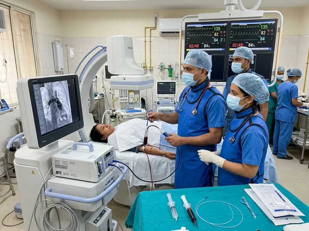
In high-risk pulmonary embolism complicated by severe anemia and active uterine bleeding, systemic thrombolysis is contraindicated. Large-bore aspiration thrombectomy provides rapid hemodynamic rescue without fibrinolysis, supported by inferior vena cava filter placement and multidisciplinary critical care.
Today

A cross-sectional study in Zanzibar reveals that 46.5% of adults with type 2 diabetes experience multidomain autonomic dysfunction on the COMPASS-31 tool, primarily orthostatic intolerance, while 45.9% exhibit cardiovascular autonomic neuropathy, highlighting the urgent need for structured clinical screening.
Today
This comparative study evaluates posterior labral tears with and without glenohumeral instability, highlighting distinct demographic patterns, MRI underestimation in stable shoulders, and successful arthroscopic repair outcomes.
Today

A recent case-control study reveals that urinary clusterin provides superior diagnostic performance for early diabetic kidney disease detection in pediatric type 1 diabetes mellitus, identifying tubular damage before conventional microalbuminuria occurs.
Today
