.webp&w=384&q=75)

Loading, please wait...
Loading, please wait...
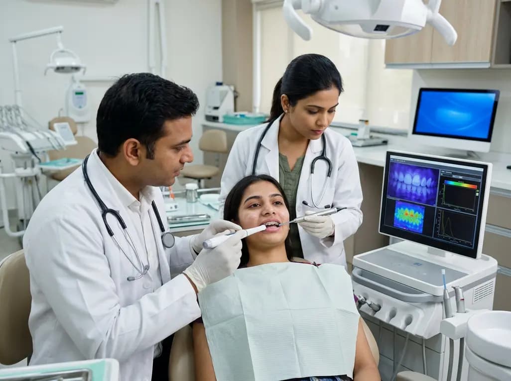
White spot lesions (WSLs) remain a significant challenge for orthodontists across India, often marring the aesthetic outcome of otherwise successful treatments. These areas of enamel demineralization appear as opaque, milky-white spots, primarily due to prolonged plaque accumulation around fixed appliances. Effective white spot lesion assessment is crucial for early intervention and monitoring. However, determining the most accurate diagnostic tool is complex. Recent research suggests that relying on a single method may lead to incomplete clinical pictures. Clinicians frequently choose between visual inspection, laser fluorescence, and light-induced fluorescence, but each technology captures different aspects of the lesion. Some tools measure the depth of mineral loss, while others focus on surface area or optical changes. Understanding how these methods correlate is essential for developing a robust protocol. Furthermore, as patient expectations for aesthetics rise, Indian dental professionals must adopt more sophisticated monitoring strategies. This study highlights the limited interchangeability of current assessment tools, emphasizing the need for a multimodal approach to ensure comprehensive patient care and successful remineralization outcomes.
The process of white spot lesion assessment involves evaluating sub-surface porosities caused by acidogenic bacteria. When plaque remains undisturbed around orthodontic brackets, organic acids dissolve enamel minerals, specifically calcium and phosphorus. This process alters the refractive index of the enamel, resulting in the characteristic white appearance. Specifically, the porosity within the enamel increases, which scatters light differently than healthy tissue. Consequently, clinicians must distinguish between active lesions, which are porous and matte, and inactive ones, which are shiny and smooth. Quantitative tools aim to remove the subjectivity of traditional visual methods like the International Caries Detection and Assessment System. However, the underlying physics of these tools varies significantly. For example, some utilize the auto-fluorescence properties of the tooth, whereas others measure light scattering or mineral density. Therefore, a clinician must understand that a high score on one device might not perfectly correlate with another. In the context of Indian clinical practice, where patient follow-up can sometimes be inconsistent, using highly sensitive tools for early detection becomes vital to prevent permanent cavitation.
Among the most popular technologies used in modern dentistry are Quantitative Light-induced Fluorescence (QLF) and DIAGNOdent. QLF works by illuminating the tooth with blue light, causing the enamel to fluoresce. Areas of demineralization appear darker because they lose this fluorescence. This method provides various metrics, such as lesion area and the percentage of fluorescence loss. On the other hand, DIAGNOdent uses a red laser to detect bacterial by-products and changes in tooth structure. While both are helpful for white spot lesion assessment, they often provide different insights. Specifically, QLF is highly sensitive to changes in the volume and area of the lesion. In contrast, DIAGNOdent is often praised for its ease of use and portability in a busy chairside environment. Recent data indicates that the correlation between these two methods is surprisingly weak immediately after lesion induction. This suggests that during the early stages of demineralization, the optical changes captured by QLF do not align perfectly with laser-detected changes. Thus, practitioners should be cautious when switching between these devices during a patient's treatment journey, as the values are not directly interchangeable.
To truly understand the severity of a white spot lesion, one must look at the mineral composition of the enamel. Scanning electron microscopy with energy-dispersive X-ray spectroscopy (SEM-EDS) is the gold standard for this, as it measures the calcium-to-phosphorus (Ca/P) ratio. High-quality white spot lesion assessment often attempts to correlate chairside tools with these laboratory-grade measurements. Research shows that intensity-based QLF metrics, such as ΔF, align more closely with mineral-related changes after remineralization treatment. Interestingly, the lesion area does not always decrease in direct proportion to mineral gain. This discrepancy occurs because a lesion might become denser without significantly shrinking in size. Furthermore, colourimetry provides another layer of information by assessing the whiteness or L* value of the lesion. While mineral gain is the biological goal, the patient’s primary concern is usually the visual disappearance of the spot. Consequently, a combined model that includes both intensity-based fluorescence and colour-based indices offers a more holistic view. For Indian orthodontists, this highlights the importance of explaining to patients that a lesion might be healing internally even if the white spot remains visible for some time.
For clinicians in India, the choice of diagnostic tool often depends on availability and cost. However, the latest evidence regarding white spot lesion assessment suggests that no single chairside measure is sufficient for all clinical scenarios. During the active phase of orthodontic treatment, monitoring the surface area of lesions might be the most practical way to track hygiene compliance. Once the braces are removed and remineralization therapy begins, focusing on the intensity of fluorescence or mineral density becomes more critical. Moreover, the integration of these tools into digital workflows is becoming increasingly common. Specifically, using QLF allows for photographic documentation, which can be shared with the patient to improve motivation. Therefore, a multimodal approach is not just a scientific necessity but also a powerful communication tool. Clinicians should consider using a combination of visual inspection, colour assessment, and at least one quantitative measure. This comprehensive strategy ensures that the clinician is not misled by the limitations of a single device. In addition, keeping detailed records of these various metrics allows for better long-term monitoring of enamel health throughout the retention phase.
The quest for a single explanatory model for lesion volume remains ongoing. Current mixed-effects models have shown that while combined models perform better than single-metric models, their overall explanatory power is still modest. This indicates that white spot lesion assessment is influenced by numerous factors, including saliva composition, fluoride exposure, and individual enamel variations. Consequently, the development of more sophisticated AI-driven models may eventually provide better predictive accuracy. For now, the takeaway for the dental community is that WSLs are dynamic and multifaceted. Furthermore, future research should focus on longitudinal studies within the diverse Indian population to account for dietary differences. In the interim, practitioners must rely on a blend of technology and clinical judgment. Notably, the transition from demineralization to remineralization involves different optical and structural pathways. Therefore, a tool that is excellent at detecting early loss might not be the best at tracking recovery. By acknowledging these nuances, orthodontists can provide more personalized and effective care, ensuring that the final aesthetic result of orthodontic treatment is not compromised by preventable enamel scarring.
While both tools are used for white spot lesion assessment, they serve different primary functions. QLF is excellent for measuring the area and volume of demineralization using blue light fluorescence, providing a visual map of the lesion. DIAGNOdent uses laser fluorescence to detect changes in tooth structure and bacterial presence. Research suggests they are not interchangeable, as their correlation is often weak, particularly during the early stages of lesion development before treatment.
White spot lesions can often be remineralized, but complete visual reversal is not always guaranteed. Remineralization treatments, such as fluoride varnishes or CPP-ACP, aim to restore the mineral content and Ca/P ratio. However, while the internal structure might improve, the optical white appearance may persist if the surface remains porous. Effective white spot lesion assessment helps clinicians determine if a lesion is active or stable, guiding the appropriate aesthetic intervention for the patient.
A multimodal approach is recommended because no single tool captures every aspect of a lesion. For instance, QLF measures fluorescence loss, colourimetry tracks aesthetic changes, and SEM-EDS monitors mineral density. Because these metrics often do not correlate perfectly, using a single method might overlook critical changes in lesion depth or area. Combining these insights allows for a more accurate white spot lesion assessment, ensuring better clinical decision-making during and after orthodontic treatment.
Disclaimer: This content is for informational and educational purposes only. It is not a substitute for professional medical advice, diagnosis, or treatment. Always seek the advice of your physician or other qualified health provider with any questions you may have regarding a medical condition. Refer to the latest local and national guidelines for clinical practice.
References
1. Firinciogullari EC et al. Intermethod Association in Orthodontic White Spot Lesion Assessment: A Multimodal-Assessment. Int Dent J. 2026 Jul 16. doi: undefined. PMID: 42462347.
2. Pretty IA. Caries detection and diagnosis: Novel technologies. J Dent. 2006;34(10):727-739.
3. Kim S, et al. Assessment of enamel demineralization during orthodontic treatment: A review. Korean J Orthod. 2013;43(6):278-286.
"
Read summarized clinical updates, watch expert medical content, and earn CME certifications right from your smartphone.
Open your smartphone camera to scan and install.





A recent study evaluates the association between various diagnostic methods for orthodontic white spot lesions (WSLs). Findings suggest that tools like QLF and DIAGNOdent provide complementary information, and no single measure is sufficient for a comprehensive assessment of demineralization and recovery.
3 months ago
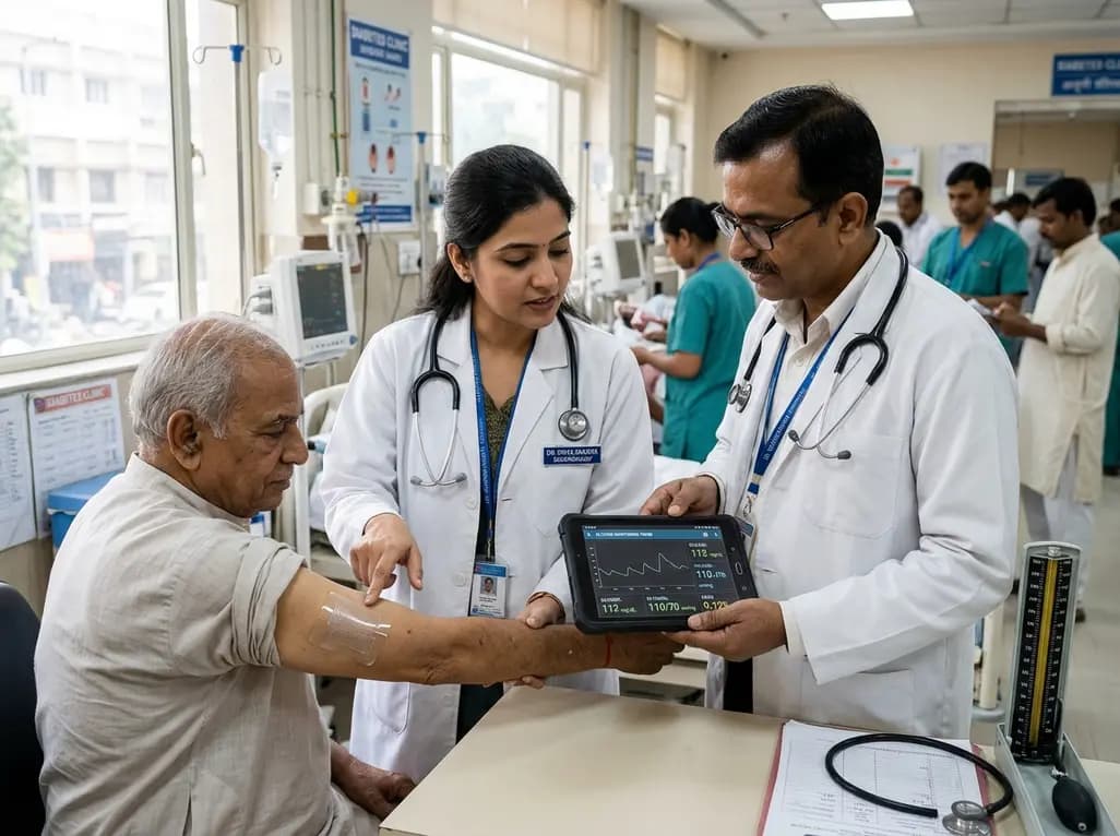
A breakthrough ultrathin multimodal sensor decouples physical hemodynamic monitoring from disposable electrochemical calibration, achieving long-term wearable glucose prediction with 81.36% accuracy and a 9.12% MARD. This offers a robust, drift-free solution for outpatient metabolic surveillance.
Today

Demanding exhaustive hormonal profiling can inadvertently discourage clinical studies involving females. Methodological standards must align with the specific scientific question to expand, not restrict, equitable evidence for women.
Today

Co-administering antiseizure medications and DOACs frequently causes pharmacokinetic interactions, subtherapeutic drug levels, and thromboembolic failures. This review highlights enzyme induction risks, unpredictable outcomes with levetiracetam, and the vital role of therapeutic drug monitoring.
Today

A comparative review of tension band wiring and intramedullary screw fixation in thumb metacarpophalangeal arthrodesis, evaluating union rates, hardware complications, and clinical outcomes.
Today

Following regulatory scrutiny from FSSAI regarding promotional representations and lean muscle preservation claims, Dr. Reddy's Laboratories and Nestlé Health Science are currently reviewing the branding and marketing strategy for their joint nutritional supplement Celevida GLP+.
Today
