.webp&w=384&q=75)

Loading, please wait...
Loading, please wait...

Increased intracranial pressure represents a life-threatening emergency in intensive care units across the globe. When intracranial pressure escalates, cerebral perfusion falls dramatically and ischemic injury accelerates rapidly. Historically, neurointensivists have relied on invasive monitors or repeated computed tomography scans to identify rising pressures. However, invasive catheter insertion carries substantial risks of hemorrhage, infection, and catheter misplacement. Consequently, point-of-care ultrasonography has emerged as a revolutionary bedside diagnostic tool. In particular, sonographic assessment of optic nerve sheath diameter provides immediate insight into intracranial dynamics without exposing fragile patients to transport hazards. While initial validation centered largely on traumatic brain injury cohorts, recent clinical breakthroughs now confirm its definitive value in nontraumatic neurocritically ill patients.
Managing acute intracranial hypertension outside traumatic brain injury presents unique diagnostic challenges for critical care teams. Nontraumatic acute brain injuries encompass diverse pathologies, such as acute ischemic stroke, intracerebral hemorrhage, central nervous system infections, and hypoxic-ischemic encephalopathy. In many of these complex presentations, clinicians cannot safely perform invasive intracranial pressure monitoring due to underlying coagulopathy or severe systemic instability. Furthermore, transferring unstable patients to radiology suites for repeated neuroimaging introduces severe physiological hazards, including accidental airway dislodgement and hemodynamic decompensation. Therefore, clinicians urgently need reliable, noninvasive bedside tools to identify dangerous intracranial shifts before irreversible brain stem herniation occurs. Bedside ultrasound offers an ideal diagnostic solution because it delivers rapid, repeatable examinations directly at the patient's bedside. In addition, point-of-care sonography eliminates radiation exposure and completely removes the risks inherent in patient transport. Nevertheless, until recently, clinicians lacked comprehensive meta-analytic validation regarding whether optic sheath measurements performed reliably across diverse nontraumatic populations. Thus, establishing robust diagnostic accuracy metrics has remained a paramount clinical objective in modern neurocritical medicine.
The optic nerve functions embryologically and anatomically as a direct extension of the central nervous system. Because the nerve originates from the diencephalon, it remains wrapped within the three meningeal layers: the dura mater, arachnoid mater, and pia mater. Crucially, the perineural space surrounding the optic nerve communicates freely with the intracranial subarachnoid space. Consequently, cerebrospinal fluid circulates continuously between the intracranial vault and the retrobulbar optic sheath. When intracranial pressure increases, fluid shifts forward into this retrobulbar sheath under hydrostatic pressure. As a result, the optic sheath expands prominently within minutes of intracranial pressure elevation. Ultrasonographers measure the external diameter of this distended sheath precisely three millimeters behind the globe. Anatomical investigations confirm that this specific retrobulbar segment demonstrates the highest compliance and the greatest physiological distension. Moreover, high-frequency linear ultrasound probes easily visualize these distinct ocular boundaries through closed eyelids using acoustic coupling gel. Physicians obtain longitudinal and transverse views to document an average sheath dimension. This rapid sonographic technique provides an immediate, real-time physiological surrogate for intracranial vault dynamics without breaching the cranial vault.
A landmark systematic review and meta-analysis authored by Melo and colleagues synthesized evidence from 18 observational studies comprising 1,484 nontraumatic patients. The included investigations evaluated diverse medical etiologies, including central nervous system infections, spontaneous intracerebral hemorrhage, ischemic stroke, aneurysmal subarachnoid hemorrhage, and hypoxic-ischemic brain injury. The pooled statistical analysis revealed remarkable diagnostic accuracy across this diverse population. Specifically, the pooled diagnostic odds ratio reached an impressive 44.7, underscoring the strong discriminatory capability of ocular sonography. Furthermore, the analysis demonstrated a pooled sensitivity of 0.92 and a pooled specificity of 0.90. These figures indicate that the method successfully captures true intracranial hypertension while minimizing false-positive alarms. Additionally, the positive likelihood ratio was 6.19, whereas the negative likelihood ratio was 0.148. The summary receiver operating characteristic curve yielded an outstanding area under the curve of 0.925. Consequently, these pooled metrics prove that ultrasound reliably rules in and rules out intracranial hypertension in medical intensive care units. Intensivists can now interpret enlarged sheath diameters with greater confidence during acute encephalopathy workups.
A critical question in diagnostic meta-analyses centers on the validity of comparative reference standards. Within the neurocritical care literature, investigators evaluate intracranial pressure using either invasive parenchymal catheters or noninvasive multi-modal criteria. Invasive monitoring devices, such as intraventricular drains and intraparenchymal microtransducers, represent the traditional gold standard. However, researchers frequently utilize validated radiological features on computed tomography or opening pressure on lumbar puncture when invasive devices are unavailable or contraindicated. Melo and colleagues conducted a planned subgroup analysis comparing studies that utilized invasive standards against those that employed noninvasive standards. Remarkably, the subgroup analysis demonstrated similar diagnostic accuracy across both cohorts. This pivotal finding confirms that optic sheath sonography correlates strongly with physiological pressure elevations regardless of the reference tool chosen. Moreover, it reinforces the clinical utility of sonography in centers lacking immediate neurosurgical invasive monitoring capabilities. Clinicians can therefore trust sonographic measurements as dependable indicators of intracranial hypertension. In resource-limited settings, this reliable correlation provides indispensable support for acute triage decisions and timely medical escalation.
To achieve high diagnostic accuracy in daily practice, critical care clinicians must adhere to standardized sonographic scanning techniques. First, operators should place a high-frequency linear probe gently over the closed upper eyelid using generous amounts of sterile acoustic gel. Applying excessive mechanical pressure on the orbit must be strictly avoided, as ocular compression alters orbital hemodynamics and causes patient discomfort. Next, the sonographer identifies the hypoechoic optic nerve surrounded by the hyperechoic sheath margins. Measurements must occur perpendicular to the nerve axis exactly three millimeters posterior to the retina. Most published literature recommends a diagnostic cutoff between 5.6 and 5.9 millimeters to detect pressure exceeding 20 millimeters of mercury. However, clinicians must remain vigilant regarding potential artifacts, including acoustic shadowing and optic nerve head drusen. Furthermore, conditions such as direct ocular trauma, local orbital inflammation, or optic neuritis can distort sheath anatomy. Therefore, clinicians should always interpret sonographic findings within the broader clinical context, integrating neurological examinations, pupillary responses, and systemic hemodynamics before initiating aggressive osmotic therapy.
Most clinical protocols consider an optic nerve sheath diameter exceeding 5.7 to 5.9 millimeters as a reliable indicator of increased intracranial pressure above 20 millimeters of mercury. However, clinicians must remember that slight variations exist based on patient demographics, ultrasound machine calibration, and underlying clinical conditions.
Ocular sonography serves as an exceptional noninvasive screening and triage tool rather than a complete replacement for continuous invasive monitors. While ultrasound provides rapid snapshot assessments at the bedside, invasive parenchymal microtransducers remain the reference standard for continuous, real-time pressure waveforms and calculating cerebral perfusion pressure throughout therapy.
Patients with acute ischemic stroke, intracerebral hemorrhage, meningitis, encephalitis, and hypoxic-ischemic brain injury benefit substantially from this technique. It proves exceptionally valuable when systemic coagulopathy, severe thrombocytopenia, or limited neurosurgical resources preclude the safe placement of invasive intracranial monitoring devices in emergency and critical care units.
Disclaimer: This content is for informational and educational purposes only. It is not intended to be a substitute for professional medical advice, diagnosis, or treatment. Always seek the advice of your physician or other qualified health provider with any questions you may have regarding a medical condition or treatment. Never disregard professional medical advice or delay in seeking it because of something you have read herein. Refer to the latest local and national guidelines for clinical practice.
References

Read summarized clinical updates, watch expert medical content, and earn CME certifications right from your smartphone.
Open your smartphone camera to scan and install.





A landmark systematic review and meta-analysis reveals that optic nerve sheath diameter sonography provides high diagnostic accuracy for detecting increased intracranial pressure in nontraumatic neurocritically ill patients, offering a rapid, noninvasive bedside monitoring tool.
Today

Self-performed ultrasound empowers patients to capture diagnostic-quality scans for remote clinical review. This review evaluates its accuracy, technological challenges, and integration hurdles.
Today
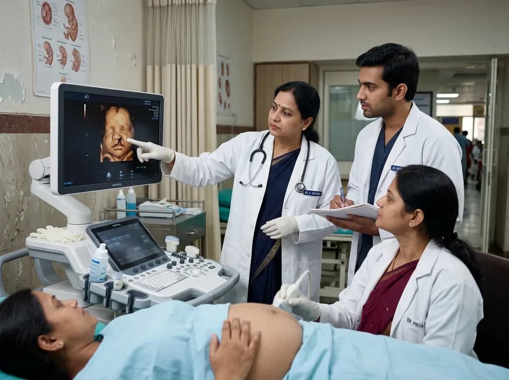
A 5-year retrospective study evaluates prenatal ultrasound detection of fetal cleft lip and palate. While overall detection reached 87%, accuracy varied from 99.3% in combined clefts to 50% in isolated cases. Combined defects showed elevated risks of concurrent structural anomalies and chromosomal abnormalities.
Today

An updated systematic review and meta-analysis of 7,875 patients reveals that routine microaxial left ventricular support during nonemergent high-risk PCI does not reduce mortality or ischemic complications, while significantly increasing major bleeding events.
Yesterday

Following Cyclone Chido, a WHO EMT-2 field hospital performed 268 surgeries in Mayotte. While acute trauma is expected initially, 98% of cases were septic wounds of the extremities requiring secondary intention healing, revealing crucial operational insights for disaster surgical preparedness.
Today

The SED-SEEN-SEEP consensus outlines an Integrated Care Pathway for preclinical type 1 diabetes, structuring target screening, autoantibody confirmation, metabolic staging, and proactive follow-up to prevent diabetic ketoacidosis and facilitate early disease-modifying intervention.
Yesterday
