

Loading, please wait...
Loading, please wait...
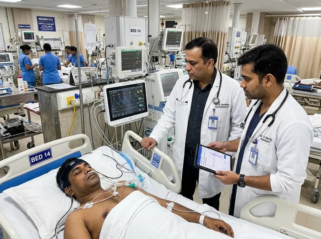
Accurate detection of elevated intracranial pressure remains an essential cornerstone in neurocritical care. Historically, clinicians have relied on invasive ventriculostomy or parenchymal microtransducers to identify elevated intracranial pressure. However, these invasive procedures carry distinct hazards, including intracerebral hemorrhage, catheter-related ventriculitis, and technical placement challenges. Consequently, noninvasive ICP monitoring has emerged as an urgent priority for modern critical care medicine. Recent multicenter data demonstrate that continuous monitoring of micro-cranial expansions reliably reveals surrogate pressure waveforms. By deriving reproducible physiological parameters, this advanced modality empowers critical care teams to rapidly assess intracranial dynamics at the bedside.
Prompt recognition of acute neurological deterioration dictates patient survival in emergency settings. In acute brain injury, expanding mass lesions or cerebral edema quickly deplete intracranial reserves. As a result, small volume increases cause dramatic pressure spikes according to the classic Monro-Kellie hypothesis. Invasive intracranial sensors provide the reference standard for pressure measurement. Nevertheless, many regional hospitals and secondary care centers in India lack round-the-clock neurosurgical coverage to perform urgent catheter placement. Furthermore, systemic coagulopathy often delays surgical cannulation, creating dangerous diagnostic blind spots.
Noninvasive ICP monitoring bridges this critical gap by providing immediate physiological insight without requiring operative intervention. Clinicians can initiate rapid risk stratification right in the emergency triage bay. Therefore, noninvasive cranial monitoring tools represent a transformative shift toward early, risk-free neuromonitoring. Additionally, noninvasive monitoring facilitates rapid serial assessments across varying clinical environments. Critical care teams can assess traumatic brain injury and ischemic stroke without exposing vulnerable patients to invasive cranial risks. Thus, reliable surrogate metrics streamline timely medical decision-making.
The cranial vault accommodates vascular pulsatility through continuous physiological compensation. Each cardiac cycle drives arterial blood volume into the intracranial space, producing a characteristic three-peak pressure waveform. Specifically, the P1 percussion wave reflects arterial pulsation transmitted directly through the choroid plexus. The P2 tidal wave denotes intracranial compliance and brain tissue elastance. Finally, the P3 dicrotic wave represents venous outflow during late systole. Under normal physiological circumstances, brain compliance remains high, and P1 amplitude surpasses P2 amplitude.
However, as cerebral perfusion compromises and tissue elastance rises, P2 progressively enlarges, eventually towering over P1. Advanced noninvasive sensors track these subtle micrometer-scale cranial expansions through external strain-gauge mechanisms. Consequently, sophisticated digital processing calculates the P2/P1 ratio and normalized time-to-peak metrics. These dynamic morphological parameters reveal evolving cerebral hypertension long before clinical signs of transtentorial herniation become evident. Furthermore, automated waveform processing eliminates subjective visual error from waveform interpretation. As a result, physiological tracking becomes standardized across intensive care teams.
A rigorous multicenter validation study recently established the clinical efficacy of this noninvasive platform across 124 acutely injured patients. Researchers analyzed concurrent invasive recordings and noninvasive cranial expansion signals to define specific diagnostic thresholds. Interestingly, the estimated P2/P1 ratio exhibited distinct operational characteristics across different cutoff values. At a low threshold of 0.8, the system achieved a 92% sensitivity alongside a 19% specificity. Because of this high sensitivity, a value under 0.8 provides exceptional rule-out capacity for intracranial hypertension exceeding 20 mm Hg.
Conversely, elevating the P2/P1 threshold to 1.4 transformed the diagnostic profile, producing a remarkable 90% specificity. Therefore, a ratio above 1.4 confirms severe intracranial hypertension with high certainty. In clinical practice, this dual-threshold approach provides a structured pathway for patient stratification. Clinicians can confidently exclude intracranial crises at low values, while escalating therapy aggressively when values exceed 1.4. Additionally, this quantitative separation enables precise risk categorization in acute settings. Thus, cutoff stratification empowers clinicians to optimize intensive care resources safely.
Alongside waveform amplitude ratios, temporal dynamics within the cardiac cycle provide vital diagnostic clarity. The multicenter study evaluated time-to-peak as an independent noninvasive biomarker of intracranial hypertension. Specifically, time-to-peak measures the latency between waveform inception and maximal pulse amplitude. When intracranial compliance deteriorates, arterial pulse propagation slows and diastolic recoil vanishes. Consequently, the noninvasive pulse contour demonstrates marked temporal widening.
The multicenter trial identified 0.3 as the optimal normalized time-to-peak cutoff, delivering a robust 92% specificity for detecting elevated pressure. Thus, temporal metrics perform exceptionally well in verifying critical hypertension. Furthermore, combining temporal measurements with the P2/P1 ratio creates an integrated physiological profile. This multimodal analysis reduces false discoveries and enhances diagnostic confidence in complex intensive care scenarios. Intensive care teams can subsequently track real-time hemodynamic shifts following hyperosmolar therapy. As a result, time-to-peak acts as a reliable window into both compliance and cerebrovascular resistance.
Indian tertiary centers and district hospitals face substantial resource constraints when managing neurotrauma. Head injury admissions represent a massive public health challenge across suburban and urban trauma centers. However, specialized neurosurgical facilities remain concentrated in major metropolitan hubs. Therefore, noninvasive cranial monitoring offers exceptional utility for resource-limited emergency departments. Medical officers can swiftly deploy noninvasive sensors to evaluate secondary brain injury risks during initial triage.
In addition, peripheral intensive care units can monitor patients awaiting inter-facility transport without performing risky invasive procedures. Serial noninvasive measurements also guide targeted mannitol and hyperosmolar therapy in intensive care wards. Consequently, hospitals conserve precious neurosurgical resources for patients who truly require surgical decompression. Furthermore, nursing staff can safely maintain noninvasive sensors without introducing catheter-associated infection risks. Thus, incorporating cranial expansion analysis elevates neurocritical care standards across diverse tiers of the Indian healthcare ecosystem.
Noninvasive cranial expansion monitors track minute skull deformations caused by arterial pulsations during each cardiac cycle. Consequently, these mechanical expansions generate a waveform contour that closely mirrors invasive intracranial pressure waveforms. Clinicians evaluate the relationship between the first and second peaks to accurately detect impaired intracranial compliance without cranial penetration.
A P2/P1 ratio below 0.8 provides high clinical utility for ruling out intracranial hypertension. Because this cutoff achieves a 92% sensitivity, emergency physicians can confidently identify patients who have preserved intracranial compliance. Therefore, clinicians can safely avoid unnecessary invasive monitoring procedures and reserve invasive catheter placement for higher-risk individuals.
A threshold of 1.4 yields a high specificity of 90%, which minimizes false-positive diagnoses of intracranial hypertension. When clinicians observe this elevated waveform ratio, cerebral compliance is severely exhausted. Thus, this specific cutoff provides reliable objective justification to promptly initiate osmotherapy, optimize cerebral perfusion pressure, or consider invasive ventricular drain placement.
Disclaimer: This content is for informational and educational purposes only. It is not intended as medical advice, diagnosis, or treatment. Always consult a qualified healthcare professional regarding any medical condition or before making healthcare decisions. The views expressed are those of the authors and do not necessarily reflect the official policy or position of any medical institution or organization. Refer to the latest local and national guidelines for clinical practice.
References

Read summarized clinical updates, watch expert medical content, and earn CME certifications right from your smartphone.
Open your smartphone camera to scan and install.





A multicenter clinical study evaluates the noninvasive brain4care system in 124 neurocritical patients. Analyzing cranial expansion waveforms, a P2/P1 ratio cutoff of 0.8 yielded 92% sensitivity to rule out intracranial hypertension, while a 1.4 cutoff provided 90% specificity, providing rapid triage insights.
Today
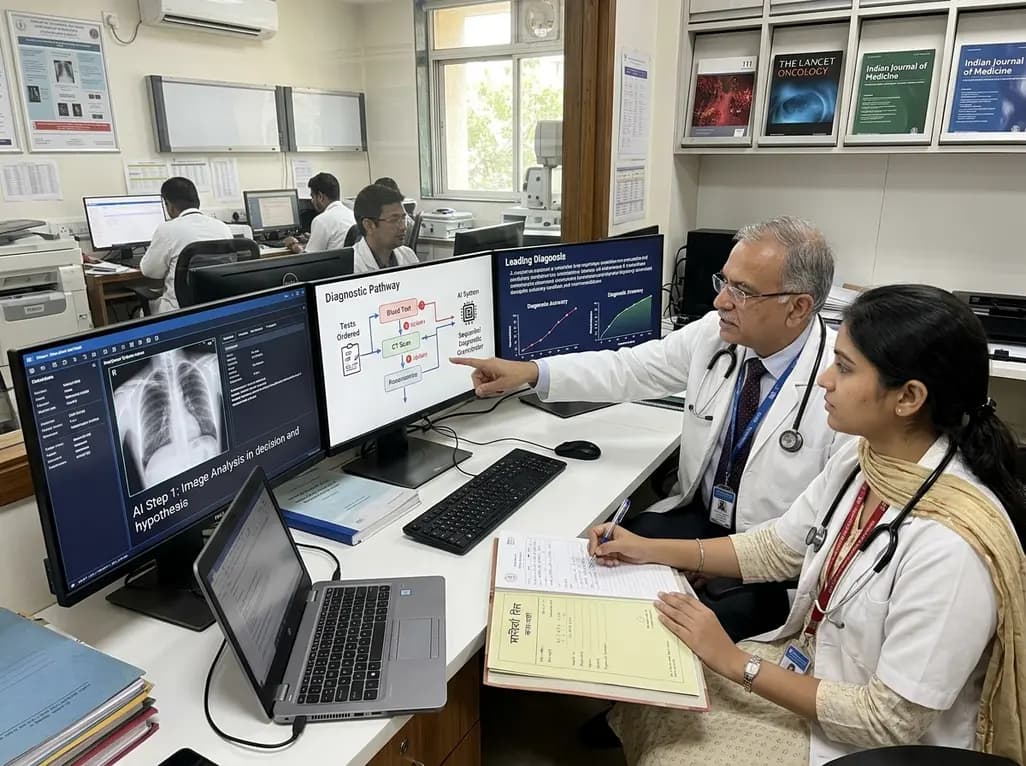
A breakthrough peer-reviewed study demonstrates that a sequential AI diagnosis system can mirror real-world clinical reasoning. By ordering stepwise tests and updating hypotheses dynamically, this multi-agent orchestrator achieved superior diagnostic accuracy while reducing investigative costs.
Today

A prenatal case highlights severe glutaric acidemia type II presenting as bilateral hyperechoic cystic kidneys at 18 weeks. Whole-exome sequencing revealed a novel homozygous ETFDH frameshift variant, demonstrating the power of rapid genetic testing in fetal renal anomalies.
Today

Review how tailored exercise programs benefit frail older patients with heart failure. Explore pathophysiological mechanisms, individualized training prescriptions, and strategies to overcome adherence barriers in clinical practice.
Yesterday

This comparative study evaluates posterior labral tears with and without glenohumeral instability, highlighting distinct demographic patterns, MRI underestimation in stable shoulders, and successful arthroscopic repair outcomes.
Yesterday

A clinical study demonstrates that early intervention with tocilizumab, either as monotherapy or bridged with corticosteroids, achieves significant proptosis reduction and resolves active inflammation in treatment-naive thyroid eye disease.
Yesterday
