

Loading, please wait...
Loading, please wait...

Emergency departments regularly admit patients with established epilepsy experiencing breakthrough convulsions. Consequently, clinicians routinely face the critical decision of whether to order urgent brain imaging. The routine application of nonindex seizures neuroimaging remains widespread across emergency units worldwide. However, empirical data demonstrate that repetitive computed tomography scans rarely alter acute clinical management. In a recent observational investigation by Espinosa-Jovel and colleagues, researchers analyzed 307 adult patients with known epilepsy who presented with nonindex seizures. Surprisingly, emergency physicians ordered non-contrast head computed tomography scans for nearly 80% of these encounters. Although physicians frequently request emergent scans out of caution, this standard defensive practice imposes substantial burdens. For instance, indiscriminate computed tomography utilization exposes patients to unnecessary ionizing radiation and inflates hospital expenditures. Moreover, repeated scanning congests radiology suites and delays care for other critically ill emergency patients. Therefore, physicians must carefully balance immediate diagnostic concerns against genuine clinical utility. In low- and middle-income healthcare systems like India, this diagnostic dilemma carries immense financial consequences for self-paying families. Understanding the real-world diagnostic yield of urgent imaging provides an essential foundation for rationalizing neuroimaging protocols.
The investigation revealed a stark contrast between baseline structural imaging changes and actionable acute abnormalities. Specifically, computed tomography identified preexisting structural defects in 43.6% of the imaged patient cohort. These common findings included established gliosis, old ischemic infarcts, focal encephalomalacia, and calcified granulomas. However, these chronic neuroimaging abnormalities did not influence acute clinical decision-making or therapeutic trajectory. In sharp contrast, only 3.7% of all scanned patients demonstrated acute pathologic lesions requiring immediate medical or surgical modification. Thus, more than 96% of the emergency computed tomography scans yielded no meaningful change in patient care. Clinicians primarily altered management only when scans detected emergent complications such as intracranial hemorrhage or acute contusion. Most patients with recurrent seizures simply required antiepileptic drug optimization, supportive care, or metabolic correction. Consequently, performing routine computed tomography for all recurrent seizures generates low clinical value. Physicians often order these scans automatically due to uncertainty regarding seizure etiology. Nevertheless, the clinical evidence demonstrates that static anatomical defects rarely explain acute breakthrough events. Therefore, healthcare teams must recognize that chronic lesions should not prompt emergent intervention without clear secondary deterioration.
Clinicians must distinguish benign breakthrough seizures from life-threatening neurological emergencies through careful bedside risk stratification. In the Bogotá study, the only clinical variable significantly associated with acute computed tomography findings was seizure-related traumatic brain injury. Specifically, acute pathologic changes occurred in 33.3% of patients with head trauma, compared to 0% in those without physical impact. In addition, established international emergency literature highlights several red flags that justify immediate neuroimaging. These critical clinical indicators include new persistent focal neurological deficits, protracted altered consciousness, and severe progressive headache. Furthermore, emergency physicians must maintain heightened suspicion for immunocompromised patients, individuals with known malignancy, or those receiving active anticoagulation. Conversely, patients who promptly return to their baseline cognitive state without traumatic injury demonstrate a true positive imaging yield approaching zero. Therefore, clinicians can safely defer neuroimaging when a reliable witness confirms a typical breakthrough seizure. Instead, healthcare teams should concentrate on identifying common reversible precipitants such as medication non-adherence, systemic infection, or sleep deprivation. This targeted diagnostic strategy accurately identifies patients requiring urgent intervention while protecting stable individuals from unnecessary imaging procedures.
In Indian healthcare environments, optimizing diagnostic resources remains an urgent clinical and economic priority. Government emergency facilities face severe resource constraints, overcrowded casualty wards, and extensive queues for diagnostic computed tomography scanners. Meanwhile, in private hospitals, out-of-pocket healthcare expenses frequently push vulnerable families into acute financial hardship. If emergency physicians order routine head computed tomography for every breakthrough seizure, they exhaust limited scanner time and delay acute stroke thrombolysis. Moreover, repeated ionizing radiation exposure from unnecessary computed tomography scans creates cumulative long-term carcinogenic risks for young epilepsy patients. Indian clinicians also encounter unique endemic etiologies of epilepsy, including calcified neurocysticercosis, tuberculomas, and perinatal hypoxic insults. While these chronic calcified lesions frequently appear on computed tomography scans, they represent stable background pathology rather than new emergencies. Therefore, documenting known neurocysticercosis or remote gliosis rarely changes acute emergency treatment. Instead, Indian emergency physicians can optimize healthcare delivery by focusing on compliance checks, serum antiepileptic drug levels, and bedside glucose monitoring. Consequently, adopting restrictive neuroimaging policies directly alleviates financial toxicity and improves emergency department throughput across tertiary institutions.
Establishing structured clinical pathways allows emergency physicians to practice evidence-based, defensive-free medicine with greater confidence. Medical leadership must align institutional practice with recommendations from the American College of Emergency Physicians and the Indian Academy of Neurology. According to these clinical guidelines, adult patients with established epilepsy do not require emergent computed tomography if they recover uneventfully. Specifically, patients should demonstrate a complete return to baseline neurological status without signs of acute head trauma or persistent focal deficits. In such uncomplicated presentations, clinicians can safely discharge patients after appropriate observation and antiepileptic drug reconciliation. However, when diagnostic ambiguity persists or physicians suspect non-accidental trauma, targeted neuroimaging becomes essential. Furthermore, if clinicians identify progressive focal deficits or failure to regain baseline consciousness within several hours, they must obtain emergent neuroimaging immediately. Outpatient magnetic resonance imaging remains the gold standard for comprehensive structural epilepsy evaluation rather than hasty emergency computed tomography scans. By implementing standardized triage checklists in emergency departments, hospitals can significantly decrease inappropriate scanner utilization. Ultimately, this evidence-based approach elevates quality of care, prevents radiation overexposure, and preserves crucial diagnostic infrastructure.
Emergency physicians should reserve urgent neuroimaging for patients exhibiting specific red-flag features. Indicated scenarios include seizure-related head trauma, prolonged altered mental status, new focal neurological deficits, or high suspicion of intracranial infection. Additionally, clinicians should scan patients taking anticoagulant medications or those with active systemic malignancy. In contrast, stable patients returning promptly to baseline cognitive function rarely benefit from acute computed tomography, as actionable diagnostic yield remains exceptionally low.
Clinicians frequently order computed tomography scans due to diagnostic uncertainty, fear of litigation, and institutional habits prioritizing defensive medicine. Furthermore, emergency teams may misinterpret postictal confusion as an acute intracranial complication before adequate clinical recovery occurs. However, evidence shows that most imaging abnormalities in established epilepsy represent preexisting, chronic structural changes. Understanding that acute management changes occur in under four percent of cases helps emergency physicians confidently avoid unnecessary scanning.
Instead of ordering automatic computed tomography, emergency physicians should prioritize evaluating medication adherence, measuring therapeutic antiepileptic drug levels, and screening for systemic triggers. Clinicians must assess serum glucose, electrolytes, and inflammatory markers to rule out metabolic derangements or concurrent systemic infections. Additionally, performing serial bedside neurological examinations ensures the patient steadily returns to baseline status. For comprehensive structural reassessment, scheduling an elective outpatient magnetic resonance imaging study offers far superior diagnostic value.
Disclaimer: This content is for informational and educational purposes only... Refer to the latest local and national guidelines for clinical practice.
References

Read summarized clinical updates, watch expert medical content, and earn CME certifications right from your smartphone.
Open your smartphone camera to scan and install.





A study in The Neurologist reveals that neuroimaging in patients presenting with recurrent nonindex seizures yields acute actionable findings in only 3.7% of cases, primarily in those with head trauma. Clinicians should adopt selective imaging protocols to reduce costs, radiation, and emergency department delays.
Today
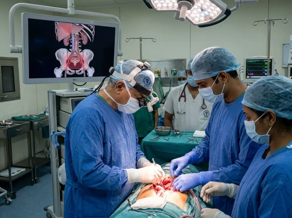
Surgical oncologists at AIIMS Delhi have mapped two critical anatomical landmarks: Ray's Zone in the deep pelvis and Ray's Hepatocaval Triangle near the liver. These predefined hazard boundaries guide dissection, dramatically reduce severe intraoperative bleeding, and preserve vital nerves and visceral organs.
Today
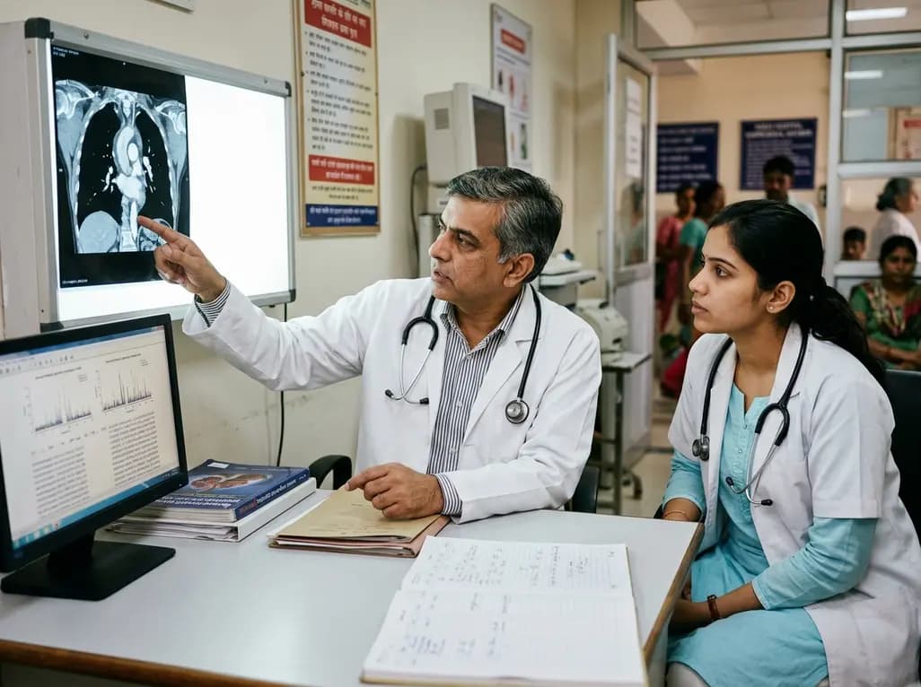
A study reveals that the MYLK c.4079G>A, p.(Gly1360Asp) variant is linked to high-penetrance heritable thoracic aortic disease and early-onset aortic dissection, showing a founder effect in Finnish cohorts. Findings emphasize early cascade screening and vigilant surveillance.
Today
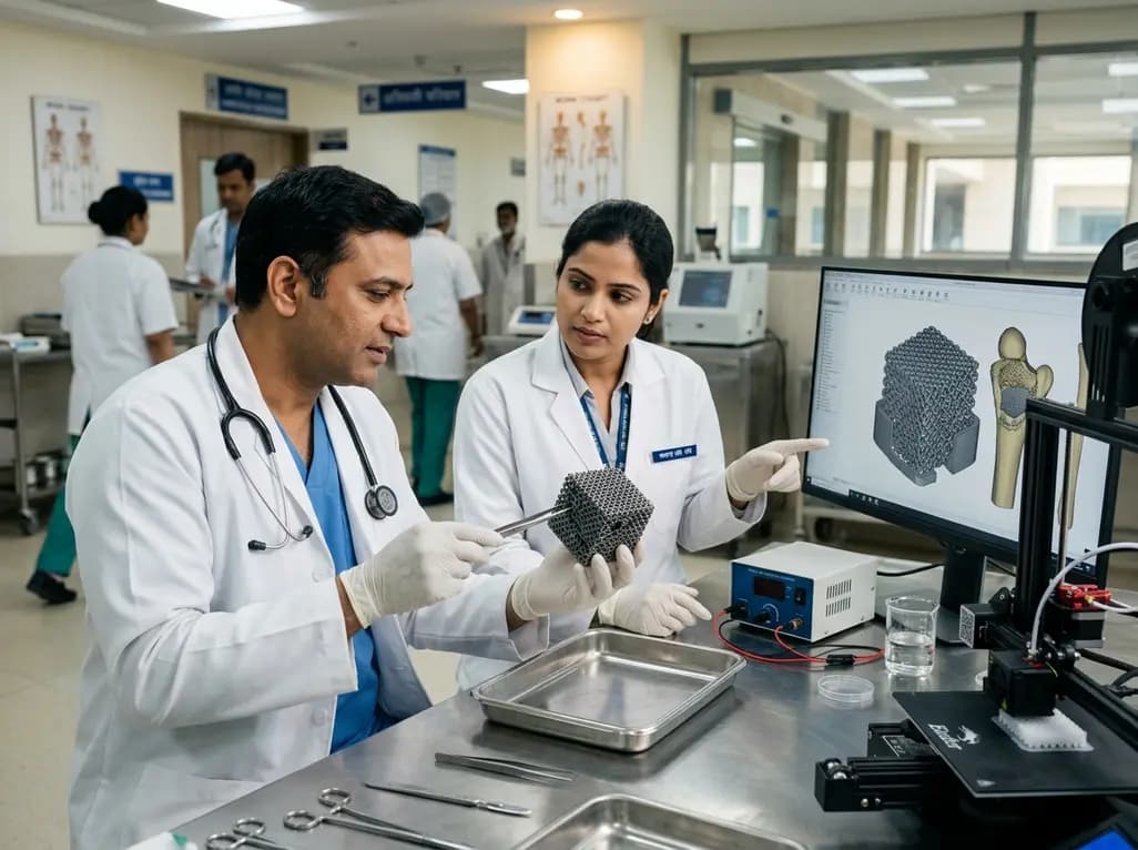
A novel sacrificial-template technique combines FDM polymer 3D printing and micro-arc oxidation to fabricate orientation-controlled porous titanium scaffolds. These bioactive constructs offer tailored stiffness, exceptional multidirectional load resistance, and robust osseointegration for bone defect repair.
Today
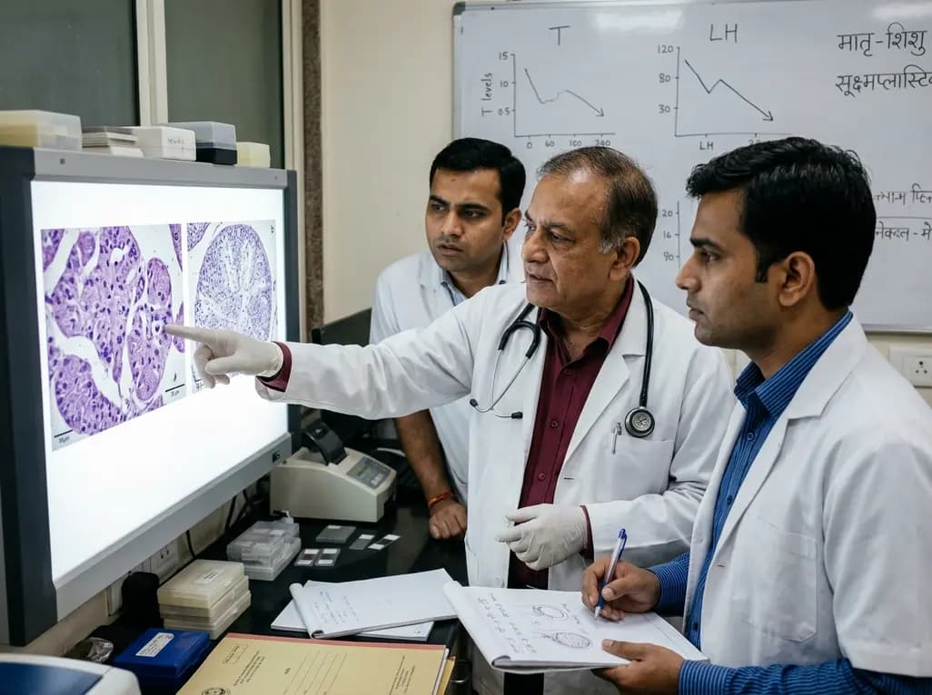
An animal model study reveals that gestational exposure to polystyrene microplastics causes persistent intrauterine growth restriction, testicular hypoplasia, Leydig cell depletion, and an atypical low-testosterone, low-LH endocrine profile in male offspring from birth into adulthood.
Today
