.webp&w=384&q=75)

Loading, please wait...
Loading, please wait...

Chronic liver disease poses a growing public health burden worldwide, primarily fueled by metabolic dysfunction-associated steatohepatitic disease, viral hepatitis, and alcohol consumption. Detecting early stage liver fibrosis represents a pivotal clinical opportunity because early intervention can halt or reverse hepatic damage before irreversible cirrhosis develops. However, identifying early fibrotic changes remains remarkably difficult during standard clinical evaluations. Consequently, clinicians urgently need reliable, non-invasive diagnostic strategies to manage asymptomatic individuals effectively.
Hepatic fibrosis represents the common wound-healing response to chronic parenchymal injury. Pathologists classify fibrosis across stages from F0 to F4, with stages F1 and F2 defining early disease. During these initial stages, extracellular matrix remodeling remains dynamic and biologically reversible. Therefore, prompt therapeutic adjustments, including targeted metabolic therapies and lifestyle modifications, can normalize liver histology. In contrast, advanced fibrosis and cirrhosis carry significant morbidity, liver failure risks, and portal hypertension complications. Unfortunately, patients with early fibrotic deposition rarely manifest overt clinical symptoms or routine biochemical anomalies. Routine liver enzyme panels frequently remain normal even during progressive matrix deposition. Histological evaluation via liver biopsy has historically served as the definitive diagnostic standard. Nevertheless, biopsy carries recognized disadvantages, including procedural discomfort, bleeding risks, and substantial sampling variability. As a result, clinicians cannot use liver biopsy as a routine population-screening tool.
To overcome biopsy limitations, physicians currently utilize first-line non-invasive tests to evaluate liver stiffness and scarring. Simple scoring systems, such as the Fibrosis-4 index and the NAFLD fibrosis score, combine basic parameters like age, transaminases, and platelet counts. Furthermore, point-of-care ultrasound elastography methods measure physical shear wave speed through hepatic parenchyma. While these modalities excel at ruling out advanced cirrhosis, they perform poorly when distinguishing minimal from moderate fibrosis. Specifically, simple serum panels lack biological sensitivity for subtle architectural shifts in early stage liver fibrosis. Similarly, vibration-controlled transient elastography faces known technical limitations in patients with thoracic obesity, severe hepatic steatosis, or acute parenchymal inflammation. These confounding variables frequently generate false-positive elevations or indeterminate results. Consequently, a substantial diagnostic gray zone persists, leaving many early-stage patients without a clear clinical trajectory.
Researchers are actively developing next-generation circulating markers to overcome these historical diagnostic gaps. Unlike static markers that merely reflect downstream hepatic dysfunction, emerging biomarkers quantify active fibrogenesis and extracellular matrix turnover directly. For instance, specialized assays measure neo-epitopes derived from type III and type IV collagen synthesis and degradation. Moreover, high-sensitivity immunoassay platforms can now detect picogram quantities of pro-fibrogenic cytokines and circulating microRNAs. Alongside serum assays, advanced imaging protocols are revolutionizing functional tissue assessment. Novel molecular magnetic resonance imaging probes target specific cell-surface receptors on activated hepatic stellate cells. Therefore, these molecular tracers visualize ongoing cellular fibrogenic activity rather than passive collagen accumulation. In addition, dynamic contrast-enhanced magnetic resonance elastography provides superior diagnostic precision across heterogeneous liver tissue. These molecular tools permit earlier detection when biological remodeling remains most responsive to pharmacotherapy.
Artificial intelligence algorithms now significantly enhance standard medical imaging and laboratory datasets. Machine learning models analyze complex textural patterns on routine ultrasound, computed tomography, and conventional magnetic resonance scans that escape human visual inspection. Furthermore, algorithms integrate multidimensional clinical information to forecast disease progression with high fidelity. Beyond traditional clinical parameters, multiomics platforms present groundbreaking screening avenues. Investigators are identifying distinct diagnostic signatures within gut microbiota-derived metabolites, including circulating short-chain fatty acids and secondary bile acids. Similarly, liquid biopsy approaches interrogate small extracellular vesicles and epigenetic methylation marks to discern early fibrogenesis. Researchers are also exploring non-invasive biofluids such as exhaled breath volatile organic compounds, saliva, and urine. Consequently, multiomics panels could soon deliver high-throughput, non-invasive screening during routine outpatient consultations.
Despite impressive technical advances, translating novel diagnostic tests into real-world primary care practice presents substantial hurdles. First, emerging biomarkers require thorough prospective validation across ethnically diverse cohorts and varied underlying etiologies. Second, high technical costs and proprietary analytical platforms currently restrict specialized imaging probes to academic medical centers. Third, health systems need standardized operational cut-offs to eliminate inter-laboratory variability before clinical guideline committees endorse widespread implementation. In addition, regulatory authorities demand proven clinical utility demonstrating that early detection directly improves long-term patient outcomes. To overcome these barriers, professional societies advocate for tiered diagnostic pathways. In such algorithms, clinicians deploy simple, cost-effective serological scores in primary care to identify high-risk individuals. Subsequently, specialists reserve advanced molecular imaging, next-generation biomarkers, or elastography for ambiguous cases. This stratified paradigm ensures prudent resource utilization while maximizing early therapeutic intervention.
Early-stage liver fibrosis corresponds histologically to stages F1 and F2. In this phase, extracellular collagen matrix accumulates around portal tracts or sinusoids without extensive architectural distortion or regenerative nodule formation. Importantly, this stage remains asymptomatic and biologically reversible if clinicians identify the underlying cause and initiate lifestyle or medical therapy promptly.
Standard liver panels measure circulating levels of alanine aminotransferase, aspartate aminotransferase, and bilirubin, which reflect active hepatocyte necrosis rather than architectural matrix deposition. Consequently, patients with chronic low-grade fibrogenesis can maintain normal transaminase concentrations. Serum markers fail to quantify the slow, cumulative progression of collagen fibers during early stages.
Transient elastography measures hepatic tissue stiffness by tracking physical shear waves. While elastography reliably rules out advanced cirrhosis (F4), its accuracy diminishes significantly when distinguishing mild fibrosis (F1) from normal tissue (F0). Confounding factors like severe obesity, ongoing hepatic inflammation, recent meal ingestion, or hepatic congestion can artificially alter stiffness readings.
Disclaimer: This content is for informational and educational purposes only. Refer to the latest local and national guidelines for clinical practice.
References
Wang C et al. Non-invasive tests for early-stage liver fibrosis: Current advances and challenges. Chin Med J (Engl). 2026 Sep 15. doi: 10.1097/CM9.0000000000004311. PMID: 42740701.
European Association for the Study of the Liver. EASL Clinical Practice Guidelines on non-invasive tests for evaluation of liver disease severity and prognosis: 2021 update. J Hepatol. 2021;75(3):659-689.
Newsome PN, Cramb R, Davison SM, et al. Guidelines on the management of abnormal liver blood tests. Gut. 2018;67(1):6-19.

Read summarized clinical updates, watch expert medical content, and earn CME certifications right from your smartphone.
Open your smartphone camera to scan and install.





Early-stage liver fibrosis (F1-F2) is a reversible phase where timely detection can halt progression. This review evaluates established and emerging non-invasive tests, including molecular imaging, AI modalities, and multiomics biomarkers, highlighting clinical pathways and current implementation barriers.
Today
Hormonal shifts throughout puberty, menstruation, fertility treatments, pregnancy, and menopause cause profound glucose excursions in women with diabetes. Understanding these endocrine transitions and utilizing advanced continuous glucose monitoring enables clinicians to optimize personalized diabetes care.
Today
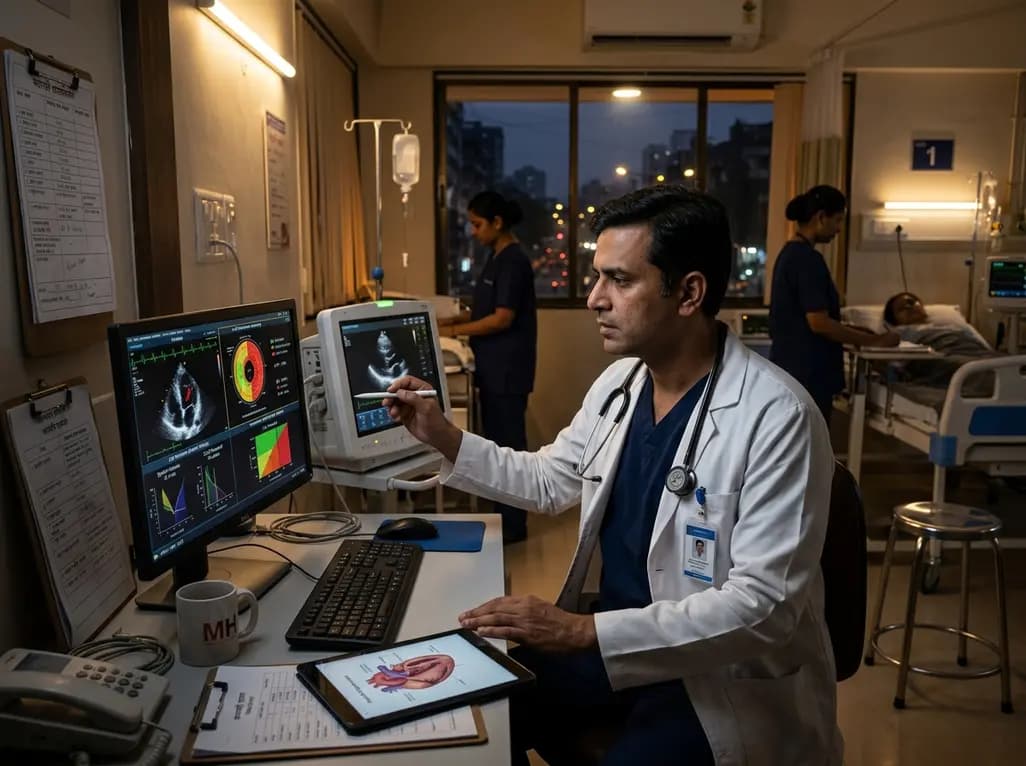
Emerging evidence shows that even dim night-time light exposure alters cardiac geometry and weakens contractility. In a landmark cohort study, nocturnal light above 3 lux triggered concentric left ventricular hypertrophy and subclinical dysfunction, highlighting nocturnal illumination as a modifiable cardiac risk factor.
Today
Cervi-AI achieves expert-level agreement in evaluating cervical spinal stenosis and spinal cord compression on MRI, while its automated imaging metrics reliably predict anterior versus posterior surgical approaches in multilevel degenerative cervical myelopathy.
Today
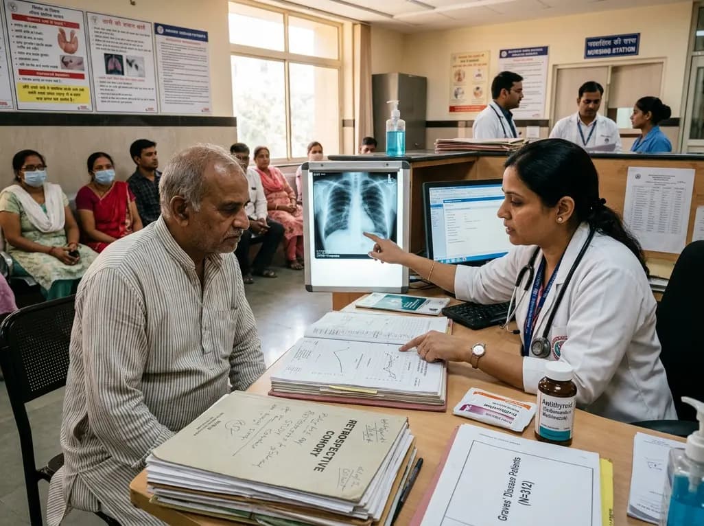
A retrospective cohort study evaluates the short- and long-term impacts of SARS-CoV-2 infection on patients with Graves' disease receiving antithyroid drug therapy, highlighting thyroid status destabilization, clinical symptom exacerbations, and post-viral sequelae.
Today
