.webp&w=384&q=75)

Loading, please wait...
Loading, please wait...

Degenerative cervical myelopathy represents the primary cause of nontraumatic spinal cord impairment in adults globally. Consequently, timely diagnostic evaluation using degenerative cervical myelopathy MRI is critical for identifying neural canal stenosis and myelomalacia. Spine surgeons face significant clinical challenges when assessing multilevel disease because degenerative pathology often spans several vertebral segments. Furthermore, clinical presentations vary widely from mild hand clumsiness to severe quadriparesis and gait disturbance. Therefore, precise radiological quantification remains essential for guiding timely surgical intervention. Traditional manual radiological evaluation requires detailed measurement of canal diameters, stenosis severity, and spinal cord deformation. However, manual assessments suffer from notable interobserver variability, subjectivity, and substantial time demands. To resolve these diagnostic hurdles, biomedical researchers have introduced deep learning architectures to automate spine imaging analysis. Cervi-AI represents an automated artificial intelligence system designed to quantify cervical cord compression objectively. Evaluating how automated models compare against experienced spine specialists offers vital clinical insight. Ultimately, objective imaging metrics can streamline diagnostic workflows and establish consistent criteria for operative triage.
A recent clinical investigation evaluated diagnostic agreement between the Cervi-AI model and expert spine surgeons. Specifically, researchers performed a retrospective analysis of 146 surgical patients treated at an academic medical center. The cohort included 77 male and 69 female patients with a mean age of 52 years. Additionally, investigators evaluated critical imaging parameters including cervical spinal stenosis grade, involved segments, and the maximum spinal cord compression index. The statistical results revealed outstanding concordance between artificial intelligence measurements and expert assessments. For instance, the intraclass correlation coefficient reached 0.970 for the number of involved stenosis levels. Furthermore, the model achieved an agreement coefficient of 0.958 for classifying maximum stenosis grade across motion segments. Most notably, agreement reached 0.984 for computing the maximum spinal cord compression index. Bland-Altman analyses also confirmed minimal systematic bias between automated readings and expert evaluations. Consequently, these findings prove that deep learning architectures can quantify complex cervical anatomy with specialist-level accuracy. Clinicians can therefore utilize automated algorithms to eliminate measurement discrepancies in spinal cord imaging.
Determining the optimal surgical approach represents a central dilemma in multilevel cervical myelopathy management. Spine surgeons frequently deliberate between anterior decompression with fusion and posterior decompression via laminoplasty or laminectomy. In the study cohort, 86 patients underwent anterior surgery whereas 60 patients received posterior surgery. Notably, both surgical groups demonstrated comparable baseline demographics and symptom duration. However, multivariable logistic regression analysis revealed that AI-derived imaging parameters strongly correlated with approach selection. Specifically, each additional involved stenotic level significantly decreased the probability of anterior surgery, demonstrating an odds ratio of 0.058. Consequently, patients with widespread multilevel disease underwent posterior decompression far more frequently. Additionally, longer symptom duration independently favored posterior intervention, exhibiting an odds ratio of 0.752 per twelve-month interval. Clinicians prefer posterior decompression for multi-segmental narrowing because posterior approaches avoid extensive anterior plating and graft complications. Conversely, anterior procedures remain favorable for focal pathology with localized disc herniation or cervical kyphosis. Receiver operating characteristic curves confirmed that combining automated metrics with clinical duration reliably predicts operative approach decisions.
Automated imaging metrics offer immediate benefits for clinical workflows in spine surgery and diagnostic radiology. For example, selecting surgical approaches currently depends on subjective visual interpretation of sagittal cord compression. Consequently, individual surgeon preference and institutional training often generate variable practice patterns across centers. In contrast, artificial intelligence provides standardized anatomical measurements that eliminate subjective grading discrepancies. Furthermore, precise calculation of the maximum spinal cord compression index identifies severe cord flattening and vulnerable ischemic areas. This precise data enables surgeons to determine whether direct ventral decompression or indirect posterior canal widening is safest. Moreover, integrating automated tools into hospital picture archiving systems enables immediate report generation prior to clinical consultations. As a result, spine specialists can spend less time performing manual measurements and more time discussing personalized operative goals. In resource-constrained healthcare settings, automated screening can also support non-specialist physicians in rapidly identifying high-risk spinal cord compression. Timely referral prevents prolonged diagnostic delays, which directly worsen permanent neurological recovery in myelopathy patients.
Although current findings highlight promising automated capabilities, several technical aspects require ongoing investigation. First, retrospective validation studies require prospective confirmation across diverse demographic populations and different MRI hardware manufacturers. Varying magnetic field strengths and patient motion artifacts can alter convolutional network performance. Therefore, developers must train computer vision algorithms on diverse multicenter datasets to guarantee clinical robustness. Second, future machine learning platforms must integrate sagittal spinal alignment parameters alongside canal stenosis measurements. Specifically, radiographic parameters such as cervical lordosis, sagittal vertical axis, and dynamic segmental instability strongly influence operative success. Posterior decompression in a kyphotic spine frequently produces inadequate cord clearance and poor neurological recovery. Additionally, neural networks should combine patient-reported outcome measures and electrophysiological assessments to guide personalized decision algorithms. By uniting morphological measurements with functional patient data, predictive algorithms can forecast surgical complications and recovery trajectories. Ultimately, artificial intelligence will not supplant surgical judgment. Instead, smart decision-support systems will augment spine surgeons, facilitating consistent, evidence-based operative care.
Cervi-AI provides rapid, automated segmentation of the cervical spine on magnetic resonance imaging scans. The platform objectively measures canal stenosis grades, counts involved cord levels, and computes the maximum spinal cord compression index. By eliminating subjective manual tracing, the algorithm delivers high measurement consistency that closely mirrors experienced spine specialists. Consequently, clinicians gain fast, reproducible anatomical insights, which streamline diagnostic workflows, minimize interobserver diagnostic variability, and support reliable surgical decision-making.
The number of compressed spinal levels directly affects surgical complexity and complication profiles. Anterior cervical decompression and fusion works exceptionally well for focal one or two-level disease, directly removing ventral pathology. However, multilevel anterior fusions spanning three or more segments carry higher risks of pseudoarthrosis, dysphagia, and construct failure. Therefore, surgeons frequently utilize posterior approaches, such as laminoplasty or laminectomy, for widespread multilevel compression to achieve indirect canal expansion while minimizing anterior approach-related morbidity.
Artificial intelligence tools cannot replace the holistic clinical judgment of an experienced spine surgeon. While automated models excel at rapid anatomical quantification and objective image segmentation, surgical decisions require synthesizing imaging with patient history, physical examination, and comorbidities. Furthermore, clinical nuance encompasses dynamic cervical alignment, bone density, and individual patient lifestyle goals. Consequently, artificial intelligence operates best as a complementary decision-support system, augmenting surgeon precision and efficiency rather than autonomously dictating complex operative care.
Disclaimer: This content is for informational and educational purposes only... Refer to the latest local and national guidelines for clinical practice.
References
Zhang WY et al. [Agreement between Cervi-AI and expert measurements of cervical MRI parameters and association of AI-derived imaging metrics with surgical approach selection in multilevel degenerative cervical myelopathy]. Zhonghua Yi Xue Za Zhi. 2026 Sep 15. doi: 10.3760/cma.j.cn112137-20260716-01771. PMID: 42736124.
Kouli O, Al-Nusair L, Basnet A, Kaiser R, Fehlings M, Wilby M, Srikandarajah N. Anterior vs posterior approaches in the management of multilevel degenerative cervical myelopathy: a systematic review and meta-analysis. Spine J. 2026 Mar;26(3):558-570. doi: 10.1016/j.spinee.2025.08.336.
Merali Z, Wang JZ, Badhiwala JH, et al. A deep learning model for detection of cervical spinal cord compression in MRI scans. Sci Rep. 2021;11:10473. doi: 10.1038/s41598-021-89848-3.

Read summarized clinical updates, watch expert medical content, and earn CME certifications right from your smartphone.
Open your smartphone camera to scan and install.





Cervi-AI achieves expert-level agreement in evaluating cervical spinal stenosis and spinal cord compression on MRI, while its automated imaging metrics reliably predict anterior versus posterior surgical approaches in multilevel degenerative cervical myelopathy.
Today
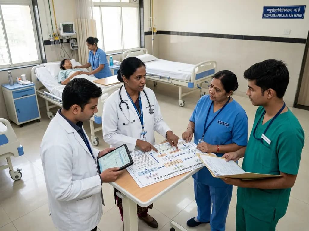
Agitation complicates neurorehabilitation and challenges clinical staff. A structured quality improvement initiative demonstrates that validated scales, clinical decision trees, and multidisciplinary education significantly enhance provider confidence and optimize patient safety during inpatient recovery.
Today
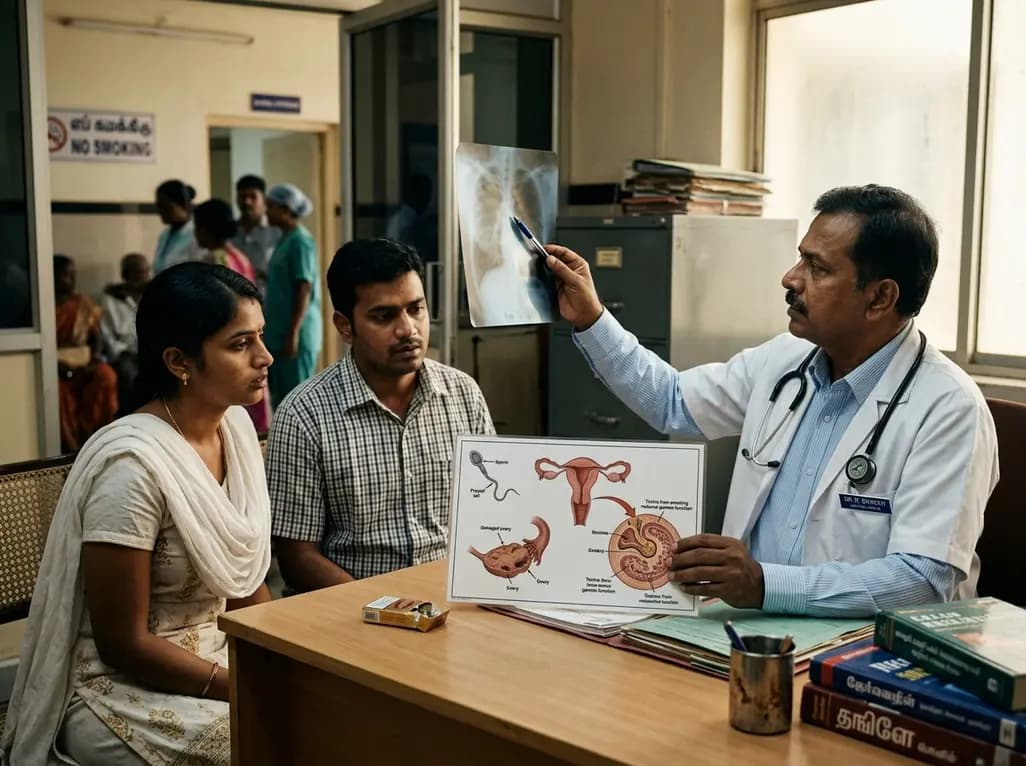
A global knowledge summary from the World Health Organization warns that tobacco smoking significantly increases infertility risks in women and causes substantial sperm and erectile dysfunction in men. Fortunately, quitting smoking substantially lowers these risks, providing clear reproductive benefits.
Today
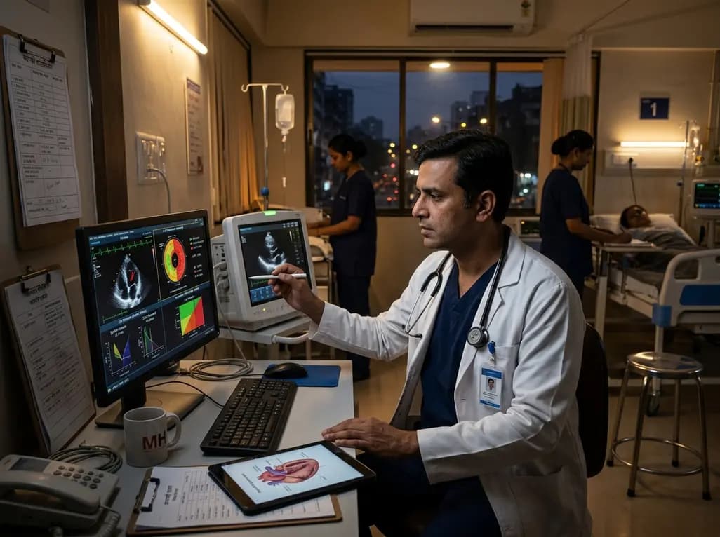
Emerging evidence shows that even dim night-time light exposure alters cardiac geometry and weakens contractility. In a landmark cohort study, nocturnal light above 3 lux triggered concentric left ventricular hypertrophy and subclinical dysfunction, highlighting nocturnal illumination as a modifiable cardiac risk factor.
Today

A retrospective cohort study evaluates the short- and long-term impacts of SARS-CoV-2 infection on patients with Graves' disease receiving antithyroid drug therapy, highlighting thyroid status destabilization, clinical symptom exacerbations, and post-viral sequelae.
Today
