

Loading, please wait...
Loading, please wait...

Suprasellar arachnoid cysts represent a rare clinical entity that accounts for a small subset of all intracranial mass lesions. Although these developmental anomalies remain benign, their critical anatomical position within the skull base often causes significant neurological morbidity. Most frequently, affected patients present with progressive obstructive hydrocephalus, visual impairment, or neuroendocrine disruption. Consequently, clinicians must evaluate surgical interventions that establish definitive cerebrospinal fluid pathway restoration while avoiding unnecessary morbidity. In this clinical review, we examine contemporary evidence regarding endoscopic fenestration techniques, focusing on procedural efficacy, long-term radiographic reduction, and operative safety.
Congenital arachnoid cysts constitute nearly one percent of all nontraumatic intracranial space-occupying lesions. Among them, suprasellar arachnoid cysts develop within the subarachnoid space near the sellar floor, the optic chiasm, and the third ventricle. Because these fluid-filled structures occupy a critical junction, they frequently disrupt normal cerebrospinal fluid hydrodynamics. Nearly ninety percent of symptomatic individuals present with hydrocephalus caused by upward displacement of the third ventricular floor or aqueductal occlusion. In pediatric cohorts, caregivers frequently report progressive macrocephaly, irritability, developmental delays, and failure to thrive. Conversely, adult patients more commonly describe intractable headaches, cognitive slowing, visual field disturbances, and chronic fatigue. In addition, upward cyst expansion often exerts mechanical pressure on the hypothalamic-pituitary axis. As a result, patients may experience precocious puberty, growth hormone deficiency, or secondary hypothyroidism. High-resolution magnetic resonance imaging remains essential for accurate preoperative characterization. Neuroimaging clearly depicts the thin cyst membrane, displaced neurovascular structures, and ventricular enlargement. Therefore, early diagnostic confirmation helps neurosurgeons intervene before irreversible optic atrophy or permanent hypothalamic injury occurs.
Historically, open microsurgical craniotomy and cystoperitoneal shunting served as standard treatments for expanding suprasellar lesions. However, extracranial shunts carry high lifelong revision rates, mechanical hardware occlusion, and recurrent infection risks. Similarly, extensive open craniotomies carry increased risks of neurovascular damage and hypothalamic injury. Consequently, neuroendoscopy has emerged as the definitive first-line standard of care across contemporary neurosurgical centers worldwide. Minimally invasive neuroendoscopic fenestration primarily utilizes two distinct approaches: ventriculocystostomy and ventriculocystocisternostomy. In ventriculocystostomy, the surgeon establishes communication exclusively between the lateral or third ventricle and the superior cyst dome. This internal marsupialization equalizes transmantle pressures and permits fluid egress into the ventricular system. In contrast, ventriculocystocisternostomy creates dual communication channels through both the superior cyst roof and the inferior cyst floor. This dual fenestration directs fluid into both the third ventricle and the prepontine or interpeduncular subarachnoid cisterns. Rigid neuroendoscopes offer superior visualization and high illumination, which enable precise bipolar coagulation and gentle mechanical balloon dilation. Moreover, creating wide fenestrations reduces the probability of spontaneous closure over time.
Recent comparative data demonstrate remarkably high procedural success rates for both endoscopic modalities in managing symptomatic cysts. Procedural success requires sustained clinical improvement accompanied by documented radiographic cyst reduction without requiring secondary interventions. Overall success rates reach approximately eighty-eight percent across modern surgical series. When comparing the two modalities, ventriculocystocisternostomy achieves a procedural success rate near eighty-nine percent, whereas ventriculocystostomy yields approximately eighty-six percent. Both approaches reliably alleviate intracranial hypertension and diminish ventricular dilatation. Furthermore, serial magnetic resonance examinations demonstrate steady reduction in cyst volume over twenty-four months of follow-up. This radiological improvement directly correlates with the resolution of compressive symptoms, including visual field deficits and headaches. Nevertheless, neurosurgeons observe that cysts rarely disappear entirely following endoscopic fenestration. Instead, internal decompression equalizes hydrostatic pressures across the subarachnoid spaces, rendering the remaining cyst physiologically inactive. Therefore, clinical symptom resolution and ventricular stability remain superior markers of treatment success compared to complete volumetric obliteration.
Although neuroendoscopy offers a remarkably safe surgical profile, potential intraoperative and postoperative complications demand vigilant perioperative management. Critical neurovascular structures border the suprasellar region, including the basilar artery bifurcation, posterior communicating arteries, and oculomotor nerves. Therefore, surgeons must meticulously identify anatomical landmarks before performing blunt or thermal fenestration of the basal membrane. Reported postoperative complications include temporary cerebrospinal fluid rhinorrhea, wound leakage, and transient central diabetes insipidus. Fortunately, permanent endocrine deficiencies or neurological deficits occur infrequently in experienced surgical centers. When endoscopic fenestration fails to control intracranial pressure, patients typically demonstrate persistent ventricular enlargement or worsening headache patterns. In such cases, secondary ventriculoperitoneal shunt placement provides a reliable rescue option. Approximately twelve percent of treated patients eventually require shunting due to secondary arachnoid scarring or impaired distal subarachnoid absorption. Importantly, prior endoscopic intervention simplifies subsequent shunt management by converting complex, multicompartmental hydrocephalus into a unicompartmental system. Consequently, primary endoscopic fenestration avoids initial shunt dependence while preserving standard salvage options.
In Indian healthcare settings, managing suprasellar lesions requires careful consideration of resource availability, follow-up reliability, and socioeconomic realities. Many patients from rural or semi-urban regions travel extensive distances to access tertiary care neurosurgical institutions. Consequently, implanting permanent ventriculoperitoneal shunts poses major long-term management challenges due to the threat of shunt infection or sudden hardware malfunction. If a shunt malfunctions in remote regions, emergency neurosurgical intervention may not be immediately accessible. Therefore, neuroendoscopic fenestration offers immense value in India by providing a durable, shunt-free solution in most patients. Tertiary neurosurgical centers across India routinely utilize high-definition rigid neuroendoscopes and intraoperative navigation systems to perform safe fenestrations. However, surgical candidates must meet strict clinical criteria before surgeons proceed to the operating theatre. Asymptomatic, incidentally discovered lesions without ventricular obstruction generally warrant conservative observation with serial neuroimaging. Conversely, patients presenting with progressive hydrocephalus, severe headaches, or visual deterioration require timely surgical decompression. Multidisciplinary collaboration between neurosurgeons, neurologists, neuro-ophthalmologists, and pediatric endocrinologists ensures optimal preoperative staging and comprehensive endocrine assessment.
Ventriculocystostomy creates a single surgical opening between the cyst cavity and the third or lateral ventricle. In contrast, ventriculocystocisternostomy creates dual fenestrations, opening both the superior cyst dome into the ventricle and the inferior cyst floor into the prepontine or interpeduncular basal cisterns, facilitating bilateral cerebrospinal fluid redistribution.
Surgical intervention is indicated primarily for symptomatic patients or those presenting with progressive hydrocephalus, intractable headaches, developmental delay, or visual impairment. Conversely, incidentally discovered, asymptomatic cysts without mass effect or ventricular dilatation should be managed conservatively using serial neurological examinations and scheduled magnetic resonance imaging surveillance.
Clinicians assess recurrence through longitudinal neurological assessments and serial magnetic resonance imaging over several years. Surveillance focuses on symptomatic relief, stable ventricular dimensions, and persistent stoma patency using cine phase-contrast flow studies. Persistent ventricular enlargement or returning headaches may indicate stoma closure, which warrants timely revision or shunt placement.
Disclaimer: This content is for informational and educational purposes only and does not constitute medical advice, diagnosis, or treatment. Healthcare professionals should exercise independent clinical judgment. Refer to the latest local and national guidelines for clinical practice.
References

Read summarized clinical updates, watch expert medical content, and earn CME certifications right from your smartphone.
Open your smartphone camera to scan and install.





A comparative study evaluates endoscopic ventriculocystostomy versus ventriculocystocisternostomy for suprasellar arachnoid cysts. Both modalities provide high success rates and safe alternatives to shunting in hydrocephalus.
Today

Novel glucose-responsive mPEG-PSA nanocarriers deliver insulin aspart on demand, achieving dual postprandial and basal glycemic control without hypoglycemia in diabetic models. This biomaterial breakthrough offers promising clinical implications for physiologic diabetes management.
Today

A novel folate-conjugated tecto-dendrimer nanoplatform successfully delivers siRNA to achieve targeted HK2 silencing, disrupting ovarian cancer glycolysis and suppressing lactate output.
Today
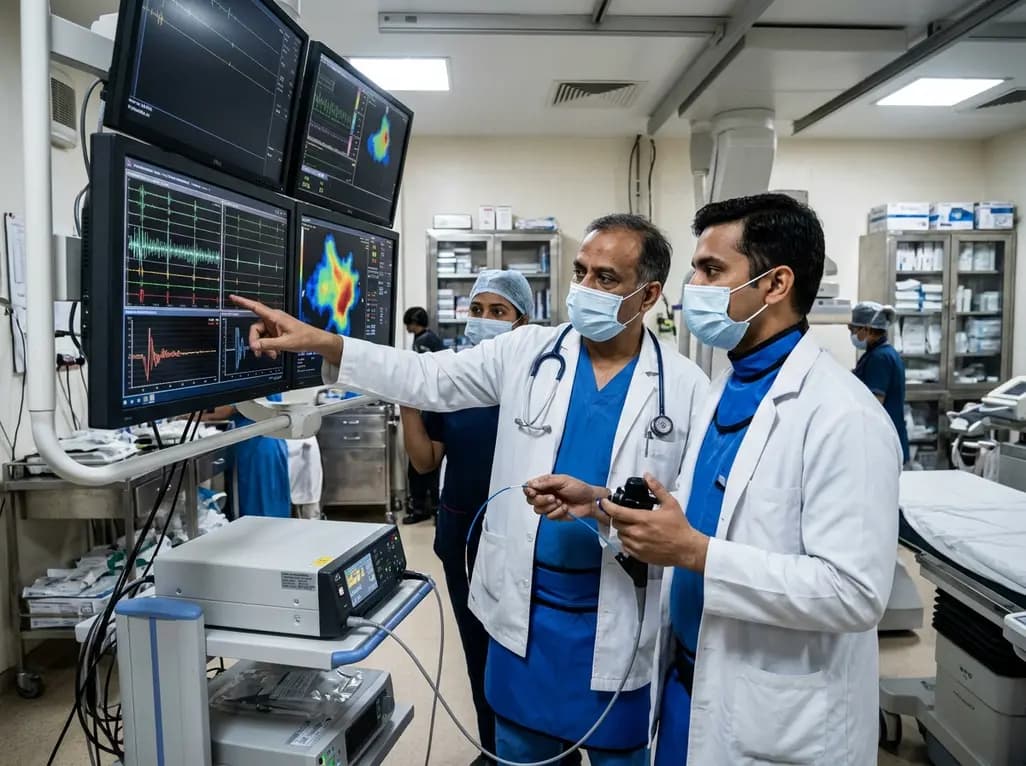
A comparative non-contact study reveals that high-power radiofrequency delivery with the QDOT MICRO catheter induces an artifactual contact force rise. Electrophysiologists must understand this power-dependent thermal and electrical phenomenon to avoid inadvertent catheter withdrawal and ineffective ablation.
Today

A novel proof-of-concept study presents a six-degree-of-freedom articulated manikin neck equipped with linear potentiometers. The system provides real-time kinematic motion feedback during cervical immobilisation, delivering sub-degree tracking accuracy to improve pre-hospital trauma training and patient safety.
Today

A retrospective comparative cohort study evaluated genitourinary safety in HFpEF between dapagliflozin and empagliflozin over six months. Rates of symptomatic UTI and asymptomatic bacteriuria showed no statistically significant differences, supporting comparable safety profiles across both SGLT2 inhibitors.
Today
