

Loading, please wait...
Loading, please wait...

Clinicians frequently encounter complex overlaps in systemic autoimmunity, but the emergence of neuro-renal syndrome represents a critical diagnostic frontier. This neuroimmunomediated condition couples acute nephrotic syndrome—most commonly membranous glomerulonephritis—with severe subacute or chronic peripheral neuropathies. Historically, practitioners misclassified these neuromuscular presentations as atypical chronic inflammatory demyelinating polyneuropathy (CIDP). However, recent scientific advances separate autoimmune nodopathies and paranodopathies into a distinct disease class. In these disorders, circulating autoantibodies disrupt specialized microdomains along myelinated axons. Concurrently, identical or cross-reactive epitopes expressed within podocytes trigger glomerular basement membrane injury. Consequently, patients manifest marked proteinuria, peripheral edema, sensory ataxia, and profound muscle weakness. Recognizing this clinical triad early remains imperative because delayed diagnosis often leads to irreversible axonal loss and advanced renal failure requiring hemodialysis.
The molecular architecture connecting peripheral nerve junctions and the renal filtration barrier explains this syndrome. Autoimmune nodopathies and paranodopathies target specific cell adhesion molecules. These targets include contactin-1 (CNTN-1), neurofascin-155 (NF155), neurofascin-140/186, and contactin-associated protein 1 (CASPR-1). Normally, CNTN-1 and NF155 form axoglial junctions that anchor the terminal myelin loops to the axonal membrane. When pathogenic IgG4 or IgG3 autoantibodies bind these proteins, they disrupt nodal voltage-gated sodium channel clustering without inducing classic complement-mediated demyelination. Similarly, podocyte foot processes express contactin-1 and related surface proteins. Circulating IgG4 antibodies traverse the fenestrated endothelium and deposit subepithelially along glomerular capillary walls. Therefore, these antibodies disrupt both paranodal adhesion and the slit diaphragm filtration barrier simultaneously, driving severe neuromuscular deficits alongside heavy nephrotic-range proteinuria.
Patients with this neuro-renal entity typically display distinct symptoms that contrast sharply with classical autoimmune neuropathy. Neurologically, patients experience a rapid or subacute onset of symmetric weakness that predominantly affects distal muscles. Furthermore, prominent sensory ataxia and postural tremors frequently emerge due to severe proprioceptive fiber disruption. Many affected individuals rapidly lose independent ambulation and become wheelchair-dependent within months. Nephrologically, systemic signs of nephrotic syndrome appear concurrently or shortly after neuropathic symptoms develop. Patients present with refractory peripheral edema, hypoalbuminemia, hypercholesterolemia, and recurrent heavy proteinuria. While some individuals preserve baseline glomerular filtration, others develop acute kidney injury. In severe situations, rapidly progressive renal failure necessitates emergency dialysis. Thus, clinicians must maintain high suspicion when nephrotic syndrome accompanies sudden motor-sensory collapse.
Achieving a definitive diagnosis requires specialized neuroimmunological assays and comprehensive renal investigation. Transfected cell-based assays (CBA) represent the gold standard method to detect serum antibodies directed against CNTN-1, NF155, and CASPR-1. However, access to commercial or validated in-house CBAs remains restricted in many regional laboratories across India. Clinicians must therefore identify surrogate clinical warnings when definitive serological testing faces logistical hurdles. Electrodiagnostic studies offer crucial clues, frequently demonstrating severe conduction block, prolonged distal motor latencies, and marked axonal attenuation. Simultaneously, nephrologists should perform urine protein electrophoresis and renal biopsy. Histopathological examination characteristically reveals membranous glomerulonephritis with subepithelial IgG4 immune deposits. When specialized autoantibody testing remains inaccessible, the concurrent demonstration of membranous nephropathy and an aggressive, tremor-predominant polyradiculoneuropathy justifies presumptive diagnosis and aggressive intervention.
Selecting appropriate immunotherapy represents the most critical determinant of long-term prognosis. Standard first-line treatments for CIDP, such as intravenous immunoglobulin (IVIg), typically fail in this cohort. Because IgG4 antibodies lack complement-fixing capacity and act through steric hindrance of cell-adhesion molecules, IVIg yields high rates of therapeutic refractoriness. Furthermore, large IVIg infusions can occasionally exacerbate renal overload in nephrotic patients. Conversely, therapeutic plasma exchange rapidly removes circulating pathogenic IgG3 and IgG4 autoantibodies, providing prompt acute stabilization. For sustained remission, clinicians must promptly institute targeted B-cell depletion using rituximab. Anti-CD20 therapy effectively suppresses antibody-secreting B-cell clones, arresting glomerular podocyte injury and preventing irreversible axonal degradation. Corticosteroids provide adjunctive bridge suppression, but targeted monoclonal antibodies remain the definitive foundation of lasting clinical recovery.
Managing this condition in resource-variable settings requires active interdisciplinary vigilance between neurologists and nephrologists. Because routine dipstick urinalysis is inexpensive and universally available, all neurologists treating patients with CIDP-like presentations should perform baseline urine protein screening. Conversely, nephrologists evaluating atypical membranous nephropathy must deliberately assess patients for distal paresthesias, gait ataxia, and diminished deep tendon reflexes. When antibody assays require long turnaround times, physicians must not delay therapeutic decisions while awaiting outside reference confirmation. Rapid initiation of plasma exchange followed by generic rituximab regimens provides a cost-effective, limb-saving, and kidney-sparing strategy. By fostering bidirectional cross-specialty referrals, Indian clinicians can identify these vulnerable individuals early, prevent permanent motor disability, and avert end-stage renal disease.
This disorder combines autoimmune peripheral nodopathies or paranodopathies with acute nephrotic syndrome, particularly membranous glomerulonephritis. Patients develop subacute sensorimotor ataxia, distal muscle weakness, and heavy proteinuria. Pathogenic IgG4 or IgG3 antibodies targeting shared neuronal and podocyte adhesion molecules drive this simultaneous neurological and renal dysfunction.
Most autoimmune nodopathies involve non-complement-fixing IgG4 autoantibodies that cause functional disruption rather than macrophage-mediated demyelination. Because intravenous immunoglobulin functions primarily by blocking complement activation and Fc receptors, it fails to restore disrupted cell adhesion. Consequently, affected individuals require plasma exchange or anti-CD20 therapy.
When specialized cell-based assays are inaccessible, clinicians should establish the diagnosis through clinical and histopathological findings. The combination of treatment-refractory sensorimotor polyradiculoneuropathy, distal tremors, and biopsy-confirmed membranous nephropathy strongly suggests this syndrome. Clinicians should initiate plasma exchange and rituximab without waiting for external antibody confirmation.
Disclaimer: This content is for informational and educational purposes only. It is not intended to be a substitute for professional medical advice, diagnosis, or treatment. Always seek the advice of your physician or other qualified health provider with any questions you may have regarding a medical condition. Refer to the latest local and national guidelines for clinical practice.
References
Gonçalves MVM et al. Neuro-renal syndrome (NRS) in autoimmune nodopathies and paranodopathies: A warning to consider the diagnosis when anti-node and paranode antibody tests are difficult to perform. J Neurol Sci. 2026 Oct 15. doi: 10.1016/j.jns.2026.126104. PMID: 42497634.
Le Quintrec M, Teisseyre M, Bec N, et al. Contactin-1 is a novel target antigen in membranous nephropathy associated with chronic inflammatory demyelinating polyneuropathy. Kidney Int. 2021;100(6):1240-1249.
Cortese A, Lombardi R, Briani C, et al. Antibodies to neurofascin, contactin-1, and contactin-associated protein 1 in CIDP: Clinical relevance of IgG isotype. J Peripher Nerv Syst. 2020;25(1):16-24.

Read summarized clinical updates, watch expert medical content, and earn CME certifications right from your smartphone.
Open your smartphone camera to scan and install.





Neuro-renal syndrome links autoimmune nodopathies and paranodopathies with membranous nephropathy. Driven by IgG4 and IgG3 autoantibodies targeting contactin-1 and neurofascin, this condition shows refractoriness to IVIg but responds well to rituximab and plasma exchange, requiring prompt dual-organ recognition.
Today
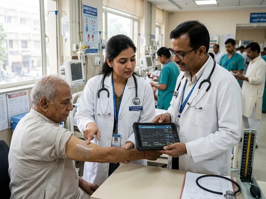
A breakthrough ultrathin multimodal sensor decouples physical hemodynamic monitoring from disposable electrochemical calibration, achieving long-term wearable glucose prediction with 81.36% accuracy and a 9.12% MARD. This offers a robust, drift-free solution for outpatient metabolic surveillance.
Today

A clinical review of menopause and neurology highlights the neurobiological impacts of midlife hormonal shifts on cognition, epilepsy, migraine, multiple sclerosis, and stroke, emphasizing safe hormone therapy prescribing.
Today

Co-administering antiseizure medications and DOACs frequently causes pharmacokinetic interactions, subtherapeutic drug levels, and thromboembolic failures. This review highlights enzyme induction risks, unpredictable outcomes with levetiracetam, and the vital role of therapeutic drug monitoring.
Today

A comparative review of tension band wiring and intramedullary screw fixation in thumb metacarpophalangeal arthrodesis, evaluating union rates, hardware complications, and clinical outcomes.
Today

Following regulatory scrutiny from FSSAI regarding promotional representations and lean muscle preservation claims, Dr. Reddy's Laboratories and Nestlé Health Science are currently reviewing the branding and marketing strategy for their joint nutritional supplement Celevida GLP+.
Today
