

Loading, please wait...
Loading, please wait...

Suicide remains a grave global public health crisis, claiming hundreds of thousands of lives every year. Consequently, clinicians often struggle to differentiate lethal intent from non-lethal self-harm. In clinical psychiatry, evaluating self-directed violence requires objective neurobiological markers to supplement subjective patient reports. Furthermore, emerging neuroimaging tools offer unprecedented opportunities to investigate these complex behavioral pathways. Recently, researchers have utilized resting-state functional magnetic resonance imaging to explore functional connectivity alterations across distributed brain networks. Therefore, mapping these subcortical and cortical circuits allows investigators to identify distinct psychiatric risk profiles.
Understanding how suicidal behavior diverges from non-lethal injury is critical for clinical decision-making. Historically, clinicians have categorized non-suicidal self-directed violence as a potent predictor of prospective suicide attempts. However, the exact neurobiological mechanisms separating these presentations have remained poorly understood. To address this fundamental gap, a breakthrough study analyzed resting-state brain connectivity using data from the UK Biobank. The investigation evaluated 1,070 individuals who reported a confirmed history of self-directed violence. Researchers stratified participants into two discrete cohorts: individuals with previous suicide attempts and those with non-suicidal self-directed violence. Subsequently, investigators acquired resting-state scans across 90 predefined whole-brain regions. They applied rigorous general linear models to identify distinct functional connectivity signatures between the cohorts. Moreover, the authors conducted exploratory analyses to track how self-harm frequency and temporal recency influenced brain networks. This rigorous approach generated pivotal insights into subcortical circuit alterations. Consequently, these results provide a concrete neuroimaging framework to separate distinct dimensions of self-injurious behavior. Ultimately, the connectome findings demonstrate that suicide attempts involve unique neural vulnerabilities rather than simple emotional extremes.
The limbic system plays an essential role in modulating affective intensity and behavioral inhibition. Notably, the study revealed significant functional connectivity reductions in the bilateral amygdala among participants with past suicide attempts compared to those with non-suicidal self-harm. The amygdala governs acute threat processing, fear conditioning, and emotional salience. Therefore, attenuated amygdalar connectivity may disrupt normal emotional appraisal during intense crisis states. Patients experiencing severe psychological distress often encounter profound cognitive bottlenecks. Consequently, reduced amygdala connectivity may diminish the natural emotional aversion associated with lethal physical injury. In addition, frontolimbic uncoupling impairs cognitive reappraisal, leaving patients unable to downregulate acute despair. In contrast, individuals who engage solely in non-suicidal behavior maintain higher amygdalar integration. This preserved connectivity might maintain an instinctual protective threshold against complete self-annihilation. Furthermore, these limbic disruptions demonstrate that suicidal attempts represent distinct pathophysiological states rather than mere continuations of superficial self-injury. Thus, neuroimaging objectively confirms distinct neurobiological pathways between suicidal intent and non-suicidal crisis coping. Clinicians must recognize these distinct limbic signatures when evaluating severe self-harm presentations.
Striatal circuits regulate reward anticipation, motivation, and motor initiation. Interestingly, the UK Biobank investigation unveiled divergent striatal alterations that differentiated suicide attempters from non-suicidal individuals. Specifically, the suicide attempt cohort demonstrated significantly decreased functional connectivity within the right putamen. The putamen regulates habitual action selection and motor planning. Therefore, diminished putamen connectivity may impair adaptive motor responses during emotional turbulence. Conversely, the analysis identified elevated functional connectivity involving the right caudate in the suicide attempt group. The caudate plays a central role in goal-directed decision-making and reward integration. Accordingly, hyperconnectivity within caudate networks might facilitate rigid cognitive fixation on suicide as a viable solution to unbearable emotional distress. Furthermore, this striatal dissociation indicates that reward system dysfunction drives lethal behavioral execution. In contrast, individuals with non-suicidal self-directed violence did not exhibit this specific imbalance. As a result, altered basal ganglia connectivity appears to serve as a meaningful biomarker for suicide vulnerability. These reward circuit shifts illuminate why certain individuals transition from suicidal thoughts to lethal actions.
Beyond categorical diagnoses, the study examined continuous dimensions such as the recency and frequency of self-injurious episodes. Importantly, these exploratory findings highlighted prominent alterations centered on the orbitofrontal cortex. The orbitofrontal cortex serves as an essential hub for impulse control and value-based decision-making. Researchers observed that higher lifetime frequency of self-directed violence correlated directly with increased connectivity between the orbitofrontal cortex and occipital visual regions. Consequently, heightened visual-orbitofrontal coupling may reflect enhanced sensory rumination or intrusive mental imagery related to self-injury. Meanwhile, individuals who engaged in self-harm within the past twelve months displayed decreased connectivity between the orbitofrontal cortex, the rectus gyrus, and temporal lobes. This acute disruption suggests that recent self-directed violence temporarily blunts frontotemporal cognitive control. Thus, clinicians must recognize that recent behavioral episodes carry an elevated neurobiological risk profile. In summary, network connectivity fluctuates dynamically based on episode recency and repetitive behavioral patterns. Identifying these temporary network vulnerabilities provides medical teams with objective windows for intensive clinical intervention.
These neuroimaging findings provide clinicians with invaluable conceptual perspectives on suicide prevention. Currently, psychiatrists rely predominantly on clinical interviews and rating scales to assess immediate suicidal intent. However, self-reported metrics carry inherent vulnerabilities, including patient concealment and cognitive minimization. By demonstrating that suicide attempts involve unique limbic and striatal disruptions, this study validates the biological reality of suicidal behavior. Furthermore, identifying specific frontolimbic and basal ganglia signatures could eventually support algorithmic risk stratification tools. Clinicians can also utilize these insights to select tailored psychotherapeutic interventions. For example, interventions targeting emotion regulation, such as dialectical behavior therapy, directly address frontolimbic dysregulation. Similarly, understanding striatal hyperconnectivity could guide neuromodulation strategies, such as repetitive transcranial magnetic stimulation. Hospital emergency departments frequently struggle to triage ambiguous self-injury presentations safely. Ultimately, bridging neuroimaging science with bedside evaluation will enable earlier identification of high-risk individuals. Consequently, personalized psychiatric medicine moves closer to preventing fatal outcomes before catastrophic behavioral escalation occurs.
Patients with suicide attempts demonstrate decreased functional connectivity in the bilateral amygdala and right putamen, coupled with increased connectivity in the right caudate. In contrast, individuals engaging in non-suicidal self-directed violence preserve higher amygdala connectivity, suggesting that distinct limbic and striatal circuits mediate lethal intent versus non-lethal crisis behavior.
The orbitofrontal cortex regulates impulse suppression and value appraisal. Repetitive self-harm enhances orbitofrontal-occipital connectivity, reflecting persistent visual or sensory rumination. Conversely, recent self-harm reduces orbitofrontal connectivity with the rectus gyrus and temporal lobes, indicating acute disruptions in top-down cognitive control that temporarily increase vulnerability to behavioral escalation during acute emotional distress.
Resting-state neuroimaging cannot yet replace thorough psychiatric interviews, clinical rating scales, or comprehensive suicide risk evaluations. Currently, functional connectivity findings illuminate the underlying neurobiology of self-harm and help validate clinical presentations. However, future clinical translation may ultimately incorporate neuroimaging biomarkers into multimodal prediction algorithms to enhance objective risk stratification in psychiatric medicine.
Disclaimer: This content is for informational and educational purposes only... Refer to the latest local and national guidelines for clinical practice.
References

Read summarized clinical updates, watch expert medical content, and earn CME certifications right from your smartphone.
Open your smartphone camera to scan and install.





A UK Biobank rs-fMRI study reveals distinct functional connectivity alterations in limbic and striatal circuits that separate suicide attempts from non-suicidal self-directed violence, highlighting bilateral amygdala, putamen, and caudate involvement.
Today

Review how tailored exercise programs benefit frail older patients with heart failure. Explore pathophysiological mechanisms, individualized training prescriptions, and strategies to overcome adherence barriers in clinical practice.
Today
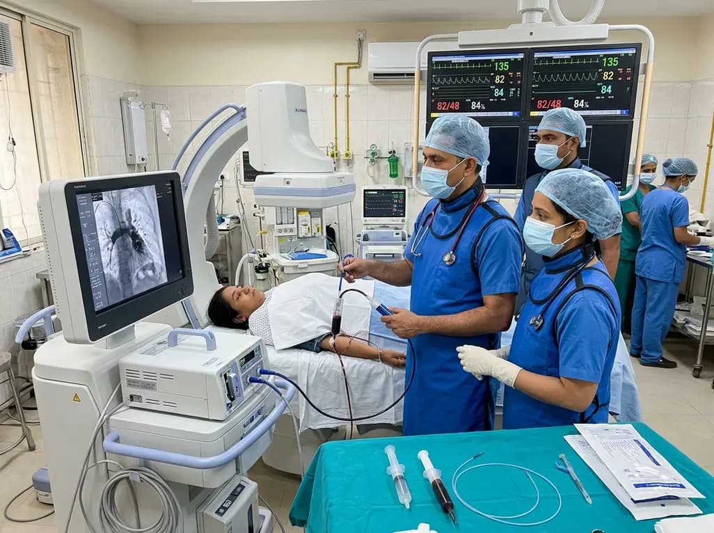
In high-risk pulmonary embolism complicated by severe anemia and active uterine bleeding, systemic thrombolysis is contraindicated. Large-bore aspiration thrombectomy provides rapid hemodynamic rescue without fibrinolysis, supported by inferior vena cava filter placement and multidisciplinary critical care.
Today

A cross-sectional study in Zanzibar reveals that 46.5% of adults with type 2 diabetes experience multidomain autonomic dysfunction on the COMPASS-31 tool, primarily orthostatic intolerance, while 45.9% exhibit cardiovascular autonomic neuropathy, highlighting the urgent need for structured clinical screening.
Today

This comparative study evaluates posterior labral tears with and without glenohumeral instability, highlighting distinct demographic patterns, MRI underestimation in stable shoulders, and successful arthroscopic repair outcomes.
Today

A recent case-control study reveals that urinary clusterin provides superior diagnostic performance for early diabetic kidney disease detection in pediatric type 1 diabetes mellitus, identifying tubular damage before conventional microalbuminuria occurs.
Today
