.webp&w=384&q=75)

Loading, please wait...
Loading, please wait...
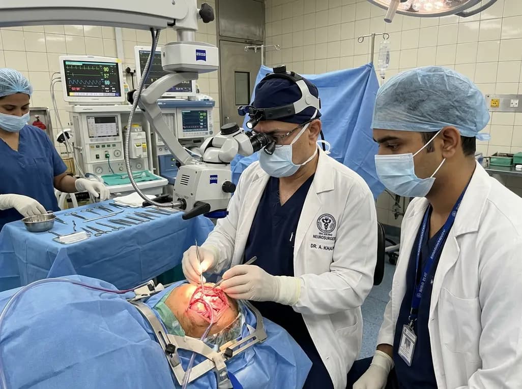
Geniculate neuralgia represents one of the most agonizing yet frequently misdiagnosed cranial neuropathies encountered in neurosurgical and otolaryngological practice. Clinicians often describe it as an electric, lancinating otalgia that radiates deep within the auditory canal. Because conservative medical therapy often fails to provide lasting relief, neurosurgeons increasingly evaluate invasive interventions. Among these surgical modalities, nervus intermedius sectioning has emerged as a definitive operative strategy for refractory cases. This delicate microvascular procedure targets the sensory root of the facial nerve to interrupt aberrant peripheral signals driving excruciating pain. Consequently, understanding its indications, operative anatomy, and clinical efficacy remains essential for treating clinicians navigating complex craniofacial pain syndromes.
Geniculate neuralgia generates severe paroxysms of lancinating pain located deep inside the external ear canal, pinna, and surrounding retroauricular tissues. Patients frequently report distressing triggers such as swallowing, chewing, light touch, or cold airflow. Historically, physicians attributed these episodic symptoms exclusively to primary vascular compression of the nervus intermedius. However, contemporary neurosurgical investigations reveal significant overlap with other cranial neuralgias. Indeed, clinical records indicate that a vast majority of patients present with concurrent trigeminal neuralgia, while nearly one-third simultaneously experience glossopharyngeal neuralgia. This anatomic convergence occurs because cranial nerves five, seven, nine, and ten share sensory innervation across the auricle and tympanic membrane. Consequently, neurovascular conflict may irritate multiple adjacent nerve roots at the cerebellopontine angle simultaneously. Therefore, clinicians must conduct detailed diagnostic evaluations to distinguish isolated geniculate pain from broader multi-nerve syndromes. High-resolution magnetic resonance imaging with constructive interference in steady state sequences helps identify offending arterial loops. Moreover, diagnostic local nerve blocks often assist in confirming the precise peripheral source of pain. Ultimately, accurate diagnosis prevents unnecessary invasive procedures and guides tailored surgical intervention for refractory suffering.
Neurosurgical teams approach nervus intermedius sectioning through a tailored retrosigmoid craniotomy under continuous neurophysiological monitoring. After opening the dura and carefully evacuating cerebrospinal fluid, the surgeon gently retracts the cerebellum to expose the cerebellopontine angle. Visualizing the seventh and eighth cranial nerve complex requires meticulous microsurgical dissection of the surrounding arachnoid membranes. The nervus intermedius typically courses between the motor root of the facial nerve and the vestibulocochlear nerve bundle. However, anatomical variations, including multi-fascicular roots or dense arachnoid adhesions, can complicate operative identification. Surgeons often employ micro-bipolar stimulation to confirm the absence of motor facial nerve responses before transection. Once verified, the operator sharply divides the nervus intermedius near the brainstem using sharp micro-scissors. In addition, surgeons routinely perform concurrent microvascular decompression whenever offending vessels contact the trigeminal or glossopharyngeal roots. Teflon felt pledgets are positioned between the compressive vessels and the neural parenchyma to ensure complete neurovascular isolation. Consequently, this combined operative strategy relieves localized mechanical compression while permanently eliminating the afferent pain pathway. Postoperatively, meticulous dural closure prevents cerebrospinal fluid leakage and facilitates smooth recovery.
Recent clinical evidence demonstrates robust efficacy for surgical rhizotomy targeting the intermediate nerve. In a pivotal institutional review comprising forty-seven procedures, over eighty percent of patients achieved meaningful long-term pain relief. Specifically, these individuals attained Barrow Neurological Institute pain scores below grade four, indicating substantial functional improvement. Furthermore, complete pain freedom without the requirement for daily pain medications occurred in over sixty-eight percent of treated patients. These numbers reflect significant success when treating a condition historically resistant to standard pharmacotherapy like carbamazepine or gabapentin. Additionally, patients undergoing concurrent microvascular decompression alongside rhizotomy demonstrated superior symptom reduction compared to those undergoing solitary procedures. This synergistic outcome highlights the therapeutic benefit of addressing multifaceted neurovascular compression simultaneously. Long-term registries also confirm that durable relief persists across years of follow-up with minimal late recurrence. As a result, early surgical evaluation offers patients a predictable pathway to restore their baseline quality of life. Clinicians therefore recommend timely operative referral when first-line anticonvulsant regimens trigger toxic side effects or lose therapeutic efficacy.
Microsurgical sectioning within the cerebellopontine angle requires strict vigilance to preserve adjacent neurological structures. Because the nervus intermedius lies adjacent to critical motor and sensory bundles, inadvertent injury can precipitate serious deficits. Fortunately, comprehensive retrospective analyses confirm an exceptionally low complication profile when experienced skull base teams perform the procedure. Most importantly, recent cohorts report zero cases of permanent facial paralysis, highlighting the reliability of intraoperative facial motor monitoring. However, mild sensory alterations occasionally arise due to the specialized function of the intermediate nerve. For instance, approximately six percent of surgical patients experience mild, persistent loss of taste over the anterior two-thirds of the tongue. Additionally, transient or unexpected sensorineural hearing loss occurs in roughly four percent of cases, typically secondary to labyrinthine artery spasm. Vestibular dysfunction remains similarly uncommon, with permanent imbalance reported in only two percent of individuals. Therefore, preoperative counseling should thoroughly explain these distinct risks while emphasizing the overwhelming safety record of modern micro-neurosurgery. Rigorous neurophysiological monitoring remains the cornerstone for averting inadvertent acoustic and facial nerve trauma throughout dissection.
Optimizing outcomes for geniculate neuralgia requires seamless collaboration among neurologists, otolaryngologists, and neurosurgeons. Initial clinical evaluations must exclude non-neuralgic sources of otalgia, including temporomandibular joint dysfunction and infections. Once physicians confirm neuralgia, they initiate pharmacotherapy using membrane-stabilizing sodium channel blockers. However, adverse pharmacological effects such as cognitive clouding and profound lethargy frequently limit long-term compliance. In addition, patients frequently develop pharmacological tolerance, prompting repeated dosage escalations without adequate relief. When conservative treatments fail, skull base teams should evaluate operative candidacy promptly. Neurosurgeons assess patient age, comorbidities, high-resolution vascular imaging, and concurrent cranial nerve symptoms during planning. Furthermore, engaging patients in shared decision-making ensures realistic expectations regarding postoperative recovery. Following surgery, multidisciplinary follow-up facilitates systematic medication weaning and tracks functional restoration. Consequently, an integrated care pathway significantly improves long-term outcomes and patient quality of life.
Geniculate neuralgia arises from irritation or neurovascular compression of the nervus intermedius, a sensory branch of the facial nerve. Patients experience excruciating, sharp, stabbing paroxysms of pain deep inside the ear canal. Common physiological triggers include swallowing, chewing, light cutaneous touch, and exposure to cold wind or water.
Many patients present with concurrent trigeminal or glossopharyngeal neuralgia due to shared regional neurovascular compression at the cerebellopontine angle. Combining nervus intermedius rhizotomy with microvascular decompression addresses multi-nerve conflict simultaneously. This synergistic operative strategy significantly enhances overall pain relief and reduces the risk of persistent or recurrent craniofacial pain.
The surgical procedure carries a very low overall complication rate when performed under neurophysiological monitoring. Permanent facial paralysis is exceedingly rare. However, potential risks include partial loss of taste on the anterior tongue, dry eye symptoms, and rare instances of transient or permanent sensorineural hearing loss and vestibular imbalance.
Disclaimer: This content is for informational and educational purposes only... Refer to the latest local and national guidelines for clinical practice.
References
Ren AL et al. Nervus intermedius sectioning for the treatment of geniculate neuralgia. J Neurosurg. 2025 Jul 01. doi: 10.3171/2024.11.JNS241141. PMID: 40085931.
Holste KG, et al. Pain-free and pain-controlled survival after sectioning the nervus intermedius in nervus intermedius neuralgia: a single-institution review. J Neurosurg. 2019;131(2):352-359.
Kenning TJ, Kim CS, Bien AG. Microvascular decompression and nervus intermedius sectioning for the treatment of geniculate neuralgia. J Neurol Surg B Skull Base. 2019;80(Suppl 3):S316-S317.

Read summarized clinical updates, watch expert medical content, and earn CME certifications right from your smartphone.
Open your smartphone camera to scan and install.





A comprehensive clinical review of nervus intermedius sectioning for geniculate neuralgia, detailing surgical nuances, multineuralgia overlap, long-term pain outcomes, and neurofunctional safety.
last month

Novel diimide and hydrazone scaffolds, particularly compound #C4, synergistically reactivate latent HIV-1 when combined with JQ1 or PKC activators. Demonstrating donor-matched proviral transcription ex vivo and non-canonical signaling, these findings advance shock and kill strategies toward reservoir eradication.
Today

A comparative cohort study evaluates vNOTES vs TLH hysterectomy for benign conditions, demonstrating shorter operative duration, faster return to daily activities, and equivalent female sexual function scores without increased dyspareunia.
Today

Review how tailored exercise programs benefit frail older patients with heart failure. Explore pathophysiological mechanisms, individualized training prescriptions, and strategies to overcome adherence barriers in clinical practice.
Yesterday

This comparative study evaluates posterior labral tears with and without glenohumeral instability, highlighting distinct demographic patterns, MRI underestimation in stable shoulders, and successful arthroscopic repair outcomes.
Yesterday

A clinical study demonstrates that early intervention with tocilizumab, either as monotherapy or bridged with corticosteroids, achieves significant proptosis reduction and resolves active inflammation in treatment-naive thyroid eye disease.
Today
