.webp&w=384&q=75)

Loading, please wait...
Loading, please wait...

Neonatal respiratory failure often stems from common pulmonary etiologies. However, mechanical cardiovascular compression presents an uncommon yet life-threatening clinical emergency. A neonatal pericardial teratoma represents a rare primary cardiac neoplasm that causes massive cardiomegaly. Consequently, the expanding intrapericardial mass rapidly compresses the cardiac chambers and pulmonary vasculature. When clinicians encounter sudden neonatal shock and profound cardiomegaly, they must consider this unusual congenital tumor. Prompt recognition allows immediate multidisciplinary intervention. Furthermore, early diagnostic imaging helps clinicians differentiate this lesion from fatal structural defects and facilitates life-saving operative correction.
Intrapericardial germ cell tumors arise from displaced multipotent embryonic cells during early development. Specifically, these tumors typically originate near the aortic root, pulmonary trunk, or pericardial reflection. Most pediatric pericardial teratomas remain histologically benign. However, immature variants contain primitive neuroectodermal, mesenchymal, or endodermal components. Regardless of histological grade, the rapid physical expansion poses the greatest physiological danger to the neonate. Therefore, even benign tumors can trigger catastrophic hemodynamic instability.
Additionally, the tumor secretes serosanguinous fluid into the pericardium. This process creates massive pericardial effusion and precipitates acute tamponade physiology. The non-compliant neonatal pericardium cannot accommodate sudden volume expansion. As a result, the external pressure prevents ventricular diastole and drastically lowers cardiac output. Furthermore, the burgeoning mass displaces normal mediastinal structures and compresses adjacent tracheobronchial airways. Consequently, the infant experiences both mechanical airway collapse and progressive circulatory shock. Understanding these pathological mechanisms helps clinicians recognize why rapid clinical deterioration occurs. Clinicians must maintain a high index of suspicion whenever a term newborn exhibits unexplained cardiomegaly accompanied by massive pericardial effusion.
Newborns with intrapericardial tumors exhibit dramatic clinical signs shortly after birth. In most instances, term neonates present with severe respiratory distress, marked tachypnea, grunting, and cyanosis. Furthermore, physical examination typically reveals muffled heart sounds, weak peripheral pulses, and delayed capillary refill. Chest radiography consistently displays massive cardiomegaly that often obscures the lung fields entirely. Because the physical mass mimics cardiogenic shock, clinicians may mistakenly suspect congenital cardiomyopathy or primary pulmonary pathology.
Nevertheless, vigilant clinicians identify key physiological clues that point to mechanical compression. First, the neonate frequently exhibits refractory hypoxia despite positive-pressure ventilation. This persistent hypoxia occurs because the expanding mass causes extrinsic bronchial compression and severe pulmonary atelectasis. Second, elevated systemic venous pressures lead to rapid hepatomegaly and peripheral edema. If intrauterine compression was extensive, the newborn might already show signs of fetal hydrops. Moreover, acute cardiac tamponade can trigger sudden circulatory collapse during routine neonatal care. Therefore, intensive care teams must stabilize the infant immediately with inotropic support while preparing for urgent diagnostic investigations. Rapid bedside evaluation prevents irreversible ischemic organ injury and stabilizes vital organ perfusion.
Accurate anatomical visualization remains crucial for managing intrapericardial masses in neonates. Transthoracic echocardiography represents the initial diagnostic modality of choice at the bedside. Through multiplane imaging, echocardiography demonstrates a heterogeneous, multiloculated mass situated within the pericardial space. In addition, ultrasound delineates the extent of pericardial effusion and visualizes hemodynamic tamponade, such as diastolic chamber collapse. Color Doppler interrogation evaluates whether the lesion compresses the superior vena cava, pulmonary veins, or outflow tracts.
However, definitive surgical planning requires advanced cross-sectional chest tomography. Contrast-enhanced chest computed tomography rapidly maps the exact tumor origin and vascular attachments. Specifically, computed tomography reveals characteristic features of a teratoma, including mixed cystic and solid components alongside focal calcifications. Alternatively, cardiac magnetic resonance imaging offers superior soft-tissue differentiation without ionizing radiation. Nonetheless, the rapid scan times of modern computed tomography make it the safer choice for unstable, ventilated newborns. Furthermore, pre-procedural imaging distinguishes pericardial teratomas from bronchogenic cysts, vascular malformations, and thymic masses. Multimodal imaging provides pediatric cardiac surgeons with the precise structural blueprint necessary for complete tumor resection.
Complete surgical excision represents the definitive treatment for symptomatic intrapericardial teratomas. Because conservative medical therapy cannot relieve mechanical obstruction, surgical intervention must occur urgently. Preoperative management focuses on stabilizing cardiopulmonary mechanics in the neonatal intensive care unit. In patients with impending tamponade, bedside pericardiocentesis temporarily restores hemodynamic stability before formal operative intervention. However, surgeons prefer definitive primary excision over repeated drainage procedures to reduce procedural risks.
Surgical teams access the anterior mediastinum through a median sternotomy. Upon opening the pericardium, the surgeon immediately relieves intracardiac compression and aspirates the surrounding effusion. Typically, a single vascular pedicle connected to the adventitia of the ascending aorta or pulmonary artery feeds the teratoma. Surgeons meticulously dissect this vascular stalk while protecting adjacent coronary arteries and phrenic nerves. Although most resections proceed without cardiopulmonary bypass, surgeons keep bypass equipment ready in the operating suite. Cardiopulmonary bypass becomes necessary if the tumor firmly adheres to cardiac myocardium or major vessels. Ultimately, total extirpation of the mass achieves immediate hemodynamic normalization and lung re-expansion.
Histological examination of the resected specimen provides critical prognostic information. Grossly, the lesion presents as an encapsulated, lobulated tumor containing variable cystic cavities filled with mucinous or serous fluid. Microscopically, the mass comprises tissues derived from ectoderm, mesoderm, and endoderm. While mature teratomas contain fully differentiated adult-type tissues, immature pericardial teratomas contain primitive, embryonic elements such as neuroepithelial tubules. Importantly, the presence of immature neuroepithelial tissue does not signify clinical malignancy in neonates.
Consequently, complete surgical resection is considered curative for benign and immature pericardial teratomas alike. Oncologists generally avoid postoperative adjuvant chemotherapy when the surgical margin remains clear. Instead, clinical teams follow the infant with serial echocardiography and serum alpha-fetoprotein monitoring. Alpha-fetoprotein serves as a reliable circulating tumor marker because developing germ cell tumors frequently synthesize this oncofetal protein. Serial measurements confirm disease eradication and assist in early detection of rare recurrences. Fortunately, term infants who undergo prompt complete resection demonstrate excellent long-term survival and normal myocardial functional recovery. Collaborative care between pediatric intensivists, cardiologists, and surgeons guarantees optimal developmental outcomes.
Acute deterioration results primarily from physical mechanical compression and progressive pericardial effusion. As the intrapericardial tumor expands rapidly within the non-compliant neonatal pericardium, it compresses the low-pressure cardiac chambers and pulmonary veins. Consequently, ventricular filling decreases significantly, leading to decreased stroke volume and acute cardiac tamponade. Additionally, external pressure on the main airways worsens hypoxia, precipitating severe metabolic acidosis and rapid circulatory collapse.
Clinicians differentiate pericardial teratomas using multimodality imaging and biochemical markers. Transthoracic echocardiography clearly identifies an intrapericardial, multiloculated lesion arising near the great arteries, accompanied by significant pericardial fluid. Furthermore, contrast-enhanced chest tomography reveals pathognomonic heterogeneous internal architecture containing cystic spaces, soft-tissue elements, and distinct focal calcifications. Finally, serum alpha-fetoprotein testing supports the germ cell tumor diagnosis and helps exclude vascular malformations, bronchogenic cysts, and thymic neoplasms.
The long-term prognosis after complete surgical excision is extraordinarily favorable for most neonates. Although histopathology may reveal immature embryonic components, these lesions rarely demonstrate malignant clinical behavior in newborns. Therefore, complete resection without residual tumor achieves an excellent cure rate without requiring adjuvant chemotherapy. Long-term surveillance involves regular clinical follow-up, periodic echocardiography, and serial serum alpha-fetoprotein measurements to confirm normal cardiovascular development and rule out recurrence.
Disclaimer: This content is for informational and educational purposes only... Refer to the latest local and national guidelines for clinical practice.
References

Read summarized clinical updates, watch expert medical content, and earn CME certifications right from your smartphone.
Open your smartphone camera to scan and install.





A term neonate with severe respiratory distress and massive cardiomegaly was diagnosed with a giant immature pericardial teratoma. Multimodal imaging guided successful complete surgical resection, relieving life-threatening cardiopulmonary compression and leading to an excellent clinical outcome.
Today

A new clinical trial demonstrates that structured high-protein diets and enteral supplementation significantly decrease malnutrition and disease activity in inflammatory bowel disease patients, offering clear guidance for gastroenterologists and nutritionists.
Today
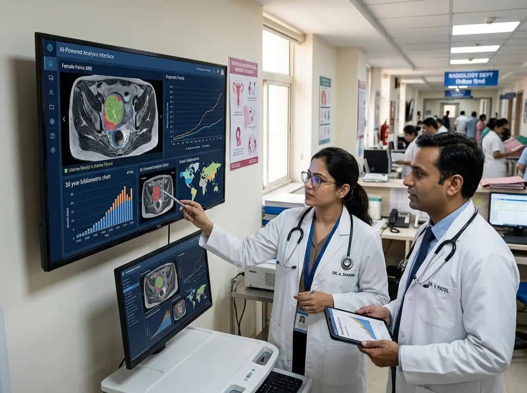
A 30-year bibliometric analysis highlights key research trends in AI in women's health from 1992 to 2024. Discover how evolving computational models, global institutional collaborations, and clinical decision support tools are shaping maternal, oncological, and reproductive care worldwide.
Today

A Level I trauma center study shows a brief bedside mental health intervention boosts 30-day follow-up screening adherence by 84% in nonviolent injury patients. However, outcomes for violence survivors reveal critical care gaps, underscoring the urgent need for specialized community violence intervention programs.
Today

A preclinical study demonstrates that the SGLT2 inhibitor remogliflozin attenuates cyclophosphamide-associated peripheral neurotoxicity in rats. Remogliflozin restores antioxidant balance via Nrf2, suppresses NF-κB-driven neuroinflammation, activates PI3K/Akt survival signaling, and preserves structural myelin integrity.
Today
