.webp&w=384&q=75)

Loading, please wait...
Loading, please wait...

Successful necrotizing fasciitis reconstruction requires proactive surgical planning right from the initial emergency debridement. Historically, acute management focused solely on life-saving debridement, but contemporary surgical algorithms demand early plastic surgical collaboration. Consequently, modern surgeons evaluate reconstructive pathways while eradicating infection. Wound bed readiness and patient physiological stability must strictly dictate operative timing rather than surgeon convenience. Therefore, clinical teams must balance infectious source control against reconstructive feasibility during early resuscitation. When invasive bacterial toxins destroy expansive fascial planes, severe systemic inflammation rapidly compromises patient hemodynamics. Because critical organ dysfunction frequently accompanies fulminant sepsis, surgeons cannot attempt premature tissue coverage. Instead, reconstructive surgeons work closely with intensivists to optimize metabolic indices, manage glycemic control, and stabilize microvascular perfusion. Furthermore, clinical experience demonstrates that attempting immediate wound coverage in physiologically unstable hosts precipitates disastrous flap failure. Conversely, establishing clear physiological criteria guarantees durable soft tissue restoration once septic shock resolves completely. Thus, the foundational thesis of this timing-based algorithm emphasizes staged intervention. Plastic surgeons must respect biological prerequisites, ensuring that durable wound closure matches each patient's physiological trajectory seamlessly.
Radical initial debridement serves as the undisputed cornerstone of patient survival during acute necrotizing soft tissue infections. However, complete infection control rarely occurs after a single operative procedure. Therefore, surgeons must schedule mandatory re-explorations at strict twelve to twenty-four hour intervals. During these planned second-look operations, surgical teams reassess fascial viability and excise lingering necrotic margins aggressively. Furthermore, adopting skin-sparing debridement techniques significantly preserves salvageable cutaneous architecture without compromising life-saving infection clearance. Although fascia and subdermal fat undergo widespread necrosis, superficial cutaneous perfusion often remains surprisingly intact across peripheral zones. Consequently, discerning surgeons meticulously preserve marginally viable skin flaps during secondary washouts. This conservative strategy markedly reduces the ultimate wound surface area that eventually requires skin graft coverage. In addition, preserving viable skin margins substantially increases opportunities for delayed primary closure later in the hospital course. Repeated inspections also confirm the complete arrest of bacterial necrosis across deep muscle compartments. Thus, thorough serial debridement defines the definitive reconstructive margin while safely eradicating all pathogenic microorganisms. Once viable, bleeding tissue beds emerge, the surgical team can confidently transition toward constructive wound management.
Following complete surgical debridement, modern plastic surgeons utilize negative pressure wound therapy as an indispensable temporizing bridge. This advanced therapeutic modality applies subatmospheric pressure to macroscopically clear wound beds, thereby accelerating secondary granulation tissue formation. Furthermore, negative pressure dressings continuously evacuate infectious exudate and decrease interstitial edema across the compromised zone. Consequently, local microvascular perfusion improves dramatically, creating a vascularized foundation for subsequent reconstructive procedures. Moreover, surgeons increasingly employ negative pressure wound therapy with automated instillation and dwell time. This sophisticated variant delivers topical antiseptic or saline solutions directly into deep wound crevices, effectively disrupting residual microbial biofilms. Therefore, instillation therapy further decreases bacterial bioburden while maintaining ideal tissue hydration. Additionally, mechanical foam contraction significantly reduces wound dimensions over several dressing changes. This progressive tissue approximation simplifies definitive reconstruction, often converting complex defects into easily manageable wounds. Nevertheless, clinicians must monitor the wound bed vigilant against occult secondary infections under closed vacuum dressings. Regular dressing exchanges every forty-eight to seventy-two hours confirm continuous tissue proliferation and rule out renewed necrosis. Consequently, negative pressure therapy reliably bridges unstable open defects toward timely, definitive tissue coverage.
Definitive closure requires a tailored algorithm matching specific reconstructive techniques to declared anatomic defects. For broad planar defects across the trunk or extremities, split-thickness skin grafting remains the workhorse modality. However, clinicians often combine skin grafts with delayed primary closure to minimize donor site morbidity whenever skin laxity permits. In contrast, perineal and scrotal defects following Fournier gangrene demand specialized regional solutions. Local advancement flaps, gracilis muscle flaps, and pudendal fasciocutaneous flaps provide excellent functional and cosmetic restoration in sensitive perineal regions. Furthermore, these regional pedicled flaps deliver well-vascularized tissue that protects vulnerable pelvic structures and resists urinary or fecal contamination. When debridement exposes denuded tendons, bone, or hardware lacking periosteum, surgeons cannot rely on simple skin grafts alone. Instead, reconstructive surgeons apply acellular dermal matrices or engineered dermal templates over poorly vascularized wound beds. These biocomposite matrices stimulate robust neovascularization, subsequently supporting successful delayed epidermal grafting. Additionally, selecting the correct reconstructive ladder rung avoids unnecessary surgical complexity while restoring structural integrity. Thus, regional anatomic algorithms guide plastic surgeons toward safe, robust, and functional reconstructive outcomes.
Complex wounds presenting massive volume loss or critical neurovascular exposure frequently necessitate microvascular free tissue transfer. Historically, surgeons hesitated to perform free flaps after necrotizing fasciitis because intense local thrombogenic vasculitis threatened microvascular anastomoses. However, accumulating retrospective evidence indicates that free tissue transfer achieves high success rates once patients achieve complete physiological stabilization. Reconstructive teams must verify normal inflammatory markers, negative microbiological cultures, and stable hemodynamic parameters before embarking on microvascular reconstruction. Furthermore, surgeons must select recipient blood vessels well outside the previous zone of active tissue infection. Careful recipient vessel dissection minimizes anastomotic thrombosis risks and ensures durable flap survival. Moreover, free muscle or perforator flaps restore substantial soft tissue contours, which greatly improves long-term functional recovery and limb salvage. Nevertheless, current literature relies primarily on small, retrospective case series vulnerable to institutional selection bias. Therefore, plastic surgical societies emphasize the urgent need for prospective multicenter registries and standardized outcome reporting metrics. Standardized registries will eventually refine patient selection criteria and clarify optimal intervention windows. Ultimately, combining meticulous surgical timing with advanced reconstructive techniques delivers superior functional outcomes for survivors of necrotizing soft tissue infections.
Surgeons initiate reconstruction only after achieving complete infection source control and physiological stability. Specifically, patients must demonstrate resolution of septic shock, normalized lactate levels, and negative wound cultures. Furthermore, serial debridements must confirm entirely healthy, non-infected wound margins. Once these critical biological and systemic criteria are met, the reconstructive team can safely perform definitive wound closure or flap transfer without risking life-threatening recurrent infection or flap loss.
Negative pressure wound therapy serves as a vital bridge between aggressive debridement and definitive reconstruction. The subatmospheric dressing removes inflammatory fluid, reduces tissue edema, and enhances local microvascular perfusion. Additionally, instillation variants clear residual biofilms from irregular wound beds. Consequently, the therapy stimulates robust granulation tissue growth while mechanically contracting wound edges. This significant reduction in overall wound dimensions simplifies future coverage, enabling delayed primary closure or smaller skin grafts.
Yes, microvascular free tissue transfer is safe and effective when performed after complete acute inflammation resolves. Although persistent peri-wound vasculitis elevates thrombosis risk, selecting recipient vessels outside the injured zone prevents anastomotic failure. Furthermore, free flaps provide extensive vascularized tissue to cover exposed tendons, bones, and neurovascular structures. Therefore, once the patient is hemodynamically stable and infection-free, free flap reconstruction achieves high survival rates and excellent long-term functional recovery.
Disclaimer: This content is for informational and educational purposes only... Refer to the latest local and national guidelines for clinical practice.
References

Read summarized clinical updates, watch expert medical content, and earn CME certifications right from your smartphone.
Open your smartphone camera to scan and install.





Reconstruction following necrotizing fasciitis requires careful staging and physiological stabilization. A timing-based algorithm prioritizes radical debridement, negative pressure wound therapy, and defect-specific tissue coverage to optimize functional and aesthetic outcomes.
Today

Systemic amyloidosis management is undergoing a therapeutic revolution. This clinical review synthesizes updates from the International Society of Amyloidosis, detailing novel plasma cell therapies, TTR stabilizers, silencers, translational models, and artificial intelligence in drug development.
Today

A retrospective study of 824 patients undergoing over 2,200 intradetrusor onabotulinumtoxinA injections identifies sex- and etiology-specific predictors of 2-week post-void residual, highlighting the influence of baseline residual volumes, toxin dosage, and distinct urodynamic parameters on urinary retention risk.
Today
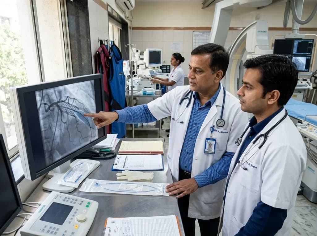
The US FDA has granted approval to the SELUTION SLR drug-eluting balloon for treating coronary in-stent restenosis. This milestone introduces a sustained-release sirolimus platform that eliminates the need for additional permanent metallic stents while maintaining robust vascular healing and luminal patency.
Today

Posttraumatic pisotriquetral instability is a rare wrist injury causing severe hypothenar pain and weakness. Explore clinical examination, imaging, and pisiformectomy.
Today

A comprehensive review analyzes periconceptional GLP-1 receptor agonist exposure, revealing reassuring data on congenital anomalies and adverse perinatal outcomes while emphasizing the need for cautious patient counseling.
Today
