

Loading, please wait...
Loading, please wait...
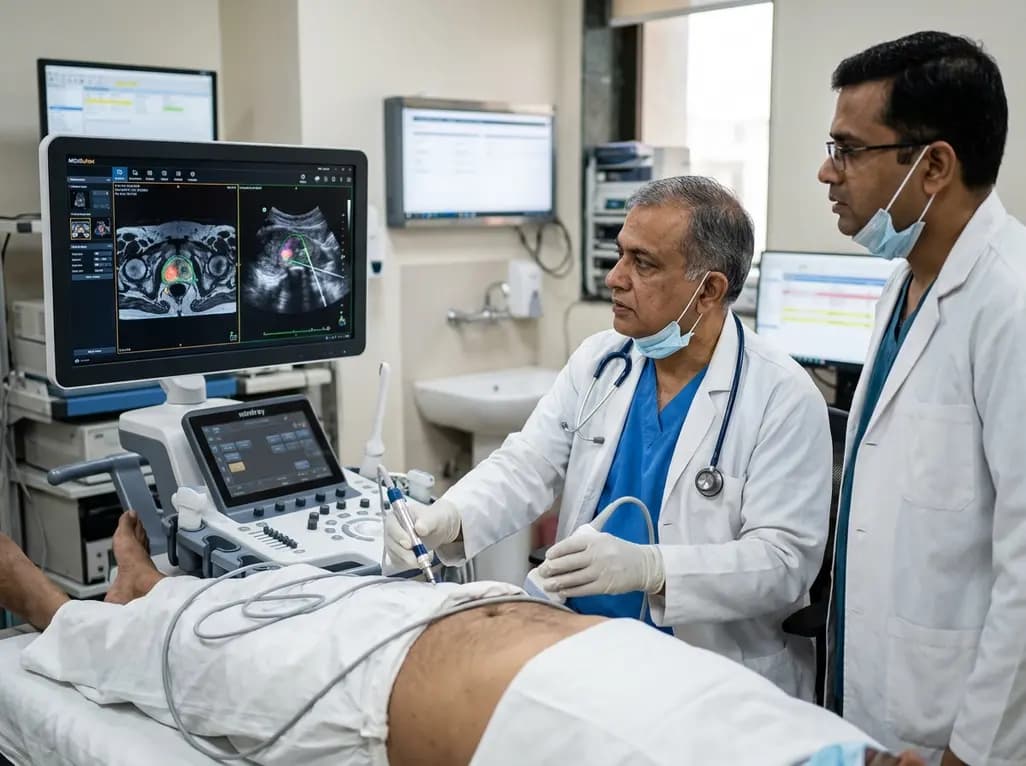
Accurate prostate cancer detection remains a pivotal cornerstone in modern urological oncology. Clinicians routinely depend on multiparametric magnetic resonance imaging and transrectal ultrasound fusion to identify clinically significant disease. However, tissue deformation and spatial discrepancies frequently cause biopsy needles to miss index lesions. To resolve this challenge, investigators created the MRUSuNet platform, a novel multimodal deep learning model designed to enhance spatial localization during targeted prostate interventions.
Conventional targeted prostate biopsies combine pre-procedural multiparametric magnetic resonance images with real-time transrectal ultrasonography. Consequently, this fusion workflow allows urologists to navigate toward suspicious prostatic zones with structured guidance. Nevertheless, significant technical challenges persist in routine clinical environments. Radiologists first identify regions of interest on static magnetic resonance images using standardized reporting criteria. Subsequently, urologists map these designated targets onto ultrasound coordinates during live interventions. However, the insertion of the endorectal probe inherently deforms the pliable gland, causing substantial mechanical tissue displacement. Therefore, rigid and non-rigid software registration often fails to compensate for dynamic anatomical changes. This registration discrepancy leads to needle trajectory errors and missed malignant foci. Furthermore, standard cognitive or software-based co-registration depends heavily on operator skill and individual radiologist expertise. When interobserver variability arises, clinicians may misclassify aggressive malignancies or biopsy benign stroma inadvertently. Consequently, patients risk repeated negative biopsies, diagnostic delay, or inappropriate active surveillance placement. Because current targeting platforms display persistent spatial errors, researchers require automated computational models that correct tissue distortion dynamically.
To eliminate registration discrepancies, researchers engineered MRUSuNet, a specialized dual-input convolutional network architecture. Specifically, this neural network processes three-dimensional multiparametric magnetic resonance volumes alongside co-registered transrectal ultrasound sequences simultaneously. The model incorporates encoder-decoder feature extractors connected by skip pathways that preserve granular anatomical contours. Furthermore, the multi-branch design extracts distinct radiological features from high-contrast magnetic resonance scans while extracting echogenic tissue boundaries from ultrasound frames. The computational system then projects these fused representations into a unified three-dimensional spatial coordinates matrix. Consequently, the network predicts voxel-level lesion probability maps directly within the live ultrasound field of view. Additionally, the algorithmic loss function emphasizes clinically significant tumor margins, penalizing false-negative predictions in high-risk zones. By maintaining spatial feature continuity across both image streams, MRUSuNet eliminates manual boundary adjustments during procedural execution. Therefore, the system automatically corrects for gland displacement without requiring extensive manual contouring by busy proceduralists. Ultimately, this deep learning framework establishes a robust pipeline for real-time visualization of malignant lesions directly inside transrectal ultrasound guidance displays.
The diagnostic power of MRUSuNet became evident during rigorous multicenter comparative evaluations. Specifically, researchers trained the multimodal platform on two large biopsy cohorts encompassing three thousand clinical cases. Subsequently, they evaluated the system on an independent testing cohort comprising seventeen hundred patients and a validation cohort of radical prostatectomy specimens. Notably, MRUSuNet demonstrated an impressive sensitivity of 0.80 for clinically significant disease, markedly outperforming single-modality magnetic resonance models that achieved a sensitivity of 0.73. Furthermore, the multimodal algorithm attained a lesion-level Dice similarity score of 0.42, which significantly surpassed the 0.30 score produced by magnetic resonance imaging alone. In addition, the network exceeded the diagnostic localization performance demonstrated by experienced radiologist interpretations. By evaluating whole-mount histopathology as the ground truth reference, the investigators confirmed that multimodal artificial intelligence localized aggressive index tumors with exceptional anatomical fidelity. Moreover, the model demonstrated superior resilience against image noise, acoustic shadows, and variable acquisition parameters. Consequently, these robust statistical outcomes demonstrate that integrating dual imaging streams provides a distinct diagnostic advantage over traditional single-modality evaluations in men with suspected malignancies.
Mechanical misalignment between static pre-operative scans and intra-operative ultrasound represents a major obstacle during needle deployment. However, MRUSuNet actively bridges this technical divide by mapping complex volumetric features straight into live procedural coordinate space. Because magnetic resonance images provide superior soft tissue contrast, they excel at differentiating cellular density and tumor cellularity. Conversely, transrectal ultrasound delivers exceptional temporal resolution and real-time needle visualization, despite exhibiting lower tissue contrast and speckle artifacts. Therefore, the fusion of both modalities allows MRUSuNet to leverage complementary strengths while neutralizing individual diagnostic vulnerabilities. Furthermore, the model adapts dynamically to probe-induced gland distortion, allowing proceduralists to visualize tumor boundaries clearly during needle insertion. Consequently, urologists no longer need to mentally infer lesion positions from misaligned overlay software. Instead, clinicians receive instantaneous, high-confidence guidance directly on their primary monitoring monitors. Moreover, this automated spatial synchronization shortens overall procedural durations by eliminating cumbersome manual re-registration steps. Ultimately, resolving mechanical misalignment minimizes procedural trauma, lowers anesthesia requirements, and prevents repeated patient re-biopsies due to missed diagnostic targets.
Integrating advanced computational tools into everyday urological practices presents both immense potential and practical considerations. Currently, wide disparities exist regarding access to specialized uro-radiology expertise, particularly across community and regional medical centers. Therefore, deploying robust automated tools like MRUSuNet can standardize prostate cancer care by providing consistent, expert-level target delineation regardless of location. Moreover, the implementation of automated segmentation significantly diminishes inter-reader variability, which frequently confounds multiparametric magnetic resonance evaluation. Clinicians can execute targeted biopsies with heightened precision, thereby reducing the acquisition of unnecessary non-targeted systematic cores. Consequently, this targeted paradigm decreases infection rates, hematuria episodes, and rectorrhagia complications associated with extensive tissue sampling. Furthermore, accurate tumor localization facilitates emerging focal ablation modalities, including high-intensity focused ultrasound and cryotherapy, which demand exacting spatial precision. As artificial intelligence architectures continue their clinical progression, regulatory validation and prospective multi-institutional trials will ensure patient safety and software reliability. In conclusion, multimodal artificial intelligence models hold transformative promise for modern interventional urology, optimizing cancer detection while preserving patient quality of life.
MRUSuNet significantly improves biopsy needle placement by aligning pre-biopsy magnetic resonance imaging directly with live transrectal ultrasound sequences. Because the model maps three-dimensional tumor margins directly into the procedural field of view, urologists can target suspicious lesions with high anatomical accuracy. Furthermore, this dynamic registration compensates for probe-induced prostate deformation, preventing needle deflection and targeting errors. Consequently, clinicians sample aggressive malignancy foci reliably while avoiding superfluous cores from benign prostatic parenchyma.
Single-modality models fail during fusion biopsies because each individual imaging method possesses inherent diagnostic limitations. Magnetic resonance imaging provides superb cellular contrast but lacks real-time guidance during needle deployment. Conversely, transrectal ultrasound offers continuous operative visualization but struggles to delineate subtle tumor margins through background tissue speckle. Consequently, single-modality models cannot reconcile organ deformation during needle puncture. In contrast, multimodal artificial intelligence integrates both data streams simultaneously, ensuring precise lesion localization throughout the entire biopsy procedure.
Multimodal artificial intelligence does not replace radiologists or urologists; rather, it functions as an advanced clinical decision support tool. Radiologists provide essential qualitative evaluations, interpret incidental pelvic pathology, and establish clinical context across complex imaging series. Furthermore, experienced physicians supervise machine learning outputs to prevent algorithmic misclassifications and guide complex biopsy trajectories. Therefore, artificial intelligence empowers clinicians by streamlining tedious manual segmentation tasks, elevating diagnostic accuracy, and optimizing procedural outcomes for men with suspected malignancies.
Disclaimer: This content is for informational and educational purposes only... Refer to the latest local and national guidelines for clinical practice.
References

Read summarized clinical updates, watch expert medical content, and earn CME certifications right from your smartphone.
Open your smartphone camera to scan and install.





A multimodal deep learning architecture, MRUSuNet, combines pre-biopsy MRI with real-time transrectal ultrasound to enhance prostate cancer detection and localization, significantly outperforming single-modality models and conventional radiologist readings during fusion biopsies.
Today
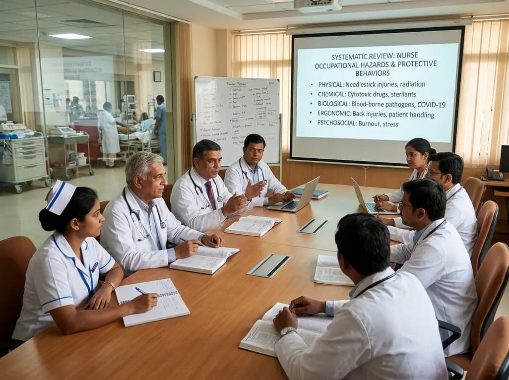
A systematic review of 56 studies classifies hospital nurse occupational hazards into physical, chemical, biological, ergonomic, and psychosocial domains. It outlines crucial protective behaviors and institutional guidelines needed to ensure clinician safety, operational resilience, and optimal patient care.
Today
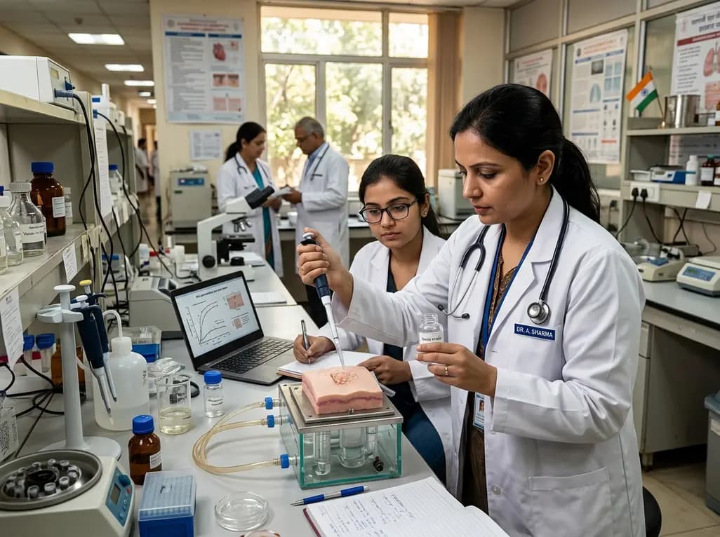
A novel high-permeation vesicle-in-gel platform enhances transdermal fulvestrant delivery, achieving sustained release, robust intradermal retention, and superior antitumor efficacy in breast cancer models while circumventing the drawbacks of painful intramuscular depot injections.
Today
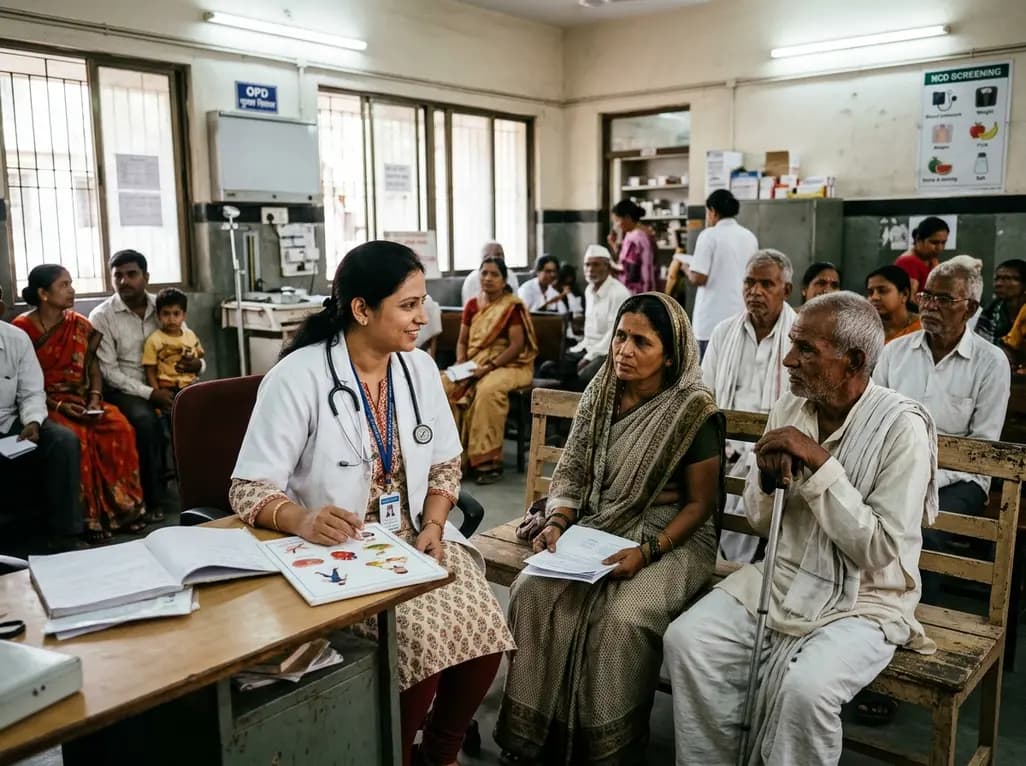
A landmark ten-year WHO report reveals how socioeconomic status, geography, and gender disparities profoundly aggravate chronic disease risks, diagnosis delays, and therapeutic outcomes globally. Discover actionable primary care strategies to bridge screening divides and optimize vulnerable patient management.
Today
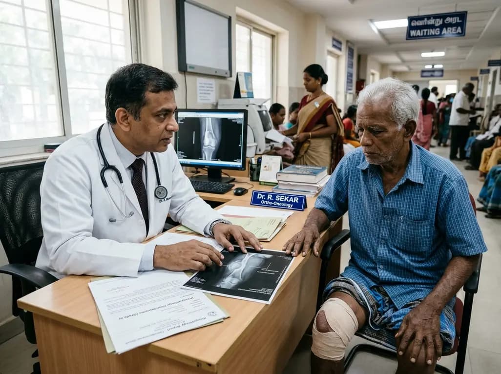
A recent single-institution analysis identifies advanced patient age and progressive systemic disease as primary negative prognostic factors for five-year survival in chondrosarcoma, underscoring the urgent necessity of precise risk stratification, clear surgical margins, and dedicated oncologic follow-up.
Today

A critical review evaluates whether starch-polyphenol complexes meet structural and metabolic criteria to qualify as type 5 resistant starch. The proposed tiered evidence framework distinguishes true inclusion complexes from simple mixtures, clarifying mechanisms for glycemic modulation and gut fermentation.
Today
