.webp&w=384&q=75)

Loading, please wait...
Loading, please wait...

Pediatric high-grade gliomas represent one of the most devastating oncological challenges in pediatric neurology and neurosurgery. These aggressive intracranial malignancies account for a substantial proportion of pediatric cancer-related deaths worldwide. Unlike adult high-grade gliomas, which feature distinct biological trajectories, childhood tumors exhibit unique epigenetic landscapes, driver mutations, and therapeutic responses. Consequently, clinicians cannot simply extrapolate adult treatment protocols or prognostic schemas to children. Traditional risk stratification still relies heavily on surgical pathology, molecular assays, and basic cross-sectional neuroimaging. However, invasive surgical biopsies carry significant neurological risks, especially when lesions sit adjacent to eloquent cortical networks. Furthermore, conventional MRI interpretation often fails to capture microscopic intratumoral heterogeneity and spatial variation in cellular density. Because therapeutic responsiveness varies widely across this patient population, oncologists urgently need non-invasive, objective risk-stratification tools. Advanced computational analytics, specifically quantitative image-based profiling, offer an unprecedented opportunity to bridge this clinical diagnostic gap. Recent investigative efforts have therefore focused on high-throughput quantitative imaging to decode hidden physiological markers embedded within routine brain imaging sequences.
To address this diagnostic challenge, a collaborative multi-center investigation evaluated whether MRI radiomics could reliably predict overall survival. Researchers gathered clinical and imaging data from 77 pediatric patients across five academic medical centers. The cohort presented with treatment-naïve, non-midline hemispheric tumors and a mean age of 140 months. Before initiating therapy, all subjects underwent standardized brain imaging, including axial gadolinium-enhanced T1-weighted and T2-weighted MRI sequences. Subsequently, investigators extracted 1,800 quantitative radiomic features from each tumor volume. Crucially, the analytical pipeline complied with Image Biomarker Standardisation Initiative (IBSI) benchmarks, ensuring strict mathematical reproducibility across diverse institutional scanners. These high-dimensional variables captured complex tumor geometry, voxel intensity distributions, and fine microstructural texture patterns. Furthermore, standardized feature extraction minimized inter-scanner variability and prevented computational artifacts from biasing downstream prognostic modeling. By examining both enhancing and non-enhancing tumor compartments simultaneously, the multi-parametric approach comprehensively characterized spatial biological diversity. Therefore, this rigorous standardization protocol established a robust, generalizable foundation for computational risk assessment across multiple academic pediatric care facilities.
Following extensive data extraction, the investigators applied machine-learning techniques to determine meaningful prognostic patterns. They utilized k-fold cross-validation alongside multivariable Cox proportional hazards regression to prevent algorithmic overfitting. This statistical modeling pipeline isolated the most predictive imaging features associated with overall patient survival. Subsequently, researchers calculated an individualized radiomic risk score for every child by weighting selected features with regression coefficients. The study team then stratified the cohort into high-risk and low-risk subgroups based on the cohort median score. Notably, patients in the high-risk group demonstrated a median overall survival of only 21.7 months. In sharp contrast, children in the low-risk category achieved a median overall survival of 44.6 months. Statistical analysis confirmed this survival disparity was highly significant, yielding a hazard ratio of 2.42 and a log-rank P-value of 0.007. Additionally, the standalone radiomics model demonstrated a concordance index of 0.75, substantially outperforming clinical variables alone. These compelling mathematical outcomes confirm that computational neuroimaging effectively disentangles clinical heterogeneity in aggressive pediatric brain tumors.
While radiomic features alone provided robust risk stratification, combining them with demographic variables yielded even greater predictive accuracy. Clinical parameters such as patient age and biological sex produced an isolated concordance index of only 0.61. However, integrating these baseline clinical characteristics with MRI-derived radiomic markers elevated the overall concordance index to 0.78. This integrated model demonstrated superior prognostic discrimination compared to either data stream evaluated independently. Specifically, multivariable synthesis allowed the computational model to calibrate tumor image phenotypes against age-dependent developmental biology. Because young children display varying intracranial anatomy and baseline tissue water content, combining clinical age with texture metrics mitigates imaging interpretation bias. Moreover, this combined scoring system provides treating clinicians with a unified, objective risk estimate at the time of initial presentation. Pediatric oncologists can immediately incorporate these multi-dimensional scores into multidisciplinary tumor board deliberations. As a consequence, medical teams can pinpoint patients requiring intensified therapeutic regimens while sparing lower-risk children from excessive treatment-related morbidity. Thus, multimodal integration represents a decisive step toward truly individualized neuro-oncological management.
The successful clinical implementation of standardized MRI radiomics could revolutionize pediatric neuro-oncology workflows in tertiary referral centers. Currently, obtaining tissue biopsies from hemispheric tumors involves surgical risks, and tissue samples may fail to capture total tumor diversity. In contrast, non-invasive computational phenotyping evaluates the entire three-dimensional tumor architecture simultaneously. Clinicians can potentially apply these quantitative risk metrics to guide surgical resection margins and plan targeted radiotherapy volumes. Furthermore, radiomic scoring may substantially refine clinical trial design by facilitating balanced patient stratification. By identifying high-risk individuals early, trial coordinators can enroll poor-prognosis cohorts into innovative targeted therapies or immunotherapy trials. Nevertheless, widespread clinical adoption requires prospective validation across diverse healthcare systems, including resource-limited hospital settings. Future studies must also evaluate how radiomic profiles correlate with underlying molecular mutations, such as histone alterations and kinase fusions. Ultimately, embedding automated, IBSI-compliant radiomic software into routine hospital picture archiving systems will empower neuro-oncologists to make faster, data-driven decisions that improve pediatric survival.
Radiomics extracts hundreds of standardized quantitative metrics from conventional MRI scans that remain imperceptible to human visual inspection. By quantifying microstructural texture, tumor heterogeneity, and spatial geometry, radiomic models capture biological aggression. When combined with clinical factors, these computational features accurately stratify patients into distinct overall survival categories.
The multi-center study utilized treatment-naïve axial gadolinium-enhanced T1-weighted and axial T2-weighted MRI brain sequences. Investigators extracted 1,800 IBSI-compliant radiomic features across these multiparametric scans. This imaging combination enabled the algorithm to comprehensively evaluate both active vascularized tumor components and surrounding edema or non-enhancing infiltration.
Radiomics currently serves as an adjunctive prognostic biomarker rather than an outright replacement for surgical biopsy. While computational MRI successfully stratifies survival risk and quantifies tumor phenotype non-invasively, definitive diagnosis still requires histopathological and molecular characterization. However, radiomics guides biopsy site selection and assists surgical planning.
Disclaimer: This content is for informational and educational purposes only and is not intended as medical advice. It should not be used as a substitute for professional medical advice, diagnosis, or treatment. Always seek the advice of your physician or other qualified healthcare provider with any questions you may have regarding a medical condition. Never disregard professional medical advice or delay in seeking it because of something you have read here. The authors and publishers are not responsible for any adverse effects or consequences resulting from the use of any suggestions, preparations, or procedures discussed in this article. Refer to the latest local and national guidelines for clinical practice.
References

Read summarized clinical updates, watch expert medical content, and earn CME certifications right from your smartphone.
Open your smartphone camera to scan and install.





A multi-center study demonstrates that MRI-derived radiomics, when combined with clinical data, significantly predicts overall survival in pediatric patients with non-midline high-grade gliomas, offering a promising non-invasive tool for individual risk stratification.
Today
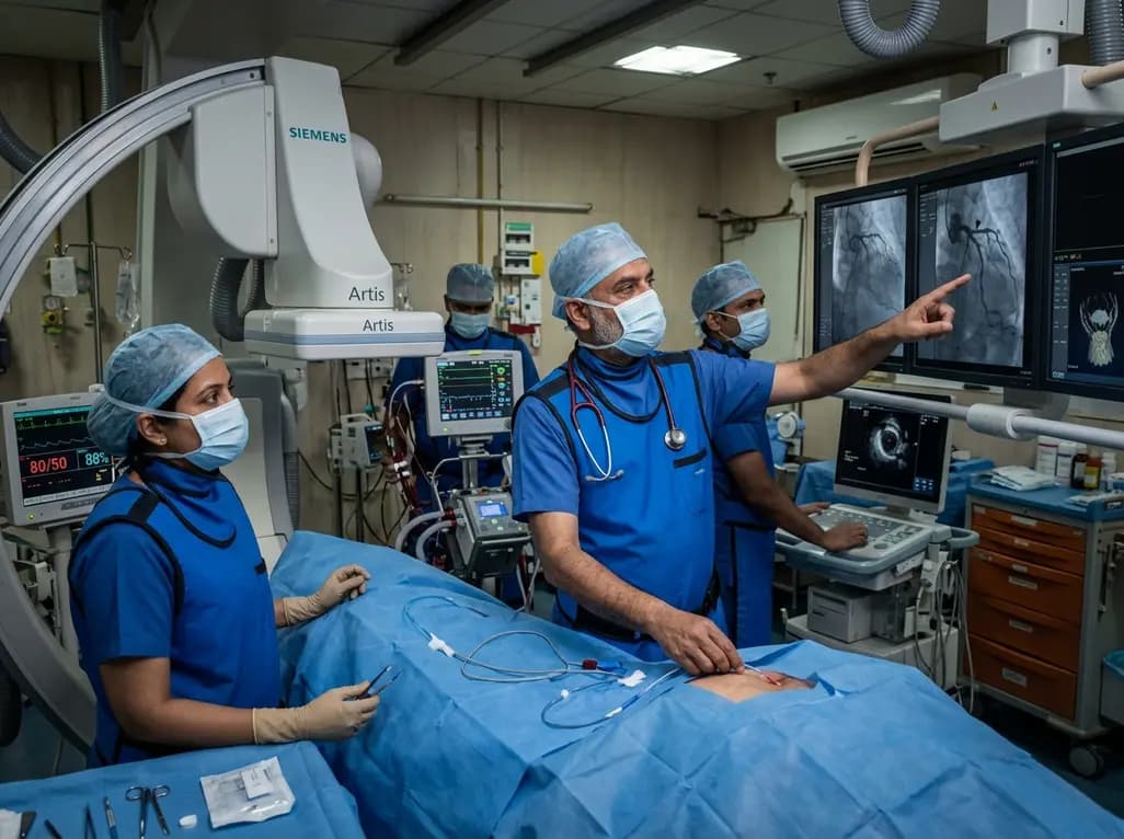
A rare case of acute coronary obstruction during TAVR caused by suspected calcified amorphous tumor embolization underscores the critical role of intracardiac imaging, emergency ECMO, and the paradox of enlarged-aperture valve geometry.
Today
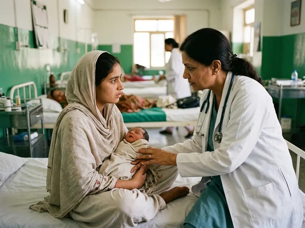
Prelacteal feeding persists in South Asian communities despite known clinical risks. This review examines determinants from Chitral, pathophysiological hazards, and pediatric strategies to eliminate harmful feeds and support exclusive breastfeeding.
Today
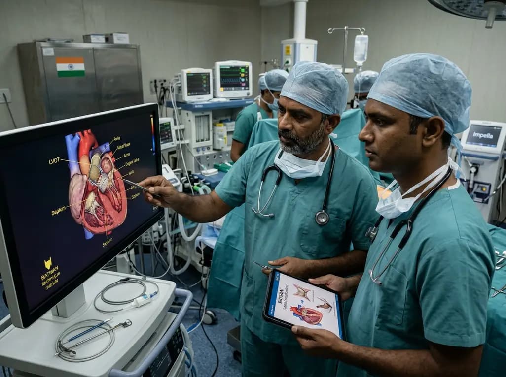
Transcatheter mitral valve replacement in patients with degenerated bioprostheses carries a substantial risk of LVOT obstruction. Combining leaflet modification via the BATMAN technique with mechanical circulatory support offers a viable, life-saving solution for anatomically challenging surgical candidates.
Today

A comparative cohort study demonstrates that patients undergoing total hip arthroplasty after prior hip arthroscopy achieve comparable functional recovery, low complication rates, and equivalent implant survivorship when conversion occurs after six months.
Today
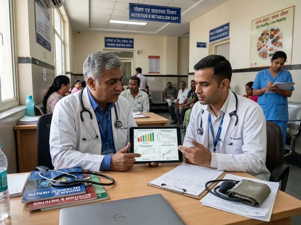
A cross-sectional study of 320 adults evaluated the link between habitual fasting duration and dietary quality using Nutrition Quotient scores, highlighting the clinical necessity of balancing time-restricted eating with nutrient density and meal quality in routine metabolic practice.
Today
