

Loading, please wait...
Loading, please wait...

Glioblastoma remains the most aggressive primary malignant brain tumor in adults. Standard post-operative management combines maximal safe surgical resection with concurrent chemoradiotherapy and adjuvant temozolomide. Historically, radiation oncologists relied solely on post-operative or pre-treatment planning magnetic resonance imaging scans to delineate radiation fields. However, these static treatment plans fail to account for continuous anatomical alterations occurring throughout the six-week therapeutic regimen. Consequently, clinicians frequently deliver high radiation doses to regions where tissue geometry has fundamentally changed. The integration of MR-guided glioblastoma radiotherapy provides a transformative solution to this longstanding clinical hurdle. Advanced hybrid magnetic resonance linear accelerator systems enable serial on-table visualization with exceptional soft-tissue resolution. Therefore, medical teams can now track daily and weekly structural shifts directly on the treatment couch. Recent clinical evidence demonstrates that both macroscopic residual disease and post-surgical cavities undergo dramatic geometric variations during fractionated radiotherapy. Understanding these continuous interfractional dynamics is critical for neuro-oncologists and radiation oncologists seeking to optimize targeting precision. By monitoring interfraction alterations, clinicians can effectively distinguish true disease evolution from transient post-surgical shifts. Ultimately, real-time visualization opens unprecedented opportunities to refine margins and enhance therapeutic ratios.
Recent prospective evaluations utilizing a 0.35T magnetic resonance workflow have quantified these dynamic anatomical shifts. In a landmark preliminary investigation, researchers followed patients undergoing concurrent chemoradiation with weekly post-contrast magnetic resonance imaging. The investigators delineated target volumes rigorously across two critical parameters. First, they mapped T2 fluid-attenuated inversion recovery sequences to define the broader hyperintense tissue envelope. Second, they evaluated gadolinium-enhanced T1-weighted sequences to capture active, contrast-enhancing macroscopic disease. Across serial imaging assessments, the investigators discovered marked volumetric contractions over the standard radiation course. Specifically, the median volume of the broader T2-defined regions decreased by forty percent across the patient cohort. Similarly, contrast-enhancing tumor volumes showed a median reduction of thirty-seven percent. These substantial reductions demonstrate that brain tumors rarely maintain fixed geometric dimensions during concurrent chemoradiotherapy. In contrast to historical assumptions of static target contours, tumor boundaries shift continuously in multiple spatial planes. Therefore, delivering an entire therapeutic course based solely on a single pre-treatment planning scan introduces significant geometric inaccuracy. Recognizing these progressive dimensional shifts helps radiation teams understand how tumor beds deform under daily therapeutic stress.
A particularly vital observation from dynamic neuroimaging studies concerns the timing of tissue alterations. The investigators demonstrated that the bulk of volumetric changes occurs early during treatment. In fact, significant contractions manifested within the first three weeks of the six-week radiation protocol. For instance, initial weekly scans revealed an immediate median reduction of thirty-two percent in peritumoral edema and non-enhancing margins. Contrast-enhancing volumes simultaneously decreased by nearly eighteen percent after merely five treatment fractions. Furthermore, post-surgical resection cavities exhibited rapid mechanical collapse during this early therapeutic window. Fluid drainage, resolving edema, and mechanical re-expansion of surrounding brain parenchyma drive these dramatic structural shifts. Consequently, the surgical bed shrinks substantially while clinicians are still administering initial phase radiation fractions. When surgical cavities contract rapidly, normal brain parenchyma naturally migrates into the high-dose radiation target space. Static planning margins fail to protect this encroaching functional brain tissue from incidental high-dose radiation exposure. Thus, early target tracking becomes imperative to document exactly when geometric shrinkage stabilizes. Identifying these early kinetic trends allows radiation oncology teams to time potential adaptive plan adjustments effectively.
The documentation of continuous anatomical shifting provides a compelling biological rationale for online adaptive radiotherapy. Historically, radiation oncologists applied wide planning target volume margins around resection cavities to account for setup inaccuracies. While wide margins ensure tumor coverage, they inadvertently subject considerable volumes of healthy neuroanatomy to destructive radiation doses. However, weekly magnetic resonance guidance enables clinicians to redefine gross tumor volumes and clinical target volumes dynamically. When significant cavity collapse or tumor regression occurs, the radiation team can re-optimize dose distributions on the treatment console. Consequently, adaptive re-planning eliminates geometric target misses while actively pruning unnecessary irradiated margins. Moreover, serial low-field magnetic resonance guidance delivers superior soft-tissue visualization without exposing patients to repeated diagnostic computed tomography radiation. Physicians can precisely verify whether tissue contraction reflects true therapeutic response or pseudoprogression. In addition, adaptive strategies ensure that prescription doses remain tightly focused on viable malignant clonogens. Tailoring radiation portals to shrinking targets allows teams to escalate local tumor control while preserving uninvolved neural networks. These strategic adaptations represent a fundamental evolution in contemporary neuro-oncology.
Preserving neurocognitive function represents a primary clinical priority for patients facing high-grade gliomas. Radiation exposure to critical intracranial organs at risk directly correlates with subsequent neurocognitive decline. Specifically, high radiation doses delivered to the bilateral hippocampi, corpus callosum, and frontal subcortical white matter tracts impair memory and executive processing. When a post-surgical resection cavity collapses, healthy functional tissue often displaces into regions originally encompassed by high-dose isodose lines. In conventional static treatments, this adjacent normal parenchyma receives inadvertent, full-dose radiation exposure. In contrast, adaptive replanning accommodates cavity shrinkage and actively pulls radiation lines away from adjacent brain structures. Simulated replanning investigations show substantial dose reductions to uninvolved hippocampi and global brain parenchyma when adjusting plans weekly. Furthermore, reducing collateral neurotoxicity decreases chronic fatigue, improves functional independence, and supports patient quality of life. Indian oncology centers adopting high-precision radiotherapy platforms can leverage these principles to minimize late treatment toxicities. Therefore, monitoring interfractional geometry directly translates into tangible functional protection for long-term brain tumor survivors.
Integrating adaptive magnetic resonance workflows into routine clinical oncology demands rigorous coordination among multidisciplinary teams. Radiation oncologists, medical physicists, and radiation therapists must collaborate closely to ensure rapid plan generation and quality assurance. Modern hybrid linear accelerators feature specialized on-table workflows, including adapt-to-shape and adapt-to-position protocols. However, daily online re-contouring of complex intracranial boundaries requires substantial physician time and computational resources. To overcome these logistical barriers, emerging artificial intelligence algorithms and automated deformable image registration tools offer promising solutions. These automated platforms can accelerate target contouring and streamline online verification workflows. Additionally, ongoing clinical trials are evaluating whether routine target reduction affects local progression-free survival. Clinicians must balance the benefits of normal tissue sparing against the theoretical risk of marginal recurrence from microscopic infiltrating disease. Prospective multicenter studies will help define standardized consensus guidelines regarding exact trigger points for plan re-optimization. As adaptive technology becomes more widely accessible across advanced cancer centers, individualized image-guided radiation will become standard neuro-oncological practice.
MR-guided glioblastoma radiotherapy utilizes onboard magnetic resonance imaging to capture high-contrast soft-tissue scans before each treatment fraction. This real-time imaging allows clinicians to visualize structural shifts, identify resection cavity collapse, and track tumor volume contractions accurately, enabling timely adaptive planning without extra radiation exposure.
Resection cavities shrink rapidly during early chemoradiotherapy because post-operative edema resolves and trapped seroma fluids drain. Additionally, healthy surrounding brain parenchyma expands back into the surgical void. Consequently, the cavity volume contracts significantly within the first three weeks, altering anatomical relationships established during initial treatment planning.
Yes, adaptive re-planning protects critical neurocognitive structures. When clinicians re-contour shrinking cavities and adjusting margins during therapy, they pull high-dose radiation contours away from expanding normal brain tissue. Therefore, adaptive plans substantially reduce accidental radiation delivery to the hippocampi and cortex, preserving patient memory and executive function.
Disclaimer: This content is for informational and educational purposes only... Refer to the latest local and national guidelines for clinical practice.
References
La Rosa A et al. Temporospatial tumor dynamic changes in glioblastoma during radiotherapy. J Neurooncol. 2025 Sep. doi: 10.1007/s11060-025-05060-7. PMID: 40299247.
Cullison K et al. Dynamics of Daily Glioblastoma Evolution During Chemoradiotherapy on the 0.35T MRI-Linear Accelerator. Int J Radiat Oncol Biol Phys. 2024. doi: 10.1016/j.ijrobp.2024.09.028. PMID: 39357789.
Kretzschmar L et al. Early target volume dynamics during MR-guided radiotherapy for glioblastoma: first results from the prospective MARGA-I trial. Radiother Oncol. 2026. doi: 10.1016/radonc.2026.111792. PMID: 42830173.

Read summarized clinical updates, watch expert medical content, and earn CME certifications right from your smartphone.
Open your smartphone camera to scan and install.





A prospective study evaluates temporospatial tumor dynamics in glioblastoma using 0.35T MR-guided radiotherapy. Findings reveal rapid tumor and surgical cavity shrinkage within the first three weeks of treatment, emphasizing the clinical value of adaptive planning to spare healthy neuroanatomy.
Today
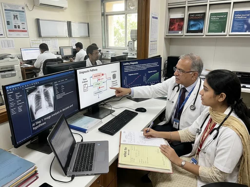
A breakthrough peer-reviewed study demonstrates that a sequential AI diagnosis system can mirror real-world clinical reasoning. By ordering stepwise tests and updating hypotheses dynamically, this multi-agent orchestrator achieved superior diagnostic accuracy while reducing investigative costs.
Today

A prenatal case highlights severe glutaric acidemia type II presenting as bilateral hyperechoic cystic kidneys at 18 weeks. Whole-exome sequencing revealed a novel homozygous ETFDH frameshift variant, demonstrating the power of rapid genetic testing in fetal renal anomalies.
Today

Review how tailored exercise programs benefit frail older patients with heart failure. Explore pathophysiological mechanisms, individualized training prescriptions, and strategies to overcome adherence barriers in clinical practice.
Yesterday

This comparative study evaluates posterior labral tears with and without glenohumeral instability, highlighting distinct demographic patterns, MRI underestimation in stable shoulders, and successful arthroscopic repair outcomes.
Yesterday

A clinical study demonstrates that early intervention with tocilizumab, either as monotherapy or bridged with corticosteroids, achieves significant proptosis reduction and resolves active inflammation in treatment-naive thyroid eye disease.
Yesterday
