

Loading, please wait...
Loading, please wait...

Recent neuroimaging research provides paradigm-shifting insights into the pathophysiology of primary trigeminal neuralgia. Clinicians historically conceptualized this debilitating facial pain condition as an isolated peripheral disorder driven primarily by neurovascular conflict at the root entry zone. However, advanced diffusion tractography now demonstrates that the microstructural changes extend far beyond the peripheral sensory branches. Investigators have uncovered widespread alterations in central pathways, specifically implicating motor-related subcortical networks. Consequently, these findings challenge conventional views and encourage clinicians to consider complex central reorganization when assessing chronic neuropathic facial pain presentations.
Researchers recruited 57 prospective patients with classical primary trigeminal neuralgia alongside 29 matched healthy control subjects. The study evaluated microstructural integrity across 72 distinct white matter bundles. To achieve precise reconstruction in crossing fiber regions, the authors employed constrained spherical deconvolution combined with fixel-based tractography. Permutation testing with false discovery rate correction revealed significant fractional anisotropy reductions across six specific white matter pathways in affected patients. Remarkably, all six altered tracts belong directly to motor-associated corticofugal and striato-cortical projection systems. These damaged tracts included the right fronto-pontine tract and the left corticospinal tract. Furthermore, four distinct striatal pathways showed marked compromise, including the right striato-prefrontal, right striato-fronto-orbital, right striato-occipital, and left striato-parietal tracts. Therefore, central architectural degradation prominently targets motor control networks rather than remaining restricted to trigeminal sensory axons.
Historically, traditional diffusion tensor imaging faced severe limitations in delineating complex cerebral fiber architectures. Standard tensor models often fail in regions where multiple nerve bundles cross or interdigitate within a single voxel. Consequently, earlier studies struggled to identify subtle white matter alterations beyond the cisternal trigeminal root. In contrast, modern constrained spherical deconvolution successfully disentangles overlapping orientations. By coupling this advanced technique with fixel-based quantitative metrics, the investigators isolated bundle-specific microstructural features with high fidelity. In addition, permutation testing protected the dataset against false-positive statistical discoveries across extensive brain regions. Thus, these analytical innovations explain why earlier imaging trials frequently overlooked motor system involvement. Advanced neuroimaging now offers actionable clarity regarding the systemic cerebral impact of repetitive paroxysmal pain.
Intriguingly, the study evaluated whether these microstructural abnormalities correlated with clinical pain duration or visual analog scores. The secondary analyses revealed no statistically significant correlations between fractional anisotropy reductions and symptom chronicity or severity after multiple testing corrections. Similarly, the presence or anatomical grade of neurovascular conflict did not predict the magnitude of tract disruption. These observations indicate that central motor tract alterations may develop rapidly following clinical onset. Alternatively, pre-existing subcortical connectivity variations might predispose specific individuals to persistent neuropathic syndromes. Moreover, the lack of linear correlation with pain duration suggests that central motor network degradation represents an early, stabilized structural shift. Clinicians must therefore understand that severe structural remodeling can exist even in patients presenting with relatively short disease histories.
Why do motor-related and striato-cortical pathways exhibit selective vulnerability in a facial sensory pain syndrome? Chronic paroxysmal pain triggers continuous antalgic motor suppression, facial guarding, and involuntary grimacing. Over time, these sustained motor behaviors drive maladaptive neuroplasticity throughout corticostriatal and corticofugal loops. Furthermore, the basal ganglia play critical, well-documented roles in sensory gating and pain modulation. Striatal dopamine dysregulation frequently accompanies sustained neuropathic firing, disrupting normal striato-cortical communication. Consequently, repetitive sensory hyperactivation appears to overburden interconnected subcortical motor hubs. This bidirectional relationship between sensory pain inputs and motor circuits creates widespread microstructural remodeling. Therefore, trigeminal neuralgia generates profound systemic reverberations throughout the motor architecture of the human brain.
These neuroimaging insights provide substantial value for neurosurgeons and pain specialists managing refractory facial pain. For decades, surgical microvascular decompression focused almost entirely on the peripheral interface between compressing vessels and the fifth cranial nerve. While mechanical decompression remains the gold-standard intervention, variable surgical outcomes and delayed recurrences often perplex surgical teams. Disrupted motor and striatal tracts could clarify persistent sensory-motor abnormalities or dysesthesias that survive successful peripheral neurovascular decompression. In addition, recognizing central motor network remodeling highlights potential therapeutic roles for neuromodulation. Modulating motor cortex excitability through transcranial magnetic stimulation or deep brain targeting may help normalize disrupted corticostriatal loops. Ultimately, adopting a network-level perspective will refine patient stratification, improve preoperative counseling, and encourage innovative multimodal therapeutic protocols.
Chronic trigeminal pain triggers extensive maladaptive neuroplasticity across subcortical circuits. Continuous facial grimacing, sensory overactivation, and altered basal ganglia pain gating disrupt structural connectivity. Consequently, microstructural degradation develops across striato-cortical and corticofugal projection pathways, leading to measurable fractional anisotropy reductions in motor-related white matter tracts.
Conventional diffusion tensor imaging cannot resolve regions containing crossing nerve fibers within single voxels. In contrast, constrained spherical deconvolution accurately separates complex multi-directional axonal pathways. This methodological precision allowed researchers to reconstruct 72 separate white matter tracts and identify subtle subcortical disruptions that standard imaging protocols missed.
Current clinical evidence has not yet established whether microstructural motor tract alterations fully reverse following surgical decompression. However, ongoing longitudinal imaging investigations are evaluating postoperative tract recovery. Persistent central remodeling may explain why certain patients experience residual discomfort or delayed facial pain recurrence despite complete peripheral vascular alleviation.
Disclaimer: This content is for informational and educational purposes only and should not be considered medical advice or substituted for professional clinical judgment. Diagnostic criteria, imaging protocols, and treatment decisions should be individualized to each patient. Refer to the latest local and national guidelines for clinical practice.
References
Pashkov A et al. Motor-related white matter tracts are prominently affected in primary trigeminal neuralgia. Clin Neurol Neurosurg. 2026 Nov. doi: 10.1016/j.clineuro.2026.109601. PMID: 42551242.
Cruccu G, Di Stefano G, Truini A. Trigeminal Neuralgia. N Engl J Med. 2020;383(8):754-762. doi: 10.1056/NEJMra1914484.
Bendtsen L, Zakrzewska JM, Abbott J, et al. European Academy of Neurology guideline on trigeminal neuralgia. Eur J Neurol. 2019;26(6):831-849. doi: 10.1111/ene.13950.

Read summarized clinical updates, watch expert medical content, and earn CME certifications right from your smartphone.
Open your smartphone camera to scan and install.





Advanced diffusion tractography demonstrates that primary trigeminal neuralgia involves significant microstructural disruption across motor-related corticofugal and striatal white matter tracts, expanding our understanding beyond sensory root compression.
Today

Standardizing continuous physiological waveforms in EHRs has long challenged clinical teams. The novel TIDE framework leverages HL7 FHIR R4 to decouple queryable clinical metadata from massive raw signal repositories, unlocking scalable telemetry exchange for bedside care and predictive research.
Today

A secondary analysis of the MOMS clinical trial reveals that infant sex does not influence neonatal opioid withdrawal syndrome severity in buprenorphine-exposed newborns. Maternal co-exposures and clinical factors drive outcomes, highlighting the need for objective assessment and supportive breastfeeding.
Today
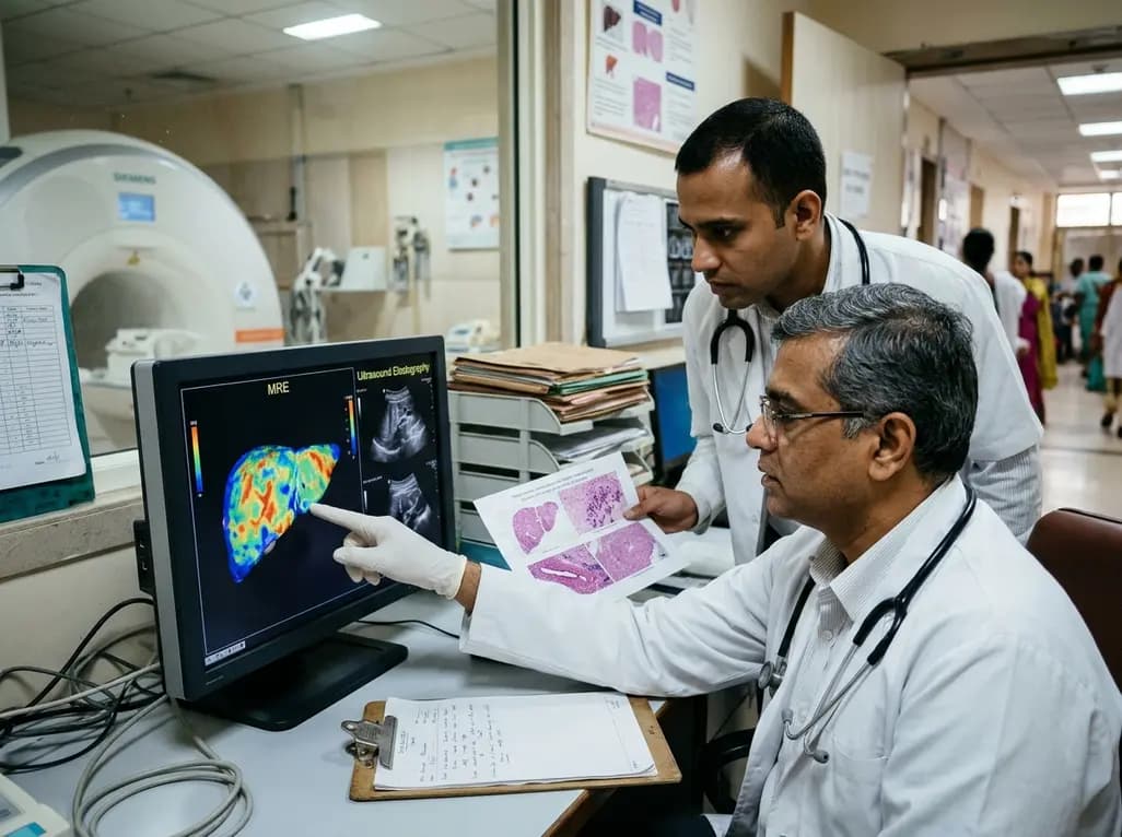
A comparative study reveals magnetic resonance elastography outperforms ultrasound elastography in assessing liver fibrosis in Fontan-associated liver disease, demonstrating stronger histological agreement and superior discrimination.
Today

A systematic review and meta-analysis highlights the substantial impact of plyometric jump training on jump performance, reactive strength, and landing kinematics in female team-sport athletes, providing evidence-based programming guidelines for sports medicine and orthopedics.
Today

A comprehensive review of the role of growth hormone in diabetic kidney disease and podocytopathy. Learn how elevated GH activates ZEB2, Notch, and TGF-beta1, driving podocyte loss and glomerulosclerosis, and explore novel targeted therapeutic approaches.
Today
