.webp&w=384&q=75)

Loading, please wait...
Loading, please wait...
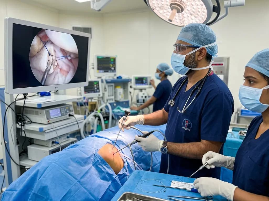
Managing an acute anterior glenoid rim fracture represents a significant challenge in modern orthopedic sports surgery. These intra-articular injuries frequently accompany traumatic shoulder dislocations when the humeral head forcibly impacts the anterior glenoid margin. Consequently, achieving anatomic restoration is crucial to prevent recurrent instability and progressive degenerative arthritis. Historically, surgeons relied on open surgical approaches to obtain direct reduction. However, open procedures carry considerable morbidity, including soft tissue trauma, subscapularis compromise, and prolonged postoperative stiffness. Therefore, minimally invasive arthroscopic techniques have become increasingly popular. Arthroscopy offers superior joint visualization and minimizes surgical morbidity. Nevertheless, balancing construct rigidity with fracture biology remains demanding. As repair strategies evolve, tissue-preserving techniques are gaining substantial momentum.
Fractures of the anterior glenoid rim compromise the vital osseous buttress preventing anterior humeral translation. When a displaced fragment exceeds fifteen percent of glenoid width, the shoulder becomes biomechanically incompetent. Furthermore, dynamic forces from the rotator cuff can perpetuate displacement of unhealed fragments. Traditional AO trauma principles advocate rigid compression screw fixation for articular reconstructions. However, applying rigid compression within the delicate glenohumeral joint poses unique obstacles. Glenoid bone fragments are frequently small, comminuted, or osteopenic. Inserting rigid metal screws into small osseous fragments can cause iatrogenic fragmentation. Moreover, screw hardware can back out or erode into articular cartilage over time. Therefore, sports surgeons question whether primary interfragmentary compression is genuinely mandatory. With appropriate postoperative immobilization, stable apposition without extreme compressive force provides sufficient mechanical security for osseous consolidation.
The balance between mechanical stability and biological preservation is central to successful fracture healing. Historically, arthroscopic strategies attempted to replicate rigid open fixation by deploying complex double-row anchor configurations. These constructs often require placing a medial row of anchors directly onto the glenoid articular face. Although these systems demonstrate robust load-to-failure strength in laboratory testing, they substantially elevate surgical morbidity. Specifically, placing medial anchors directly into the articular surface risks iatrogenic chondral damage. Moreover, drilling multiple anchor holes into fragile bone fragments compromises local vascularity. Consequently, aggressive mechanical fixation might impair fracture biology and delay union. In contrast, biology-driven techniques prioritize periosteal preservation. By utilizing tensioned high-strength sutures and low-profile implants, surgeons maintain anatomical alignment while minimizing foreign material load. Thus, respecting joint biology optimizes clinical recovery.
To eliminate the hazards associated with medial anchor placement, shoulder specialists introduced the mooring technique. This all-arthroscopic repair strategy relies on strategic suture passage to anchor the displaced bone fragment securely to the native glenoid neck. Instead of inserting hardware directly across the articular surface, the mooring technique avoids placing a medial row of anchors entirely. Consequently, it eliminates the risk of articular cartilage drilling on the glenoid face. The technique utilizes high-strength suture loops and knotless anchors placed peripherally along the rim. These sutures wrap around the fractured fragment and incorporate the robust capsulolabral sleeve. By tensioning these suture limbs, the surgeon applies continuous stabilizing forces that reduce the fragment. As a result, this construct functions similarly to a maritime mooring system, securing the fragment firmly while preserving local vascularity.
Early clinical evaluations and preliminary outcome data demonstrate encouraging results for the mooring repair method. Specifically, published series report high rates of radiographic bony union alongside remarkably low rates of recurrent anterior instability. Patients undergoing this technique experience rapid pain relief and predictable functional recovery. In addition, restoring the native bony architecture without metal screw protrusion prevents long-term hardware-related complications. Because the procedure preserves the subscapularis muscle and anterior capsule integrity, patients typically regain excellent active external rotation. In contrast, traditional open procedures often lead to internal rotation contractures. While long-term comparative clinical trials are still emerging, the mooring technique offers a compelling safety profile. Furthermore, eliminating metal screws avoids secondary implant removal procedures. Therefore, this biological fixation strategy represents a valuable surgical alternative.
Despite its notable advantages, surgeons must remain vigilant regarding potential drawbacks and patient selection criteria. One ongoing concern involves the long-term integrity of glenohumeral articular cartilage. Although the mooring technique avoids medial anchors, suture material passing near the articular margin might generate friction against the humeral head. In addition, imperfect reduction could accelerate posttraumatic chondral degeneration. Consequently, the applicability of this technique in young, high-demand collision athletes requires careful clinical judgment. High-impact contact athletes generate immense shearing forces across the anterior glenohumeral joint during athletic competition. In these high-demand individuals, suture-based soft tissue tension might theoretically carry a higher risk of late displacement. Therefore, surgeons must thoroughly evaluate fragment size, bone quality, and patient activity levels prior to surgery to ensure optimal outcomes.
Modern shoulder preservation requires a refined algorithmic approach to anterior glenoid rim injury management. Surgeons should tailor their fixation strategy to the specific morphological characteristics of the fracture. For acute, simple two-part fractures with adequate bone quality, the mooring technique provides an elegant, minimally invasive solution. It successfully marries mechanical stability with biological healing principles. Looking ahead, future innovations in bioabsorbable materials and specialized suture delivery systems will refine these arthroscopic techniques further. In addition, prospective multicenter registries are needed to evaluate long-term chondral outcomes and athletic return-to-play rates. Ultimately, the ideal surgical intervention is not always the most rigid construct. Instead, an optimal repair provides sufficient stability that respects joint biology and allows natural bone consolidation to flourish.
The primary advantage is the significant reduction in surgical morbidity and hardware-related complications. By utilizing high-strength sutures and peripheral anchors, this technique completely avoids placing rigid metal screws or medial anchors into the articular surface. Consequently, it minimizes iatrogenic cartilage injury, preserves local fragment vascularity, and prevents secondary hardware impingement. This biological approach facilitates reliable bone union while maintaining joint stability and allowing faster postoperative recovery.
Ideal candidates include active patients presenting with acute, displaced intra-articular anterior glenoid rim fractures where fragment size is sufficient for suture reduction. Patients who require joint preservation without extensive open exposure benefit greatly from this all-arthroscopic approach. However, high-demand collision athletes with severe bone loss or multi-fragmentary comminution may require more rigid constructs or bony augmentation procedures to withstand extreme biomechanical loads during contact sports.
Postoperative rehabilitation emphasizes early protected immobilization to facilitate biological bone-to-bone consolidation without introducing premature shear stresses. Patients typically wear a sling for four to six weeks while performing supervised passive range-of-motion exercises. Once radiographic evidence confirms early healing, active-assisted mobility and progressive rotator cuff strengthening begin. High-impact loading and collision sports are generally restricted until complete osseous union and functional muscle symmetry are fully established.
Disclaimer: This content is for informational and educational purposes only and does not constitute medical advice or substitute professional clinical evaluation. Refer to the latest local and national guidelines for clinical practice.
References
1. Tramer JS et al. Editorial Commentary: All Hands on Deck? The Mooring Technique Balances Biology and Biomechanics in Arthroscopic Anterior Glenoid Rim Fracture Repair. Arthroscopy. 2026 Aug 20. doi: 10.1002/arj.70473. PMID: 42624789.
2. Millett PJ, Braun S. Arthroscopic Fixation of Glenoid Rim Fractures After Reduction by Labral Repair. Arthrosc Tech. 2020;9(4):e515-e520.
3. Tauber M, Moursy M, Eppel M, Koller H, Resch H. Arthroscopic screw fixation of large anterior glenoid rim fractures. Knee Surg Sports Traumatol Arthrosc. 2008;16(3):326-332.
4. Spiegl UJ, Frosch KH, Voigt C. Arthroscopic treatment of a multifragmentary glenoid fracture. Oper Orthop Traumatol. 2021;33(2):167-176.

Read summarized clinical updates, watch expert medical content, and earn CME certifications right from your smartphone.
Open your smartphone camera to scan and install.





The mooring technique offers an innovative arthroscopic approach for anterior glenoid rim fracture repair, eliminating medial anchors to preserve cartilage and enhance biological healing while restoring glenohumeral stability.
last month
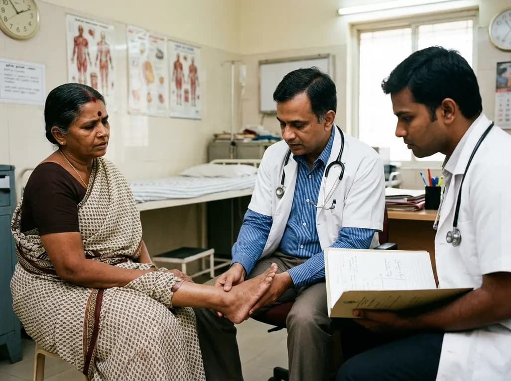
Discover how to identify superimposed entrapment neuropathy in diabetic patients. This review details clinical evaluation, surgical decompression evidence, and a structured algorithm for primary care referrals to prevent ulcers and preserve limb function.
Today

A retrospective cohort study evaluated 1,000 low-risk singleton births to assess maternal and neonatal outcomes during office-hours versus off-hours. Off-hours deliveries were significantly associated with higher odds of intrapartum cesarean sections and episiotomies, highlighting critical obstetric staffing considerations.
Today
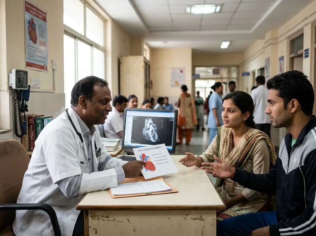
New clinical evidence demonstrates a shift away from strict exercise restrictions in genetic heart disease. While patients with channelopathies engage in more vigorous activity than those with cardiomyopathies, shared decision-making and individualized risk assessments ensure safe, beneficial exercise participation.
Today

Researchers have engineered an artificial sensory-pain integrated receptor using a multi-threshold organic synaptic transistor. The system distinguishes benign touch from noxious stimuli, enabling safer, more responsive prosthetic limbs with built-in memory and simplified hardware architecture.
Today

Medical robotics is transitioning beyond operative suites into diagnostics, cardiology, and endocrinology. Explore how innovations in tele-robotics, AI integration, and nanomedicine are overcoming geographical barriers and transforming precision healthcare delivery across India's evolving disease landscape.
Today
