.webp&w=384&q=75)

Loading, please wait...
Loading, please wait...

Scientists are currently gaining new insights into the complexities of MIH root canal morphology. Specifically, Molar-incisor hypomineralization (MIH) is a prevalent developmental enamel defect. Furthermore, it frequently leads to increased dentin permeability and pulpal inflammation. Consequently, young patients often require early endodontic intervention. Therefore, understanding the internal anatomy is vital for success. In light of this, researchers recently used microcomputed tomography (µCT) to compare hypomineralized molars with healthy teeth.
Initially, the research team analyzed sixty extracted permanent molars. In addition to scanning these samples, they applied the Sert and Bayırlı classification system. Moreover, they assessed isthmus types and recorded apical foramina numbers. Additionally, the morphometric analysis focused on canal area and cervical thickness. Notably, the team used independent t-tests to ensure statistical accuracy. However, they also looked at cervical width as a key variable. As a result, the study provides a robust comparison of structural variations. Indeed, these details help clinicians navigate complex canal systems.
Furthermore, the findings suggest that structural variations in MIH teeth may require precise endodontic techniques. For example, variations in cervical thickness might impact the safety of mechanical instrumentation. Similarly, a high frequency of minor apical foramina requires meticulous irrigation protocols. In other words, the internal environment of hypomineralized teeth is often more complex than expected. Therefore, clinicians should adapt their strategies based on these anatomical insights. Overall, understanding MIH root canal morphology ensures better long-term outcomes for pediatric patients. Subsequently, further studies will likely refine these anatomical models.
MIH is a developmental defect of enamel affecting one or more permanent molars, often resulting in sensitivity and higher caries risk.
Micro-CT provides non-destructive, high-resolution 3D images, allowing researchers to see tiny accessory canals and internal variations that traditional X-rays miss.
Detailed anatomical knowledge helps clinicians plan precise instrumentation and irrigation, which is crucial due to the increased inflammation risk in MIH teeth.
Disclaimer: This content is for informational and educational purposes only and does not constitute medical advice. Refer to the latest local and national guidelines for clinical practice.
References
Karslıoğlu H et al. Microcomputed tomography analysis of root canal morfology of hypomineralized permanent molars compared with healthy teeth: unveiling hidden anatomical variations. BMC Oral Health. 2026 Feb 17. doi: 10.1186/s12903-026-07919-1. PMID: 41703541.
Özükoç C. Examination of root canal morphology of teeth affected by Molar Incisor Hypomineralization (MIH): Frequency of accessory canals. Int Dent Res. 2021;11(1):12-15.
Neboda C, Anthonappa RP, King NM. Preliminary investigation of the variations in root canal morphology of hypomineralised second primary molars. Int J Paediatr Dent. 2018;28(3):310-318.

Read summarized clinical updates, watch expert medical content, and earn CME certifications right from your smartphone.
Open your smartphone camera to scan and install.





A micro-CT study compares the root canal morphology of hypomineralized permanent molars and healthy teeth to identify anatomical variations....
8 months ago
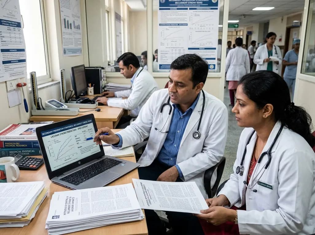
Explore vital recurrent stroke predictors in lacunar stroke patients managed with intensive blood pressure lowering. This analysis examines trial findings from SPS3 to uncover residual vascular risks, metabolic factors, and targeted secondary prevention strategies for high-risk neurological patients.
Today
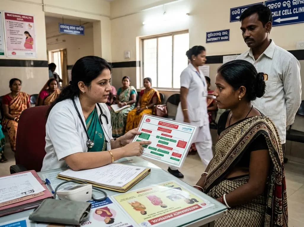
Odisha has established India's first state-level clinical protocol specifically tailored for pregnant women with sickle cell disease. Developed with NHM and UNICEF, the framework provides structured antenatal screening, crisis mitigation, standardized referral pathways, and specialized interdisciplinary care.
Today
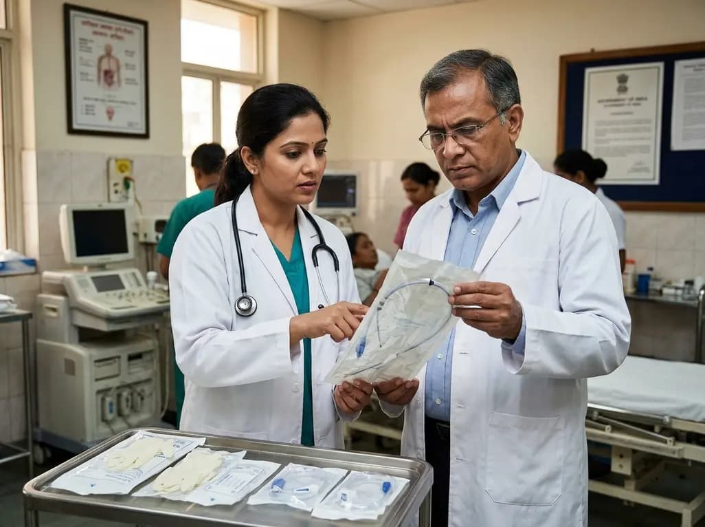
The Directorate General of Health Services directs Indian states to halt the hazardous reuse and reprocessing of single-use medical devices. Discover regulatory implications under the Drugs and Cosmetics Act, critical infection risks, legal liabilities, and operational changes required across clinical specialties.
Today

Mitochondria-targeted nanoparticles self-assembled from EGCG and L-glutamic acid effectively restore mitochondrial homeostasis, suppress synovial inflammation, and attenuate chondrocyte senescence in osteoarthritis, offering a promising disease-modifying nanotherapy.
Today

India is advancing preventive healthcare by integrating genomics, artificial intelligence, and quantum technologies. With the SANGAM programme sequencing one million Indian genomes, clinicians can unlock personalized therapies, detect cardiometabolic risks early, and curb the burden of diabetes and heart disease.
Today
