

Loading, please wait...
Loading, please wait...
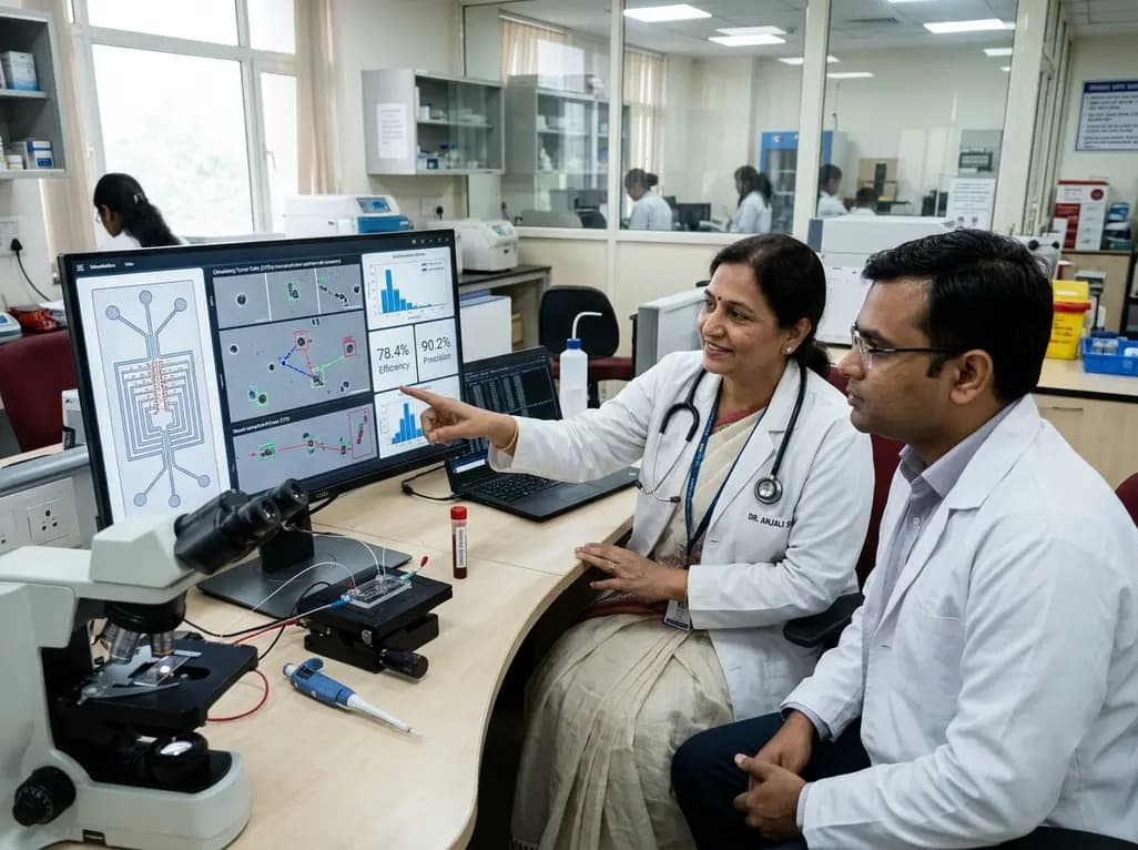
Metastatic dissemination remains the primary driver of cancer-related mortality worldwide, making early detection of disease spread a paramount oncological objective. Consequently, liquid biopsy has emerged as a revolutionary modality for real-time monitoring of therapeutic responses and disease recurrence. Within this diagnostic framework, circulating tumor cells capture provides clinicians with intact cellular substrates that reflect the evolving genomic and phenotypic landscape of primary neoplasms. However, isolating these extremely scarce cells from dense populations of peripheral blood constituents presents formidable biophysical hurdles. Standard immunoaffinity approaches frequently fail because malignant cells downregulate surface antigens during epithelial-mesenchymal transition. Therefore, biomedical researchers are turning toward label-free, biophysical separation techniques to overcome these diagnostic obstacles. A recent breakthrough platform integrates microfluidic dielectrophoresis with automated computer vision to isolate and enumerate malignant cells with remarkable precision. This innovative bioengineering paradigm establishes a practical pathway for dynamic post-treatment surveillance in routine oncology practice.
Dielectrophoresis exploits the intrinsic polarizability of biological particles subjected to non-uniform electric fields. Because malignant epithelial cells possess distinct morphological dimensions, membrane capacitances, and cytoplasmic conductivities compared to healthy leukocytes, they experience differential electrical forces. When blood suspensions traverse microfluidic channels lined with microelectrodes, high-frequency alternating current fields exert translational forces on suspended cells. Consequently, circulating malignant cells migrate toward regions of high electric field gradient, while erythrocytes and mature leukocytes continue downstream along laminar streamlines. Furthermore, this label-free methodology avoids dependence on epithelial cell adhesion molecule expression, thereby recovering heterogeneous and aggressive cell phenotypes that conventional antibody enrichment overlooks. In addition, the microscale fluid architecture minimizes shear stress, preserving cell viability for downstream transcriptomic and genomic assays. Researchers carefully regulate input frequencies to prevent thermal dissipation and cellular lysis. Thus, microfluidic platforms provide a benign yet selective microenvironment capable of segregating rare neoplastic targets from billions of normal hematologic counterparts with outstanding operational consistency.
Achieving dependable performance in biophysical cell sorting requires rigorous calibration of hydrodynamic and electrokinetic variables. Recent validation studies evaluated applied electric field strengths ranging between 12.5 and 32.5 times 10,000 volts per meter at a constant frequency of one megahertz. Simultaneously, investigators tested sample flow rates spanning from 6.25 to 31.25 millimeters per minute to evaluate hydrodynamic drag interactions. Consequently, experimental results established that an electric field strength of 25 times 10,000 volts per meter paired with a flow velocity of 18.75 millimeters per minute achieved an optimal circulating tumor cells capture efficiency of 78 percent. Elevated hydrodynamic velocities overpowered dielectrophoretic trapping forces, thereby causing target cell escape. Conversely, excessively strong electric fields increased thermal stress without enhancing separation margins. By establishing these precise electrokinetic thresholds, the microfluidic system balances processing speed with diagnostic yield. Therefore, systematic optimization of operational parameters transforms dielectrophoresis from an experimental curiosity into an efficient, reproducible platform suited for clinical processing of complex liquid biopsies.
Following electrokinetic isolation, clinicians require rapid, objective cell confirmation to eliminate operator bias. To accomplish this goal, modern microfluidic devices incorporate automated optical detection pipelines immediately following fluorescent antibody tagging. Researchers designed dedicated image processing software using the open-source OpenCV architecture to segment and classify fluorophore-labeled targets. Furthermore, the development team compared this specialized software against advanced deep learning frameworks, specifically the YOLOv8 neural network. The evaluation revealed that the OpenCV-based tool achieved an exceptional precision of 90 percent, closely mirroring the 89 percent precision recorded by YOLOv8. However, the deep learning model demonstrated superior recall, detecting 92 percent of true targets compared to 85 percent identified by traditional algorithmic thresholding. Although convolutional architectures demand greater computational resources, their heightened sensitivity minimizes false negatives in minimal residual disease assessments. Consequently, combining microfluidic sorting with machine learning algorithms creates a self-contained diagnostic loop that accelerates specimen interpretation while maintaining rigorous diagnostic fidelity.
Integrating high-efficiency cell harvesting with artificial intelligence heralds transformative changes for daily clinical oncology. Liquid biopsies enable oncologists to monitor treatment efficacy longitudinally without subjecting vulnerable patients to repeated invasive tissue biopsies. For instance, serial measurements of circulating cellular burdens provide early warning signs of systemic treatment failure months before radiographic progression manifests on computed tomography scans. Moreover, isolating viable cellular units permits functional phenotyping, such as tracking changes in hormone receptor status or assessing resistance biomarkers like human epidermal growth factor receptor 2. Pathologists can also leverage these harvested cells to cultivate patient-derived organoids or execute single-cell sequencing. Therefore, this technology empowers clinicians to tailor targeted regimens dynamically as clonal evolution occurs under selective therapeutic pressures. In addition, streamlining the enrichment pipeline significantly shortens turnaround times, allowing oncology teams to make informed decisions rapidly during critical clinical junctures.
Despite these compelling technological advantages, translating microfluidic dielectrophoresis devices into high-volume diagnostic laboratories requires overcoming practical implementation hurdles. Currently, pre-analytical variability represents a major concern, as improper phlebotomy techniques or prolonged transport intervals can trigger cell lysis and skew diagnostic results. Additionally, whole blood viscosity necessitates sample pre-dilution or red cell lysis steps, which can diminish total cellular yield. Medical centers must also establish standardized operating procedures and cross-platform quality assurance controls before deploying automated optical algorithms widely. Nevertheless, the low fabrication cost of microfluidic lab-on-a-chip cartridges offers profound economic advantages for resource-constrained health systems across India. By reducing dependence on expensive antibody cocktails and bulky flow cytometers, this integrated methodology democratizes sophisticated precision diagnostics. Consequently, collaborative initiatives bridging microengineering, computational pathology, and clinical oncology will soon drive these integrated platforms from research benchmarks into validated clinical standards.
Conventional immunoaffinity methods rely on specific antibodies targeting cell surface proteins like EpCAM. In contrast, dielectrophoresis is an active physical separation technique that utilizes intrinsic dielectric properties, membrane morphology, and cell dimensions under alternating electric fields. Consequently, dielectrophoresis isolates rare neoplastic cells regardless of phenotypic shifts, ensuring comprehensive recovery of aggressive cancer phenotypes that downregulate traditional epithelial surface markers during metastasis.
Automated image recognition eliminates subjectivity and inter-observer variability inherent to manual cytological microscopic evaluation. By coupling fluorescent labeling with advanced algorithms like OpenCV or YOLOv8, platforms instantly classify captured cells with over 90 percent precision. Furthermore, automated optical workflows process large image fields rapidly, reducing diagnostic turnaround times and preventing laboratory fatigue while analyzing scarce cellular targets within clinical samples.
Microfluidic cell capture efficiency primarily depends on the interplay between hydrodynamic drag forces and applied dielectrophoretic forces. Specifically, operators must balance sample flow velocity with electric field intensity and alternating current frequency. If the flow rate is too rapid, drag forces displace cells from traps; conversely, excessive voltage causes thermal damage to viable biological specimens.
Disclaimer: This content is for informational and educational purposes only and does not constitute medical advice, diagnosis, or treatment. Healthcare professionals should evaluate clinical decisions independently. Refer to the latest local and national guidelines for clinical practice.
References

Read summarized clinical updates, watch expert medical content, and earn CME certifications right from your smartphone.
Open your smartphone camera to scan and install.





A novel microfluidic platform combines dielectrophoresis and image recognition to capture and classify circulating tumor cells with 78% efficiency and 90% precision. This advancement highlights the growing role of automated liquid biopsy technologies in assessing minimal residual disease and therapeutic responses.
Today
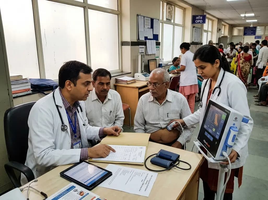
Metabolic dysfunction-associated steatotic liver disease affects nearly 70% of Indian adults with type 2 diabetes. Implementing systematic risk stratification with FIB-4 scoring, transient elastography, and patient-centric digital engagement transforms early detection into sustainable, long-term clinical care.
Today

A landmark study evaluates artificial intelligence models and prompting strategies in qualitative lupus pregnancy research, revealing that few-shot prompting in advanced LLMs reliably captures maternal experiences, clinical anxieties, and perinatal outcomes across diverse languages.
Today
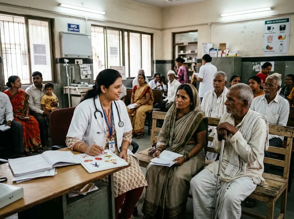
A landmark ten-year WHO report reveals how socioeconomic status, geography, and gender disparities profoundly aggravate chronic disease risks, diagnosis delays, and therapeutic outcomes globally. Discover actionable primary care strategies to bridge screening divides and optimize vulnerable patient management.
Today
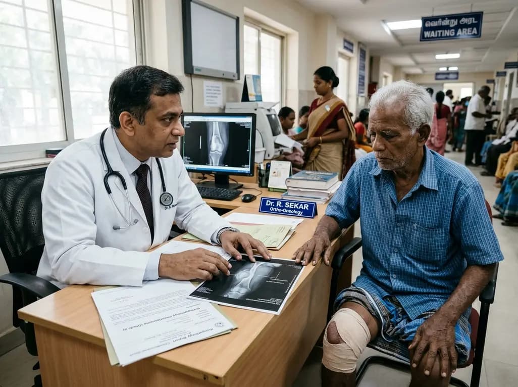
A recent single-institution analysis identifies advanced patient age and progressive systemic disease as primary negative prognostic factors for five-year survival in chondrosarcoma, underscoring the urgent necessity of precise risk stratification, clear surgical margins, and dedicated oncologic follow-up.
Today
As millions adopt GLP-1 receptor agonists, patients frequently combine unverified dietary supplements to mitigate gastrointestinal side effects. Clinicians must understand delayed gastric absorption, supplement quality risks, and evidence-based nutrition strategies to protect patient outcomes during medical weight loss.
Today
