.webp&w=384&q=75)

Loading, please wait...
Loading, please wait...
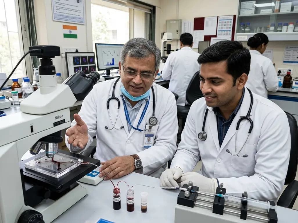
Platelets and leukocytes serve as crucial regulators of thrombo-inflammatory processes across vascular, metabolic, and infectious pathologies. Evaluating their functional activation states and intercellular interactions yields essential insights into disease progression and treatment response. However, standard laboratory workflows depend heavily on centrifugation-based blood preparation, which presents notable limitations. Conventional centrifugation requires large blood volumes, involves labor-intensive manual handling, and frequently induces shear-mediated artefactual platelet activation. Furthermore, mechanical stress during centrifugation can disrupt delicate native cell-cell interactions, including neutrophil-platelet aggregates. These operational constraints reduce translational relevance, particularly for applications requiring small blood volumes or longitudinal sampling. Advanced label-free microfluidic blood processing offers a transformative solution by directly isolating intact platelets and leukocyte complexes from minimal sample volumes without harsh mechanical disruption. By avoiding fluorescent antibody staining and density gradient centrifugation, this innovative bioengineering approach preserves native cellular physiology and bridges laboratory research with clinical diagnostics.
The microfluidic platform utilizes a two-stage label-free architecture engineered around Dean Flow Fractionation principles. Fluid flowing through curved microchannels generates counter-rotating Dean vortices that interact with hydrodynamic inertial lift forces, driving size-dependent lateral cell migration. In the primary stage, a spiral inertial channel separates larger leukocytes and aggregates from smaller platelets and abundant red blood cells. Downstream, fluid enters a serpentine channel designed for on-chip leukocyte reconcentration, eliminating off-chip centrifugation steps. The device is fabricated in poly(methyl methacrylate), commonly known as PMMA, a durable thermoplastic that enhances structural robustness and manufacturing scalability relative to traditional soft lithography materials. Moreover, the integrated microchannel geometry enables continuous fluid processing without channel clogging or cell jamming. This dual-stage design operates entirely label-free, relying solely on intrinsic cellular size and deformability to achieve efficient cell separation and consistent operational throughput across diverse blood specimens.
A primary bottleneck in traditional hematological evaluation is preventing sample-preparation-induced activation of delicate thrombocytes. Standard centrifugation subjects platelets to mechanical shear, triggering premature granule release, receptor upregulation, and morphological changes. In contrast, gentle microfluidic blood processing preserves platelets in their baseline quiescent state during separation. Maintaining resting activation profiles enables accurate measurement of true pathological hyperreactivity rather than preparation artifacts. Furthermore, thrombo-inflammatory responses rely heavily on physical cross-talk between activated neutrophils and platelets. Neutrophil-platelet aggregates participate directly in microvascular thrombosis and tissue injury during systemic inflammation and vascular disease. Conventional separation techniques routinely disrupt these fragile heterotypic complexes, yielding misleading results. The Dean Flow Fractionation platform maintains low hydrodynamic shear stress, preserving native cellular complexes for precise downstream quantification and providing an unadulterated assessment of systemic inflammatory status.
Validation studies demonstrate impressive operational efficiency and speed across diverse biological models. Utilizing a minimal starting volume of 100 microliters of whole blood, the system achieves complete separation within five minutes. The platform attained 70% to 80% leukocyte recovery alongside 45% platelet recovery. Performance was validated using blood from healthy human donors and fever patients exhibiting acute inflammatory responses. Furthermore, researchers evaluated a streptozotocin-induced diabetic mouse model. In rodent studies, small-volume sampling enabled longitudinal monitoring of platelet activation and neutrophil-platelet interactions without requiring terminal blood collection. Previously, rodent experiments required terminal cardiac puncture, preventing serial sampling from individual subjects. By allowing repeated micro-volume blood draws without inducing significant anemia, this microfluidic workflow enhances statistical rigor in translational pre-clinical research and accelerates diagnostic biochip development.
A central advantage of this label-free microfluidic processing workflow is its seamless compatibility with advanced single-cell analysis technologies. Recovered cell populations can be evaluated directly via multicolor flow cytometry to quantify surface activation markers like CD62P or CD11b. Additionally, isolated cell fractions are fully compatible with digital holographic microscopy, an advanced quantitative phase imaging modality. Digital holographic microscopy offers high-resolution three-dimensional morphological profiling of living blood cells, detecting subtle volumetric shifts associated with early activation. Combining microfluidic separation with digital holographic microscopy enables rapid, non-destructive phenotypic screening at individual cell resolution. Furthermore, because isolated cells remain functionally intact and unlabelled, downstream assays can include functional adhesion assays and aggregometry. This integrated multi-modal platform represents a vital technology for precision diagnostic workflows and clinical biomarker discovery.
Translating label-free microfluidic cell isolation into clinical practice addresses critical gaps in diagnostic hematology, cardiology, and critical care medicine. Thrombo-inflammatory dysregulation contributes to acute coronary syndromes, sepsis, autoimmune conditions, and diabetic vasculopathy. However, clinicians lack rapid point-of-care tools to quantify active platelet-leukocyte interactions in real time. Standard laboratory counters measure static cell counts but fail to evaluate cellular state or binding dynamics. By enabling micro-volume processing, microfluidic technology allows clinicians to monitor inflammatory surges directly from low-volume blood samples. This capability is especially valuable in neonatology and pediatric intensive care where blood conservation is critical. Identifying elevated neutrophil-platelet aggregates serves as an early biomarker for microvascular thrombosis or therapeutic resistance, aiding clinical decision-making and anti-thrombotic treatment optimization.
Dean Flow Fractionation separates circulating blood cells inside curved microfluidic channels utilizing hydrodynamic inertial forces and fluidic Dean vortices without external force application. Unlike traditional centrifugation, which subjects delicate blood components to high shear stress that triggers artificial platelet activation and disrupts cellular complexes, microfluidic processing operates gently. This label-free physical mechanism successfully preserves baseline platelet quiescence and retains intact neutrophil-platelet aggregates while processing micro-volume 100-microliter blood samples in five minutes.
Neutrophil-platelet aggregates serve as sensitive biomarkers for active thrombo-inflammation across cardiovascular, metabolic, and infectious disorders. When activated neutrophils bind circulating platelets, they promote microvascular thrombosis, tissue factor expression, and localized inflammatory tissue damage. Preserving these delicate heterotypic cellular complexes during blood collection allows clinicians to measure real-time intravascular immune-thrombotic activity accurately, monitor disease progression in conditions like diabetes or fever, and evaluate therapeutic responsiveness to antiplatelet regimens.
The label-free microfluidic workflow isolates functionally intact, unlabelled leukocytes and quiescent platelets that are fully compatible with diverse high-throughput downstream analytical platforms. Recovered cell populations can undergo multi-color flow cytometry for surface activation marker profiling or digital holographic microscopy for label-free three-dimensional quantitative cell phase imaging. Furthermore, the preserved cellular integrity supports functional cell adhesion assays, aggregometry, single-cell transcriptomics, and longitudinal monitoring in translational animal models.
Disclaimer: This content is for informational and educational purposes only and does not constitute medical advice, diagnosis, or treatment. Healthcare professionals should rely on their clinical judgment and local regulatory guidelines when evaluating diagnostic technologies. Refer to the latest local and national guidelines for clinical practice.
References
1. Maw KK et al. Label-free microfluidic small-volume blood processing for analysis of platelet activation and neutrophil-platelet aggregates. Lab Chip. 2026 Aug 11. doi: 10.1039/d6lc00413j. PMID: 42576810.

Read summarized clinical updates, watch expert medical content, and earn CME certifications right from your smartphone.
Open your smartphone camera to scan and install.





A novel label-free microfluidic workflow using Dean Flow Fractionation isolates intact platelets and leukocytes from 100 μL whole blood within 5 minutes. Fabricated in PMMA, it preserves platelet quiescence and neutrophil-platelet aggregates, enabling advanced flow cytometry and digital holographic microscopy.
Today

A study of hospitalized adult patients with severe eating disorders revealed that while serum leptin increases linearly with weight gain during early refeeding, thyroid hormones show a biphasic trajectory. Low T3 and undetectable leptin frequently persist at discharge, highlighting delayed endocrine recovery.
Today
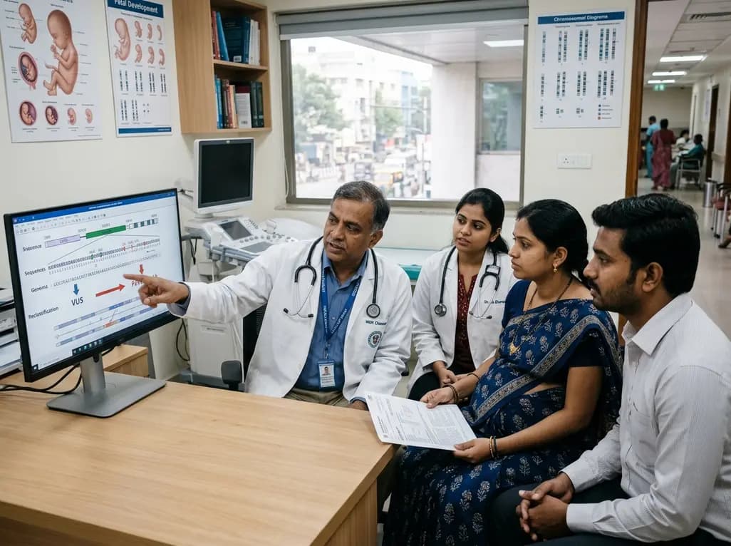
A 3-year case study demonstrates how dynamic variant reclassification between VUS and likely pathogenic states directly impacts prenatal genetic counseling, fetal diagnostic workflows, preimplantation genetic testing, and complex reproductive choices.
Today
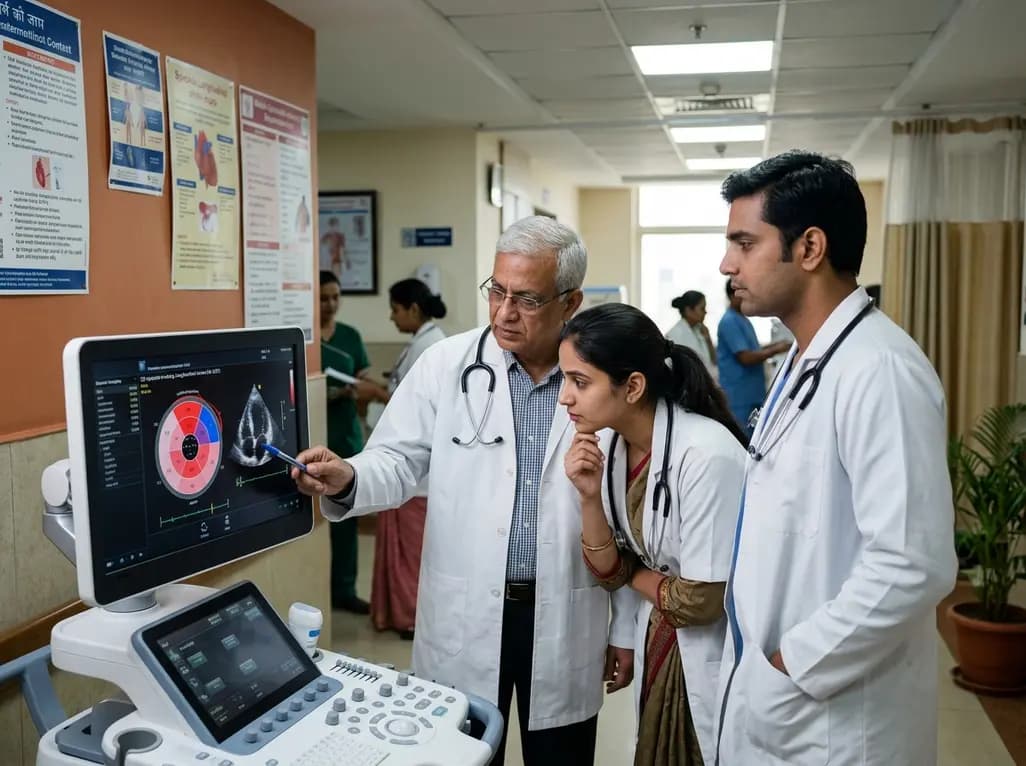
A multicenter J-CASE survey study reveals that left ventricular apical longitudinal strain (LV-apical LS) independently predicts all-cause death in patients with immunoglobulin light-chain (AL) cardiac amyloidosis, establishing an optimal prognostic cut-off threshold of 15.9%.
Today

Researchers developed a Haversian-inspired composite scaffold that addresses delayed vascularization and wet-state mechanical deterioration in critical bone defect repair. By integrating spatially programmed calcium phosphate minerals and selective silica reinforcement, the design promotes vascularized bone repair.
Today

A BMJ cohort study shows GLP-1 receptor agonists are linked to a modest rise in non-scarring hair loss in adults with type 2 diabetes compared to SGLT-2 and DPP-4 inhibitors. Although relative risk is higher, absolute risk remains low, likely driven by rapid weight loss rather than direct follicular damage.
Today
