.webp&w=384&q=75)

Loading, please wait...
Loading, please wait...
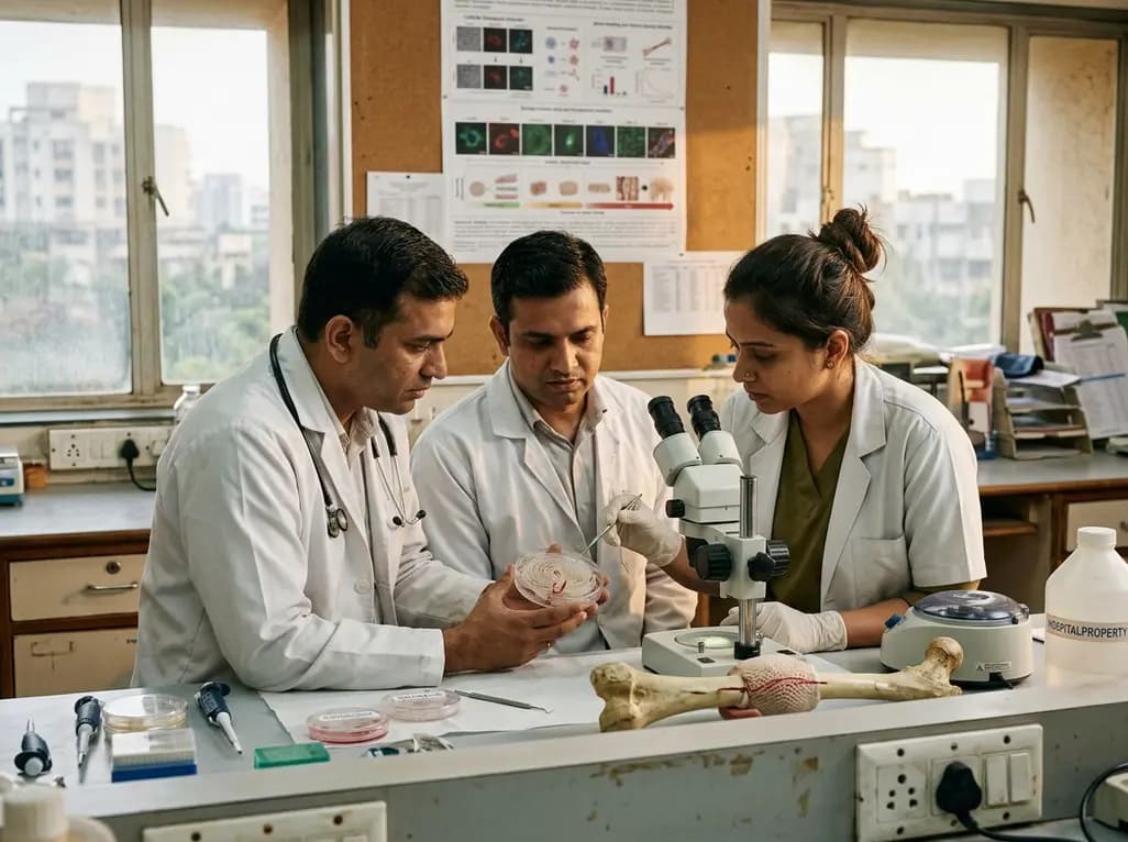
Treating critical-sized segmental bone defects remains a major clinical challenge in orthopedics. Natural bone possesses an intrinsic capacity for self-healing; however, extensive bone loss from trauma or tumor resection exceeds this threshold. Traditional autologous bone grafts represent the standard approach, yet surgeons encounter donor-site morbidity and limited graft availability. Synthetic biomimetic implants offer an alternative, but conventional designs frequently fail in large defects. Specifically, large synthetic constructs suffer from delayed vascularization within their deep central cores. Without rapid microvascular ingrowth, nutrient diffusion fails, causing cellular death and implant failure. Moreover, many polymeric materials experience severe wet-state mechanical deterioration after implantation. Body fluids weaken the structural integrity before adequate tissue infiltration occurs. Consequently, researchers focus on bio-inspired architectures to solve these challenges. Recent innovations in Haversian scaffold bone repair present a promising solution. By replicating native cortical bone microarchitecture, these systems coordinate rapid vascularization with robust mechanical support under physiological conditions.
Native cortical bone relies on the Haversian system to maintain mechanical rigidity while sustaining continuous nutrient transport. In natural bone, longitudinal Haversian canals containing blood vessels are interconnected by transverse channels. To replicate this complex structure, biomedical engineers developed a hierarchical composite matrix. The synthetic design features longitudinally interconnected primary channels surrounded by anisotropic micropores. Consequently, this interconnected network creates a directional mass transport system that mimics natural vascular pathways. Perfusion studies demonstrate that these primary channels significantly accelerate fluid movement through the scaffold core. As a result, oxygen, metabolic nutrients, and signaling molecules easily reach deeply embedded cells. Furthermore, the anisotropic microporous walls surrounding the main channels encourage rapid cellular infiltration and endothelial sprouting. Endothelial precursor cells migrate along the longitudinal axis, initiating early capillary network formation. Therefore, this structural hierarchy overcomes the mass transport limitations that previously restricted clinical tissue engineering scaffolds.
Homogeneous biomaterial compositions often fail to replicate the complex chemical microenvironment of natural bone tissue. Rather than using a uniform composition, the scaffold incorporates a spatially heterogeneous distribution of inorganic components. Initially, researchers integrated brushite and hydroxyapatite into a biocompatible chitosan matrix. Chitosan provides flexible structural support, while brushite and hydroxyapatite supply critical calcium and phosphate ions. Subsequently, a biomimetic mineralization step deposits additional calcium phosphate onto internal scaffold surfaces. Consequently, this surface mineralization creates widespread bio-interactive interfaces throughout the internal architecture. These mineralized sites promote robust initial cell adhesion, spreading, and osteogenic differentiation of progenitor stem cells. Additionally, the localized surface micro-topography enhances cell signaling pathways that drive bone matrix synthesis. By strategically positioning mineral phases at targeted structural boundaries, the scaffold effectively mimics natural osteoid formation. Therefore, osteoprogenitor cells encounter continuous osteoinductive cues, accelerating early bone matrix deposition throughout the defect area.
A major limitation of conventional biopolymer scaffolds is their rapid loss of mechanical strength in fluid environments. Exposure to physiological fluids causes swelling and severe plasticization of the polymer matrix. Consequently, the implant loses structural integrity before host bone tissue can regenerate effectively. To address this vulnerability, engineers engineered a tightly integrated silica-rich layer selectively localized along primary-channel walls. These channel walls represent the most mechanically vulnerable areas under compressive loads. By isolating silica reinforcement specifically to these critical regions, the scaffold achieves high mechanical reliability without reducing overall porosity. Furthermore, the localized silica network acts as a rigid frame resisting wet-state plasticization. When combined with calcium phosphate minerals, this reinforced wall architecture maintains compressive strength in physiological fluids. Accordingly, the scaffold provides durable mechanical support during early healing stages. Host osteoblasts can thus safely proliferate and deposit calcified matrix within a stable mechanical framework.
Beyond physical support and structural stability, effective bone repair requires continuous biochemical stimulation. The hierarchical scaffold acts as an active signaling platform through controlled constituent dissolution. Specifically, dynamic degradation of brushite, hydroxyapatite, and localized silica layers releases bioactive calcium, phosphorus, and silicon species. Dissolved calcium and phosphate ions elevate local extracellular ion concentrations, directly triggering osteogenic gene expression in progenitor cells. Simultaneously, sustained silicon ion release plays a vital role in biological signaling. Silicon species upregulate vascular endothelial growth factor expression, thereby driving endothelial cell proliferation and vessel formation. Additionally, silicon ions stimulate osteoblast maturation and structural protein synthesis. In vitro evaluations confirmed that this controlled multi-ion release profile significantly enhances cell differentiation markers. Subsequent in vivo evaluations demonstrated accelerated microvascular network formation and rapid bone volume growth. Thus, combining enhanced mass transport, localized mechanical reinforcement, and biochemical signals promotes coupled vascularized bone regeneration.
The development of this Haversian-inspired framework represents a significant advance in orthopedic biomaterials research. Critical-sized bone defects caused by trauma, oncological resections, or non-unions require advanced solutions that address mechanical and biological challenges simultaneously. Traditional scaffold designs often force a compromise between mechanical strength and high porosity. However, this spatially programmed design strategy demonstrates that targeted constituent placement reconciles permeability, wet-state mechanical support, and biological activity. Clinicians can anticipate future applications where customized 3D scaffolds match patient defect geometries while providing optimized microchannels. Furthermore, relying on well-characterized constituents like chitosan, calcium phosphate, and silica simplifies regulatory approval pathways and manufacturing scalability. As preclinical validation progresses toward clinical trials, this integrated architectural approach establishes a practical framework for bone tissue engineering. Ultimately, combining structural bio-mimicry with localized chemical reinforcement promises to shorten healing times and improve patient outcomes in complex bone reconstruction.
The Haversian system is the primary structural unit of cortical bone, comprising central longitudinal channels that carry blood vessels and nerves. Replicating this architecture in synthetic scaffolds creates interconnected micro-pathways throughout the material. Consequently, this design facilitates rapid directional nutrient transport, cellular infiltration, and early capillary growth across large implant volumes. By mimicking natural bone vascular networks, these scaffolds prevent central tissue necrosis in critical-sized bone defect repairs.
Polymeric scaffolds typically absorb fluid and lose structural strength in physiological environments. Localizing a tightly integrated silica-rich layer along primary channel walls selectively reinforces the scaffold's most vulnerable structural boundaries. Furthermore, combining silica with calcium phosphate minerals creates a water-stable matrix. This localized reinforcement maintains mechanical rigidity under compressive loads, ensuring structural stability while new bone tissue gradually infiltrates and matures within the defect site.
The composite scaffold gradually releases bioactive calcium, phosphorus, and silicon species as it degrades. Calcium and phosphate ions directly stimulate osteoblast activity and accelerate mineral matrix deposition. Meanwhile, released silicon ions promote vascular endothelial growth factor expression, which drives endothelial cell proliferation and vessel formation. Together, these dissolved ionic cues provide robust biological signals that couple angiogenesis with osteogenesis, significantly enhancing overall bone regeneration outcomes.
Disclaimer: This content is for informational and educational purposes only and does not constitute formal medical advice, diagnosis, or treatment. Refer to the latest local and national guidelines for clinical practice.
References

Read summarized clinical updates, watch expert medical content, and earn CME certifications right from your smartphone.
Open your smartphone camera to scan and install.





Researchers developed a Haversian-inspired composite scaffold that addresses delayed vascularization and wet-state mechanical deterioration in critical bone defect repair. By integrating spatially programmed calcium phosphate minerals and selective silica reinforcement, the design promotes vascularized bone repair.
last month

Population-based data demonstrate that material deprivation, ethno-racial diversity, and urban density substantially amplify peak hospitalization rates during COVID-19, influenza, and RSV surges. Clinical preparedness requires addressing socioeconomic determinants to ensure equitable resource allocation.
Today

The ClinGen Prenatal Gene Curation Expert Panel evaluated 63 disease relationships across 61 genes, establishing clinical validity for severe fetal phenotypes like hydrops and stillbirth to enhance prenatal genomic interpretation and clinical care.
Yesterday

Managing refractory hypoxemia after cardiothoracic surgery becomes perilous when prolonged air leak limits positive-pressure ventilation. This case-based review details how high-flow nasal cannula, paired with awake rehabilitation, enabled successful extubation and avoided invasive re-intubation.
Today

Idiopathic axillary web syndrome (IAWS) is a rare cause of shoulder pain and mobility restriction characterized by palpable axillary cording without prior surgery or trauma. Early diagnosis, nonsteroidal anti-inflammatory therapy, and physical rehabilitation lead to complete resolution of symptoms.
Today

SWEDEPAD-1 trial insights show paclitaxel-coated devices do not improve long-term limb salvage in patients with chronic limb-threatening ischemia and tissue loss. While one-year reinterventions decreased, the devices were linked to a higher risk of major amputation at three months.
Today
