.webp&w=384&q=75)

Loading, please wait...
Loading, please wait...

Aneurysms arising within the dural circulation represent a distinct vascular pathology that clinicians frequently misdiagnose or overlook. Although clinicians routinely encounter intracranial intradural berry aneurysms, meningeal artery aneurysms occupy a unique anatomical and physiological niche. These extra-axial vascular outpouchings develop along the branches of the middle meningeal artery, accessory meningeal channels, or posterior meningeal vessels. Historically, physicians classified them primarily by the presence or absence of preceding mechanical trauma. However, a major systematic review and illustrative case series has revealed that non-traumatic variants exhibit diverse pathophysiological mechanisms. Consequently, understanding these unique lesions is paramount for neurosurgeons, neurointerventional radiologists, and neurocritical care specialists. Timely identification and intervention can avert catastrophic neurological decline, profound morbidity, and fatal intracranial hemorrhage across diverse clinical presentations.
The vascular architecture of the dura mater differs significantly from the cerebral parenchymal vasculature. While pial and cerebral vessels course through the fluid-cushioned subarachnoid space, meningeal arteries embed tightly within the periosteal and meningeal dural layers against the calvarium. As a result, biomechanical and hemodynamic forces act distinctively upon these dural vessels. True aneurysms involve complete wall degeneration, whereas pseudoaneurysms feature disruption of the internal elastic lamina and media, creating an unstable cavity contained only by adventitia, organizing thrombus, or adjacent dura mater.
Mechanical shear forces frequently damage the middle meningeal artery during temporal bone fractures. Conversely, spontaneous lesions typically arise secondary to altered systemic flow dynamics or structural vessel wall fragility. High-flow states created by collateralized cerebrovascular occlusive diseases or hypervascular intracranial lesions can generate excessive hemodynamic stress. Over time, persistent hemodynamic shear stresses disrupt the vessel lumen and trigger aneurysmal remodeling. Additionally, connective tissue disorders and focal inflammatory vasculopathies weaken arterial wall integrity. Therefore, clinicians must appreciate that these lesions stem from distinct physical and mechanical triggers.
Researchers have proposed a comprehensive, pathophysiology-based classification system to replace outdated paradigms. The updated schema categorizes non-traumatic aneurysms into three well-defined groups: sporadic, hemodynamic or flow-related, and genetic or dysplastic. Approximately sixty-one percent of non-traumatic cases belong to the hemodynamic subtype, making it the most prevalent form. These lesions arise because pathological conditions recruit the dural circulation as high-pressure collateral conduits.
Specifically, hypervascular tumors such as angioblastic meningiomas, dural arteriovenous shunts, severe carotid occlusive atherosclerosis, and Moyamoya disease demand extensive meningeal recruitment. Sporadic aneurysms account for thirty-two percent of non-traumatic cases. These typically occur in hypertensive patients or individuals without an overt systemic vascular anomaly. The remaining seven percent constitute genetic or dysplastic etiologies, including neurofibromatosis type 1, collagen vascular disorders, and cranial Paget disease. Importantly, rupture rates vary dramatically across these cohorts. Sporadic aneurysms exhibit an alarming rupture rate of seventy-nine percent, whereas hemodynamic subtypes demonstrate a fifty-two percent rupture rate. Hence, establishing the precise subtype guides urgent prognostic stratification.
Traumatic meningeal lesions comprise the largest overall cohort of dural aneurysms, accounting for nearly seventy percent of documented occurrences. These injuries arise predominantly from blunt motor vehicle collisions, penetrating trauma, and blast injuries sustained during combat operations. When linear skull fractures traverse the sphenoparietal groove or temporal squama, the rigid bone fragments lacerate or severely contuse the adhered dural artery. This injury produces an acute pseudoaneurysm rather than a true ectasia.
Crucially, traumatic aneurysms frequently present with a treacherous, asymptomatic latency period. Systematic review data indicate that delayed rupture occurs in twenty-six percent of traumatic cases, with a median latency period of fourteen days post-injury. During this deceptive window, the primary hematoma may stabilize, and the patient may regain neurological baseline. However, as the fragile pseudoaneurysmal thrombus lyses and dynamic arterial pulsations persist, secondary rupture inevitably occurs. This secondary event precipitates massive, rapid-onset subdural, epidural, or intracerebral hemorrhage. Clinicians must maintain high clinical suspicion and perform delayed repeat vascular imaging whenever patients present with high-risk fractures crossing major meningeal grooves.
Identifying meningeal aneurysms requires meticulous radiologic evaluation because standard non-contrast head computed tomography scans often obscure small arterial outpouchings. Computed tomography scans easily demonstrate calvarial fractures and acute epidural or subdural hematomas. However, acute parenchymal bleeding or bone artifact frequently masks the primary aneurysm sac. Therefore, non-contrast scans remain insufficient for ruling out active meningeal vascular pathology.
High-resolution computed tomography angiography provides rapid, non-invasive detection of dural vessel irregularity, focal enlargement, and active extravasation. Contrast-enhanced magnetic resonance angiography and three-dimensional magnetic resonance sequences offer superior resolution for characterizing adjacent soft-tissue anatomy, dural sinus thrombosis, and underlying hypervascular neoplasms. Nevertheless, digital subtraction angiography remains the indispensable gold standard for definitive diagnosis. Selective external carotid artery catheterization with rotational angiography accurately delineates the aneurysm neck, detects associated dural arteriovenous fistulas, and maps collateral anastomoses with the ophthalmic or petrous facial pathways. Consequently, comprehensive multimodal vascular imaging prevents disastrous diagnostic delays.
The definitive management of meningeal aneurysms has experienced a profound shift over the past two decades. Historically, open craniotomy with surgical coagulation, clipping, or direct trapping represented the standard treatment protocol. Although direct surgical intervention allows concurrent evacuation of life-threatening space-occupying hematomas, opening the cranium over a friable, actively bleeding vascular sac carries severe intraoperative hemorrhage risks. Consequently, modern endovascular techniques have superseded open surgery in modern neurointerventional practice.
Recent data reveal that interventionalists implement endovascular techniques in seventy-two percent of contemporary cases. Selective catheterization permits superselective occlusion using liquid embolic systems such as ethylene vinyl alcohol copolymer, n-butyl cyanoacrylate, or microcoils. Embolization rapidly arrests flow while preserving downstream intracranial and orbitofacial branches. Clinicians must exercise extreme caution during superselective injection to prevent accidental reflux into the ophthalmic artery via the meningo-ophthalmic anastomoses or into the middle meningeal petrosal branch feeding the geniculate ganglion. Combining endovascular embolization with subsequent decompressive hematoma evacuation offers an optimal, minimally invasive dual strategy.
Patient survival and long-term functional recovery hinge decisively upon aneurysm rupture status prior to intervention. Patients presenting with ruptured aneurysms experience poor neurological outcomes in thirty-five percent of cases. In stark contrast, individuals diagnosed prior to rupture experience poor clinical outcomes in merely six percent of instances. This dramatic discrepancy underscores the profound value of proactive diagnostic screening and rapid prophylactic obliteration.
Following surgical or endovascular stabilization, neurocritical care teams must initiate targeted protocols to preserve secondary neurological function. Clinicians must maintain strict normotension to mitigate recurrent hemorrhage risks while ensuring adequate cerebral perfusion pressure. Continuous neuro-monitoring, serial pupillary assessments, and targeted sedation protocols facilitate immediate identification of intracranial pressure spikes or delayed rebleeding. Additionally, clinicians should administer prophylactic antiepileptic therapy in patients presenting with cortical hematomas or substantial subdural blood. Through meticulous hemodynamic optimization, comprehensive imaging surveillance, and multimodal neurocritical care, clinicians can achieve favorable neurological functional recovery in these critically vulnerable patients.
Traumatic aneurysms represent true pseudoaneurysms caused by arterial wall transection, typically from adjacent skull fractures or penetrating trauma. In contrast, non-traumatic aneurysms feature true or false aneurysmal remodeling secondary to chronic hemodynamic shear stresses, hypervascular intracranial tumors, arteriovenous malformations, connective tissue disorders, or severe atherosclerotic vessel stenosis.
Delayed rupture occurs because traumatic pseudoaneurysms initially tamponade with a fragile, unstable blood clot and adjacent dura. Over subsequent days, physiological fibrinolysis lyses this temporary seal while persistent arterial pulsations expand the pseudoaneurysmal pouch. Ultimately, this structural degradation triggers sudden secondary hemorrhage, typically within two weeks post-injury.
Neurointerventionalists frequently employ liquid embolic agents, such as ethylene vinyl alcohol copolymer or n-butyl cyanoacrylate, alongside detachable microcoils. Liquid agents penetrate the irregular pseudoaneurysmal cavity and provide permanent parent vessel sacrifice. Coils provide reliable structural packing, preventing recanalization while operators carefully preserve dangerous orbitofacial and cranial nerve collateral vessels.
Disclaimer: This content is for informational and educational purposes only and does not constitute medical advice, diagnosis, or treatment. Healthcare professionals should exercise independent clinical judgment. Refer to the latest local and national guidelines for clinical practice.
References
Stanishevskiy A et al. Aneurysms of the meningeal arteries: systematic review and illustrative case series. Neurosurg Rev. 2026 Sep 16. doi: 10.1007/s10143-026-04500-w. PMID: 42747682.
Bruneau M, Gustin T, Zekhnini K, Gilliard C. Traumatic false aneurysm of the middle meningeal artery causing an intracerebral hemorrhage: Case report and literature review. Surg Neurol. 2002;57(3):174-178.
Abdalkader M, Kaliaev A, Nguyen T. Endovascular Management of Ruptured Traumatic Middle Meningeal Artery Pseudoaneurysm and Arteriovenous Fistula. J Vasc Endovasc Therapy. 2019;4(2):12.
Suzuki M, Kominami S, Koketsu K, Mizunari T, Kobayashi S, Morita A. Endovascular repair of a middle meningeal artery aneurysm after cranial surgery. NMC Case Rep J. 2014;1(1):6-8.

Read summarized clinical updates, watch expert medical content, and earn CME certifications right from your smartphone.
Open your smartphone camera to scan and install.





Meningeal artery aneurysms represent rare but treacherous neurovascular pathologies. A landmark systematic review and case series establishes a novel pathophysiology-based classification, identifies significant delayed rupture risks, and highlights modern endovascular paradigms that improve patient outcomes.
Today
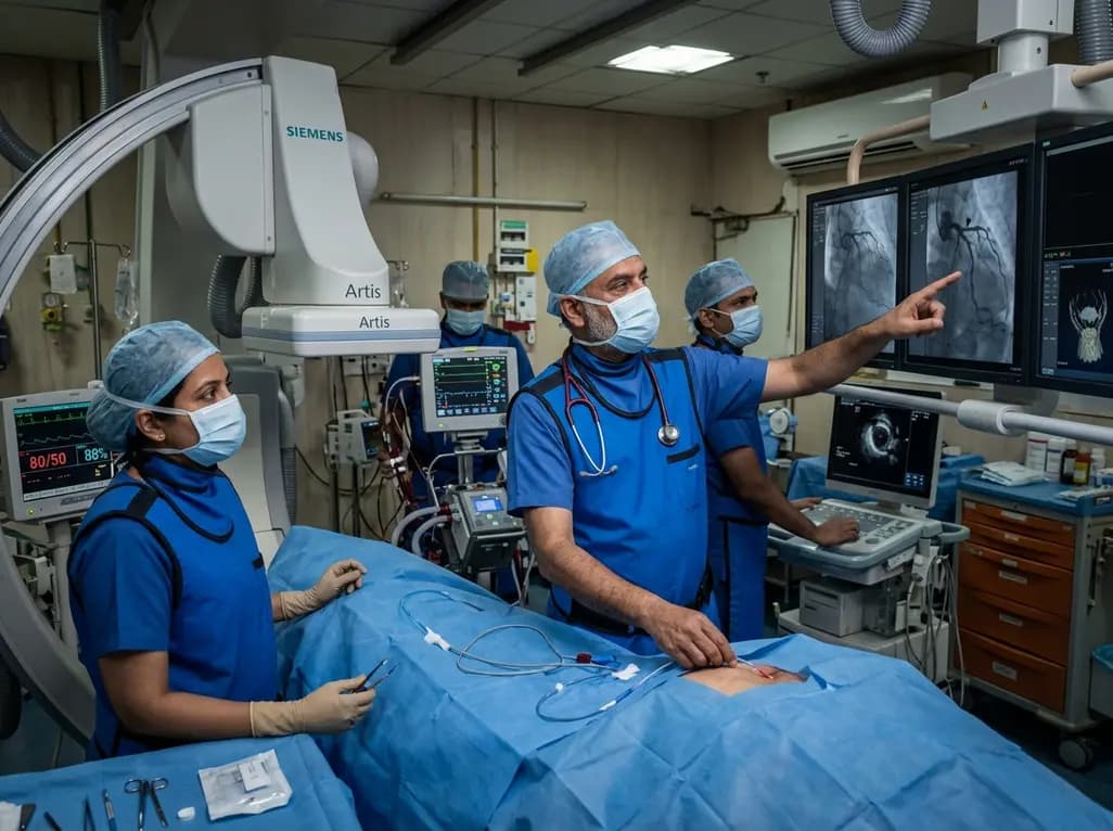
A rare case of acute coronary obstruction during TAVR caused by suspected calcified amorphous tumor embolization underscores the critical role of intracardiac imaging, emergency ECMO, and the paradox of enlarged-aperture valve geometry.
Today

Prelacteal feeding persists in South Asian communities despite known clinical risks. This review examines determinants from Chitral, pathophysiological hazards, and pediatric strategies to eliminate harmful feeds and support exclusive breastfeeding.
Today

Transcatheter mitral valve replacement in patients with degenerated bioprostheses carries a substantial risk of LVOT obstruction. Combining leaflet modification via the BATMAN technique with mechanical circulatory support offers a viable, life-saving solution for anatomically challenging surgical candidates.
Today

A comparative cohort study demonstrates that patients undergoing total hip arthroplasty after prior hip arthroscopy achieve comparable functional recovery, low complication rates, and equivalent implant survivorship when conversion occurs after six months.
Today

A cross-sectional study of 320 adults evaluated the link between habitual fasting duration and dietary quality using Nutrition Quotient scores, highlighting the clinical necessity of balancing time-restricted eating with nutrient density and meal quality in routine metabolic practice.
Today
