.webp&w=384&q=75)

Loading, please wait...
Loading, please wait...
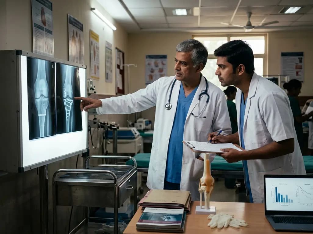
Severe medial tibial plateau fractures present a formidable challenge in orthopedic trauma care. Because these intra-articular injuries often result from high-energy collisions or severe axial loading, surgeons frequently encounter extensive articular depression, multi-segmental comminution, and substantial soft-tissue disruption. Restoring structural alignment requires clear intraoperative exposure of the weight-bearing joint surface. Furthermore, inadequate reduction increases post-traumatic osteoarthritis risks and long-term joint instability. Consequently, trauma specialists must balance aggressive surgical exposure with soft-tissue preservation. Standard medial approaches usually offer adequate visualization for simple fracture configurations. However, complex patterns involving posteromedial segments or deep depression require broader surgical access. In a recent comparative study, investigators evaluated whether an extended medial approach utilizing a medial femoral epicondyle osteotomy improves clinical and radiological outcomes compared to conventional techniques. The clinical cohort included complex high-velocity injuries, knee dislocations, and polytrauma cases requiring prolonged hospitalization. Although osteotomy provides expanded visibility of the joint, clinicians must weigh this benefit against surgical morbidity. Therefore, understanding the precise indications for epicondylar detachment remains essential for trauma surgeons striving to optimize patient recovery and restore joint congruity.
Surgical execution in high-energy proximal tibia injuries requires careful planning to prevent wound healing complications and neurovascular compromise. When tackling severe medial tibial plateau fractures, surgeons often struggle to visualize posteromedial and central articular fragments through standard surgical corridors. To address this limitation, an extended medial approach with medial femoral epicondyle osteotomy reflects the superficial medial collateral ligament proximally. Consequently, this surgical maneuver grants unprecedented access to the medial joint line and underlying articular cartilage without traumatizing delicate meniscal attachments. In contrast, standard non-osteotomy approaches rely on capsular reflection and traction, which may restrict joint visualization in highly comminuted patterns. During the procedure, surgeons osteotomize a precise bone block from the medial epicondyle, allowing controlled reflection of soft tissues. After anatomical reduction and internal fixation of the tibial plateau with locked plating, clinicians securely reattach the epicondylar fragment using tension band wiring or lag screws. Although this technique significantly expands the visual field, it demands meticulous surgical skill to avoid damaging adjacent ligamentous structures. Furthermore, surgeons must ensure robust hardware fixation of the osteotomy block to promote solid bony healing and allow early post-operative knee motion.
Achieving precise articular reduction remains the benchmark for successful intra-articular fracture management. Post-operative computed tomography scans provide detailed multiplanar evaluation of joint congruity, joint gaps, and residual articular steps. In comparative analysis, patients undergoing medial femoral epicondyle osteotomy demonstrated equivalent radiological reduction compared to those managed with standard approaches. Specifically, researchers observed medial fracture step-offs greater than two millimeters in five patients from each cohort. Additionally, residual joint gaps exceeding five millimeters appeared in three epicondyle osteotomy patients and five non-osteotomy patients, showing no statistically significant discrepancy. Anatomical alignment evaluation using the radiological Rasmussen score yielded mean values of 14.1 points in the osteotomy group and 13.4 points in the standard approach group. Consequently, both surgical cohorts achieved favorable radiographic outcomes despite the extreme severity of initial fracture displacement. These objective findings indicate that while epicondyle osteotomy provides excellent visualization, experienced surgeons can often obtain satisfactory articular reduction using conventional approaches when tissue planes permit. Therefore, radiographic success alone does not automatically justify the routine implementation of an invasive epicondylar osteotomy.
Functional recovery following complex intra-articular knee trauma relies heavily on early motion, stability, and patient-reported outcomes. Clinical follow-up spanning more than two years revealed comparable functional recovery between patients treated with epicondyle osteotomy and those managed with standard medial approaches. Patient evaluations utilized validated clinical tools, including the Knee Injury and Osteoarthritis Outcome Score and the International Knee Documentation Committee score. Specifically, the osteotomy cohort achieved a mean score of 61.3 on the functional outcome scale, whereas the non-osteotomy cohort recorded a mean score of 59.5. Similarly, documentation committee scores averaged 58.8 points in the osteotomy group and 52.6 points in the control group, demonstrating no statistically significant functional divergence. However, overall scores across both groups reflect the severe nature of these high-energy injuries. Patients frequently experienced residual stiffness, exercise limitations, and persistent mild discomfort due to extensive soft-tissue trauma and associated knee dislocations. Furthermore, concomitant injuries and prolonged hospital stays contributed to extended rehabilitation timelines. Ultimately, these clinical outcomes highlight that long-term functional recovery depends on overall injury severity rather than surgical approach choice alone.
Surgical decision-making in complex knee trauma requires balanced evaluation of visualization benefits against procedural risks. The clinical evidence demonstrates that medial femoral epicondyle osteotomy is a feasible and safe technique for severe joint injuries. However, because functional and radiographic outcomes remain comparable to standard approaches, surgeons should reserve this extended approach for specific, highly challenging fracture configurations. Specifically, osteotomy offers clear advantages in multi-fragmentary fractures where deep central depression or posteromedial displacement prevents adequate reduction through standard corridors. Conversely, routine use of epicondylar detachment adds procedural time, increases operative trauma, and introduces potential risks such as nonunion, hardware prominent discomfort, or delayed ligamentous healing. Therefore, trauma specialists must individualize their surgical strategy based on pre-operative computed tomography mapping and soft-tissue conditions. When standard medial exposure yields adequate visualization, clinicians can achieve excellent reduction without osteotomy. Ultimately, thoughtful patient selection ensures that surgeons utilize extended exposures effectively while minimizing unnecessary surgical morbidity in complex knee trauma management.
Medial femoral epicondyle osteotomy temporarily detaches the origin of the superficial medial collateral ligament along with a small bone block. This maneuver significantly expands intraoperative exposure of the medial joint line and tibial articular surface. Consequently, trauma surgeons can accurately visualize deep articular depression and comminuted posteromedial fracture fragments that are difficult to access through conventional medial surgical corridors.
Clinical studies show that epicondyle osteotomy yields comparable long-term functional scores to standard surgical approaches. Patients undergoing osteotomy achieved mean functional scores around 61 points, while non-osteotomy patients scored approximately 59 points. Because high-energy trauma causes extensive concomitant cartilage and soft-tissue damage, overall knee functional recovery is primarily determined by original injury severity rather than the surgical approach utilized.
Extended medial approaches with epicondyle osteotomy carry additional procedural risks, including nonunion of the osteotomy bone block, hardware prominence, and soft-tissue irritation. Additionally, extended surgical exposure increases overall operative time and tissue disruption. Therefore, orthopedic surgeons reserve this invasive technique for severe, highly displaced fracture patterns where adequate intra-articular reduction cannot be safely accomplished through conventional surgical access.
Disclaimer: This content is for informational and educational purposes only and does not constitute medical advice, diagnosis, or treatment. Always seek the advice of a qualified healthcare provider with any questions regarding a medical condition or treatment options. Refer to the latest local and national guidelines for clinical practice.
References

Read summarized clinical updates, watch expert medical content, and earn CME certifications right from your smartphone.
Open your smartphone camera to scan and install.





A retrospective cohort study evaluated an extended medial approach with medial femoral epicondyle osteotomy for severe medial tibial plateau fractures, demonstrating comparable radiographic and functional outcomes to standard approaches.
last month
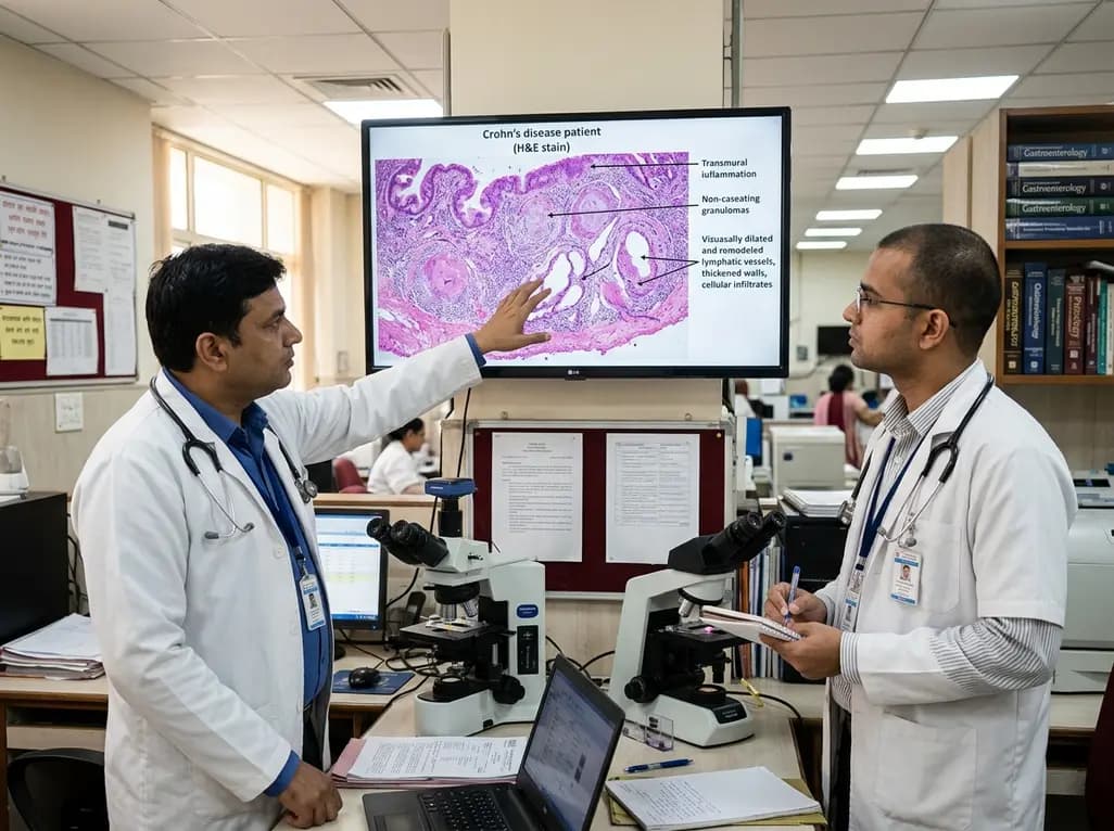
Intestinal lymphatic dysfunction is re-emerging as a fundamental driver of Crohn's disease pathogenesis. Explore how structural remodeling, lymphangitis, and defective clearance shape transmural inflammation and offer novel therapeutic targets.
Today

A school-based intervention using the Health Action Process Approach (HAPA) significantly enhanced adolescents' perceived benefits of consulting healthcare providers and promoted proactive menstrual health behaviors, offering key clinical insights for managing adolescent dysmenorrhea and endometriosis.
Today

A comprehensive analysis of the Translational Maturity Framework for nanomedicine-device combination products, evaluating regulatory complexity, manufacturing hurdles, and clinical readiness across oncology, cardiology, and orthopedics.
Today

A cross-sectional study reveals that sarcopenic obesity independently increases functional impairment and severe disc degeneration in patients with degenerative lumbar spinal stenosis, highlighting the need for body composition assessment.
Today

A landmark dose-response meta-analysis of 8.8 million adults reveals that high ultra-processed food intake significantly elevates risks for cardiovascular disease, cancer, diabetes, depression, and all-cause mortality, highlighting urgent needs for clinical lifestyle interventions.
Today
